|
Afterload
Afterload is the pressure that the heart must work against to eject blood during systole (ventricular contraction). Afterload is proportional to the average arterial pressure. As aortic and pulmonary pressures increase, the afterload increases on the left and right ventricles respectively. Afterload changes to adapt to the continually changing demands on an animal's cardiovascular system. Afterload is proportional to mean systolic blood pressure and is measured in millimeters of mercury (mm Hg). Hemodynamics Afterload is a determinant of cardiac output. Cardiac output is the product of stroke volume and heart rate. Afterload is a determinant of stroke volume (in addition to preload, and strength of myocardial contraction). Following Laplace's law, the tension upon the muscle fibers in the heart wall is the pressure within the ventricle multiplied by the volume within the ventricle divided by the wall thickness (this ratio is the other factor in setting the afterload). The ... [...More Info...] [...Related Items...] OR: [Wikipedia] [Google] [Baidu] |
Heart Systole
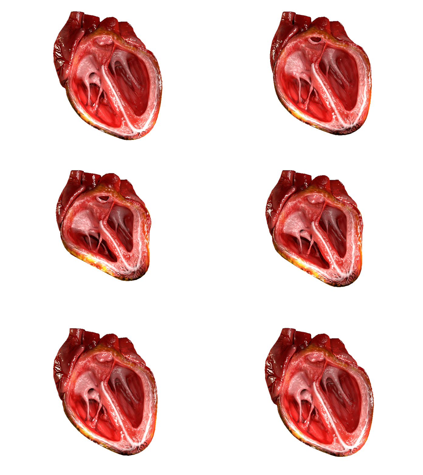
The heart is a muscular Organ (biology), organ found in humans and other animals. This organ pumps blood through the blood vessels. The heart and blood vessels together make the circulatory system. The pumped blood carries oxygen and nutrients to the tissue, while carrying metabolic waste such as carbon dioxide to the lungs. In humans, the heart is approximately the size of a closed fist and is located between the lungs, in the middle compartment of the thorax, chest, called the mediastinum. In humans, the heart is divided into four chambers: upper left and right Atrium (heart), atria and lower left and right Ventricle (heart), ventricles. Commonly, the right atrium and ventricle are referred together as the right heart and their left counterparts as the left heart. In a healthy heart, blood flows one way through the heart due to heart valves, which prevent cardiac regurgitation, backflow. The heart is enclosed in a protective sac, the pericardium, which also contains a sma ... [...More Info...] [...Related Items...] OR: [Wikipedia] [Google] [Baidu] |
Preload (cardiology)
In cardiac physiology, preload is the amount of sarcomere stretch experienced by cardiac muscle cells, called cardiomyocytes, at the end of ventricular filling during diastole. Preload is directly related to ventricular filling. As the relaxed ventricle fills during diastole, the walls are stretched and the length of sarcomeres increases. Sarcomere length can be approximated by the volume of the ventricle because each shape has a conserved surface-area-to-volume ratio. This is useful clinically because measuring the sarcomere length is destructive to heart tissue. It requires cutting out a piece of cardiac muscle to look at the sarcomeres under a microscope. It is currently not possible to directly measure preload in the beating heart of a living animal. Preload is estimated from end-diastolic ventricular pressure and is measured in millimeters of mercury (mmHg). Estimating preload Though not exactly equivalent to the strict definition of ''preload,'' end-diastolic volume ... [...More Info...] [...Related Items...] OR: [Wikipedia] [Google] [Baidu] |
End-diastolic Pressure
In cardiac physiology, preload is the amount of sarcomere stretch experienced by cardiac muscle cells, called cardiomyocytes, at the end of ventricular filling during diastole. Preload is directly related to ventricular filling. As the relaxed ventricle fills during diastole, the walls are stretched and the length of sarcomeres increases. Sarcomere length can be approximated by the volume of the ventricle because each shape has a conserved surface-area-to-volume ratio. This is useful clinically because measuring the sarcomere length is destructive to heart tissue. It requires cutting out a piece of cardiac muscle to look at the sarcomeres under a microscope. It is currently not possible to directly measure preload in the beating heart of a living animal. Preload is estimated from end-diastolic ventricular pressure and is measured in millimeters of mercury (mmHg). Estimating preload Though not exactly equivalent to the strict definition of ''preload,'' end-diastolic volume is be ... [...More Info...] [...Related Items...] OR: [Wikipedia] [Google] [Baidu] |
Millimeters Of Mercury
A millimetre of mercury is a manometric unit of pressure, formerly defined as the extra pressure generated by a column of mercury one millimetre high. Currently, it is defined as exactly , or approximately 1 torr = atmosphere = pascals.Council Directive 80/181/EEC of 20 December 1979 on the approximation of the laws of the Member States relating to units of measurement and on the repeal of Directive 71/354/EEC of the It is denoted mmHg or mm Hg ... [...More Info...] [...Related Items...] OR: [Wikipedia] [Google] [Baidu] |
Radius
In classical geometry, a radius (: radii or radiuses) of a circle or sphere is any of the line segments from its Centre (geometry), center to its perimeter, and in more modern usage, it is also their length. The radius of a regular polygon is the line segment or distance from its center to any of its Vertex (geometry), vertices. The name comes from the Latin ''radius'', meaning ray but also the spoke of a chariot wheel.Definition of Radius at dictionary.reference.com. Accessed on 2009-08-08. The typical abbreviation and mathematical symbol for radius is ''R'' or ''r''. By extension, the diameter ''D'' is defined as twice the radius:Definition of radius at mathwords.com. ... [...More Info...] [...Related Items...] OR: [Wikipedia] [Google] [Baidu] |
Aortic Insufficiency
Aortic regurgitation (AR), also known as aortic insufficiency (AI), is the leaking of the aortic valve of the heart that causes blood to flow in the reverse direction during ventricular diastole, from the aorta into the left ventricle. As a consequence, the cardiac muscle is forced to work harder than normal. Signs and symptoms Symptoms of aortic regurgitation are similar to those of heart failure and include the following: * Dyspnea on exertion * Orthopnea * Paroxysmal nocturnal dyspnea * Palpitations * Angina pectoris * Cyanosis (in acute cases) Causes In terms of the cause of aortic regurgitation, is often due to the aortic root dilation ('' annuloaortic ectasia''), which is idiopathic in over 80% of cases, but otherwise may result from aging, syphilitic aortitis, osteogenesis imperfecta, aortic dissection, Behçet's disease, reactive arthritis and systemic hypertension.Chapter 1: Diseases of the Cardiovascular system > Section: Valvular Heart Disease in: Aortic root d ... [...More Info...] [...Related Items...] OR: [Wikipedia] [Google] [Baidu] |
MmHg
A millimetre of mercury is a manometric unit of pressure, formerly defined as the extra pressure generated by a column of mercury one millimetre high. Currently, it is defined as exactly , or approximately 1 torr = atmosphere = pascals.Council Directive 80/181/EEC of 20 December 1979 on the approximation of the laws of the Member States relating to units of measurement and on the repeal of Directive 71/354/EEC of the It is denoted mmHg or mm Hg ... [...More Info...] [...Related Items...] OR: [Wikipedia] [Google] [Baidu] |
Aorta
The aorta ( ; : aortas or aortae) is the main and largest artery in the human body, originating from the Ventricle (heart), left ventricle of the heart, branching upwards immediately after, and extending down to the abdomen, where it splits at the aortic bifurcation into two smaller arteries (the common iliac artery, common iliac arteries). The aorta distributes Oxygen saturation (medicine), oxygenated blood to all parts of the body through the systemic circulation. Structure Sections In anatomical sources, the aorta is usually divided into sections. One way of classifying a part of the aorta is by anatomical compartment, where the thoracic aorta (or thoracic portion of the aorta) runs from the heart to the thoracic diaphragm, diaphragm. The aorta then continues downward as the abdominal aorta (or abdominal portion of the aorta) from the diaphragm to the aortic bifurcation. Another system divides the aorta with respect to its course and the direction of blood flow. In this s ... [...More Info...] [...Related Items...] OR: [Wikipedia] [Google] [Baidu] |
Aortic Stenosis
Aortic stenosis (AS or AoS) is the narrowing of the exit of the left ventricle of the heart (where the aorta begins), such that problems result. It may occur at the aortic valve as well as above and below this level. It typically gets worse over time. Symptoms often come on gradually with a decreased ability to exercise often occurring first. If heart failure, loss of consciousness, or heart related chest pain occur due to AS the outcomes are worse. Loss of consciousness typically occurs with standing or exercising. Signs of heart failure include shortness of breath especially when lying down, at night, or with exercise, and swelling of the legs. Thickening of the valve without causing obstruction is known as aortic sclerosis. Causes include being born with a bicuspid aortic valve, and rheumatic fever; a normal valve may also harden over the decades due to calcification. A bicuspid aortic valve affects about one to two percent of the population. As of 2014 rheumatic heart ... [...More Info...] [...Related Items...] OR: [Wikipedia] [Google] [Baidu] |
Interventricular Septum
The interventricular septum (IVS, or ventricular septum, or during development septum inferius) is the stout wall separating the ventricle (heart), ventricles, the lower chambers of the heart, from one another. The interventricular septum is directed obliquely backward to the right and curved with the convexity toward the right ventricle; its margins correspond with the anterior interventricular sulcus, anterior and Posterior interventricular sulcus, posterior interventricular sulci. The lower part of the septum, which is the major part, is thick and muscular, and its much smaller upper part is thin and membraneous. During each cardiac cycle the interventricular septum contracts by shortening longitudinally and becoming thicker. Structure The interventricular septum is the stout wall separating the Ventricle (heart), ventricles, the lower chambers of the heart, from one another. The ventricular septum is directed obliquely backward to the right and curved with the convexity t ... [...More Info...] [...Related Items...] OR: [Wikipedia] [Google] [Baidu] |
Pulmonary Hypertension
Pulmonary hypertension (PH or PHTN) is a condition of increased blood pressure in the pulmonary artery, arteries of the lungs. Symptoms include dypsnea, shortness of breath, Syncope (medicine), fainting, tiredness, chest pain, pedal edema, swelling of the legs, and a fast heartbeat. The condition may make it difficult to exercise. Onset is typically gradual. According to the definition at the 6th World Symposium of Pulmonary Hypertension in 2018, a patient is deemed to have pulmonary hypertension if the pulmonary mean arterial pressure is greater than 20mmHg at rest, revised down from a purely arbitrary 25mmHg, and pulmonary vascular resistance (PVR) greater than 3 Wood units. The cause is often unknown. Risk factors include a family history, prior pulmonary embolism (blood clots in the lungs), HIV/AIDS, sickle cell disease, cocaine use, chronic obstructive pulmonary disease, sleep apnea, living at high altitudes, and problems with the mitral valve. The underlying mechanism typ ... [...More Info...] [...Related Items...] OR: [Wikipedia] [Google] [Baidu] |
Work (physics)
In science, work is the energy transferred to or from an Physical object, object via the application of force along a Displacement (vector), displacement. In its simplest form, for a constant force aligned with the direction of motion, the work equals the Product (mathematics), product of the force strength and the distance traveled. A force is said to do ''positive work'' if it has a component in the direction of the displacement of the point of application. A force does ''negative work'' if it has a component opposite to the direction of the displacement at the point of application of the force. For example, when a ball is held above the ground and then dropped, the work done by the gravitational force on the ball as it falls is positive, and is equal to the weight of the ball (a force) multiplied by the distance to the ground (a displacement). If the ball is thrown upwards, the work done by the gravitational force is negative, and is equal to the weight multiplied by the dis ... [...More Info...] [...Related Items...] OR: [Wikipedia] [Google] [Baidu] |


