|
ATX-II
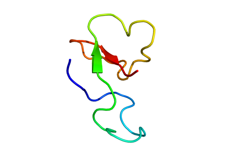
ATX-II, also known as neurotoxin 2, Av2, Anemonia viridis toxin 2 or δ-AITX-Avd1c, is a neurotoxin derived from the venom of the sea anemone ''Anemonia sulcata''. ATX-II slows down the inactivation of different voltage-gated sodium channels, including Nav1.1 and Nav1.2, thus prolonging action potentials. Sources ATX-II is the main component of the venom of Mediterranean snakelocks sea anemone, ''Anemonia sulcata''. ATX-II is produced by the nematocysts in the sea anemone's tentacles and the anemone uses this venom to paralyze its prey.Béress L. Isolation and characterisation of three polypeptides with neurotoxic activity from Anemonia sulcata. FEBS Letters. 1975;50(3):311–4. Etymology "ATX-II" is an acronym for "anemone toxin". Chemistry Structure ATX-II is a protein comprising 47 amino acids crosslinked by three disulfide bridges. The molecular mass of the protein is 4,94 kDa (calculated with ProtParam ExPASy). Family and homology ATX-II belongs to the sea anemon ... [...More Info...] [...Related Items...] OR: [Wikipedia] [Google] [Baidu] |
BcIII
BcIII is a polypeptide sea anemone neurotoxin isolated from ''Bunodosoma caissarum''. It targets the site 3 of voltage-gated sodium channels, thus mainly prolonging the inactivation time course of the channel. Sources BcIII can be isolated from the venom of the sea anemone ''Bunodosoma caissarum''. Chemistry BcIII belongs to Type 1 sea anemone neurotoxins, which target the α-subunits of voltage-gated sodium channels (Nav1.x).Oliveira, J.S., et al., ''BcIV, a new paralyzing peptide obtained from the venom of the sea anemone Bunodosoma caissarum. A comparison with the Na+ channel toxin BcIII.'' Biochim Biophys Acta, 2006. 1764(10): p. 1592-600.Malpezzi, E.L., et al., ''Characterization of peptides in sea anemone venom collected by a novel procedure.'' Toxicon, 1993. 31(7): p. 853-64.Wanke, E., et al., ''Actions of sea anemone type 1 neurotoxins on voltage-gated sodium channel isoforms.'' Toxicon, 2009. 54(8): p. 1102-11. It has 48 amino acids and a molecular mass of 4.976 atomi ... [...More Info...] [...Related Items...] OR: [Wikipedia] [Google] [Baidu] |
Neurotoxin
Neurotoxins are toxins that are destructive to nerve tissue (causing neurotoxicity). Neurotoxins are an extensive class of exogenous chemical neurological insultsSpencer 2000 that can adversely affect function in both developing and mature nervous tissue.Olney 2002 The term can also be used to classify endogenous compounds, which, when abnormally contacted, can prove neurologically toxic. Though neurotoxins are often neurologically destructive, their ability to specifically target neural components is important in the study of nervous systems. Common examples of neurotoxins include lead, ethanol (drinking alcohol), glutamate,Choi 1987 nitric oxide, botulinum toxin (e.g. Botox), tetanus toxin,Simpson 1986 and tetrodotoxin. Some substances such as nitric oxide and glutamate are in fact essential for proper function of the body and only exert neurotoxic effects at excessive concentrations. Neurotoxins inhibit neuron control over ion concentrations across the cell memb ... [...More Info...] [...Related Items...] OR: [Wikipedia] [Google] [Baidu] |
Myocyte
A muscle cell is also known as a myocyte when referring to either a cardiac muscle cell (cardiomyocyte), or a smooth muscle cell as these are both small cells. A skeletal muscle cell is long and threadlike with many nuclei and is called a muscle fiber. Muscle cells (including myocytes and muscle fibers) develop from embryonic precursor cells called myoblasts. Myoblasts fuse to form multinucleated skeletal muscle cells known as syncytia in a process known as myogenesis. Skeletal muscle cells and cardiac muscle cells both contain myofibrils and sarcomeres and form a striated muscle tissue. Cardiac muscle cells form the cardiac muscle in the walls of the heart chambers, and have a single central nucleus. Cardiac muscle cells are joined to neighboring cells by intercalated discs, and when joined in a visible unit they are described as a ''cardiac muscle fiber''. Smooth muscle cells control involuntary movements such as the peristalsis contractions in the esophagus and ... [...More Info...] [...Related Items...] OR: [Wikipedia] [Google] [Baidu] |
Neurotoxins
Neurotoxins are toxins that are destructive to nerve tissue (causing neurotoxicity). Neurotoxins are an extensive class of exogenous chemical neurological insultsSpencer 2000 that can adversely affect function in both developing and mature nervous tissue.Olney 2002 The term can also be used to classify endogenous compounds, which, when abnormally contacted, can prove neurologically toxic. Though neurotoxins are often neurologically destructive, their ability to specifically target neural components is important in the study of nervous systems. Common examples of neurotoxins include lead, ethanol (drinking alcohol), glutamate,Choi 1987 nitric oxide, botulinum toxin (e.g. Botox), tetanus toxin,Simpson 1986 and tetrodotoxin. Some substances such as nitric oxide and glutamate are in fact essential for proper function of the body and only exert neurotoxic effects at excessive concentrations. Neurotoxins inhibit neuron control over ion concentrations across the cell membrane, or commu ... [...More Info...] [...Related Items...] OR: [Wikipedia] [Google] [Baidu] |
Cardiac Muscle
Cardiac muscle (also called heart muscle, myocardium, cardiomyocytes and cardiac myocytes) is one of three types of vertebrate muscle tissues, with the other two being skeletal muscle and smooth muscle. It is an involuntary, striated muscle that constitutes the main tissue of the wall of the heart. The cardiac muscle (myocardium) forms a thick middle layer between the outer layer of the heart wall (the pericardium) and the inner layer (the endocardium), with blood supplied via the coronary circulation. It is composed of individual cardiac muscle cells joined by intercalated discs, and encased by collagen fibers and other substances that form the extracellular matrix. Cardiac muscle contracts in a similar manner to skeletal muscle, although with some important differences. Electrical stimulation in the form of a cardiac action potential triggers the release of calcium from the cell's internal calcium store, the sarcoplasmic reticulum. The rise in calcium causes the c ... [...More Info...] [...Related Items...] OR: [Wikipedia] [Google] [Baidu] |
A Delta Fiber
Group A nerve fibers are one of the three classes of nerve fiber as ''generally classified'' by Erlanger and Gasser. The other two classes are the group B nerve fibers, and the group C nerve fibers. Group A are heavily myelinated, group B are moderately myelinated, and group C are unmyelinated. The other classification is a sensory grouping that uses the terms '' type Ia and type Ib'', '' type II'', ''type III'', and ''type IV'', sensory fibers. Types There are four subdivisions of group A nerve fibers: alpha (α) Aα; beta (β) Aβ; , gamma (γ) Aγ, and delta (δ) Aδ. These subdivisions have different amounts of myelination and axon thickness and therefore transmit signals at different speeds. Larger diameter axons and more myelin insulation lead to faster signal propagation. Group A nerves are found in both motor and sensory pathways. Different sensory receptors are innervated by different types of nerve fibers. Proprioceptors are innervated by type Ia, Ib and II ... [...More Info...] [...Related Items...] OR: [Wikipedia] [Google] [Baidu] |
Myelin
Myelin is a lipid-rich material that surrounds nerve cell axons (the nervous system's "wires") to insulate them and increase the rate at which electrical impulses (called action potentials) are passed along the axon. The myelinated axon can be likened to an electrical wire (the axon) with insulating material (myelin) around it. However, unlike the plastic covering on an electrical wire, myelin does not form a single long sheath over the entire length of the axon. Rather, myelin sheaths the nerve in segments: in general, each axon is encased with multiple long myelinated sections with short gaps in between called nodes of Ranvier. Myelin is formed in the central nervous system (CNS; brain, spinal cord and optic nerve) by glial cells called oligodendrocytes and in the peripheral nervous system (PNS) by glial cells called Schwann cells. In the CNS, axons carry electrical signals from one nerve cell body to another. In the PNS, axons carry signals to muscles and glands or from sen ... [...More Info...] [...Related Items...] OR: [Wikipedia] [Google] [Baidu] |
Crayfish
Crayfish are freshwater crustaceans belonging to the clade Astacidea, which also contains lobsters. In some locations, they are also known as crawfish, craydids, crawdaddies, crawdads, freshwater lobsters, mountain lobsters, rock lobsters, mudbugs, baybugs or yabbies. Taxonomically, they are members of the superfamilies Astacoidea and Parastacoidea. They breathe through feather-like gills. Some species are found in brooks and streams, where fresh water is running, while others thrive in swamps, ditches, and paddy fields. Most crayfish cannot tolerate polluted water, although some species, such as '' Procambarus clarkii'', are hardier. Crayfish feed on animals and plants, either living or decomposing, and detritus. The term "crayfish" is applied to saltwater species in some countries. Terminology The name "crayfish" comes from the Old French word ' ( Modern French '). The word has been modified to "crayfish" by association with "fish" (folk etymology). The largely ... [...More Info...] [...Related Items...] OR: [Wikipedia] [Google] [Baidu] |
Dissociation Constant
In chemistry, biochemistry, and pharmacology, a dissociation constant (K_D) is a specific type of equilibrium constant that measures the propensity of a larger object to separate (dissociate) reversibly into smaller components, as when a complex falls apart into its component molecules, or when a salt splits up into its component ions. The dissociation constant is the inverse of the association constant. In the special case of salts, the dissociation constant can also be called an ionization constant. For a general reaction: : A_\mathit B_\mathit \mathit A + \mathit B in which a complex \ce_x \ce_y breaks down into ''x'' A subunits and ''y'' B subunits, the dissociation constant is defined as : K_D = \frac where and ''x'' B''y''are the equilibrium concentrations of A, B, and the complex A''x'' B''y'', respectively. One reason for the popularity of the dissociation constant in biochemistry and pharmacology is that in the frequently en ... [...More Info...] [...Related Items...] OR: [Wikipedia] [Google] [Baidu] |
EC50
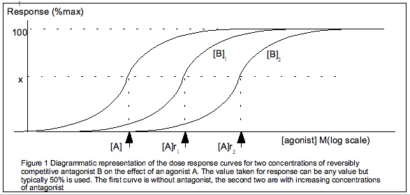
] Half maximal effective concentration (EC50) is a measure of the concentration of a drug, antibody or toxicant which induces a Stimulus%E2%80%93response_model, response halfway between the baseline and maximum after a specified exposure time. More simply, EC50 can be defined as the ''concentration required to obtain a 50% ..effect'' and may be also written as sub>50. It is commonly used as a measure of a drug's potency, although the use of EC50 is preferred over that of 'potency', which has been criticised for its vagueness. EC50 is a measure of concentration, expressed in molar units (M), where 1 M is equivalent to 1 mol/ L. The EC50 of a ''graded'' dose response curve therefore represents the concentration of a compound where 50% of its maximal effect is observed. The EC50 of a ''quantal'' dose response curve represents the concentration of a compound where 50% of the population exhibit a response, after a specified exposure duration. For clarification, a gra ... [...More Info...] [...Related Items...] OR: [Wikipedia] [Google] [Baidu] |
Sodium Channel
Sodium channels are integral membrane proteins that form ion channels, conducting sodium ions (Na+) through a cell's membrane. They belong to the superfamily of cation channels and can be classified according to the trigger that opens the channel for such ions, i.e. either a voltage-change ("voltage-gated", "voltage-sensitive", or "voltage-dependent" sodium channel; also called "VGSCs" or "Nav channel") or a binding of a substance (a ligand) to the channel ( ligand-gated sodium channels). In excitable cells such as neurons, myocytes, and certain types of glia, sodium channels are responsible for the rising phase of action potentials. These channels go through three different states called resting, active and inactive states. Even though the resting and inactive states would not allow the ions to flow through the channels the difference exists with respect to their structural conformation. Selectivity Sodium channels are highly selective for the transport of ions across cell memb ... [...More Info...] [...Related Items...] OR: [Wikipedia] [Google] [Baidu] |


