|
Lingual Nerve
The lingual nerve carries sensory innervation from the anterior two-thirds of the tongue. It contains fibres from both the mandibular division of the trigeminal nerve (CN V3 ) and from the facial nerve (CN VII). The fibres from the trigeminal nerve are for touch, pain and temperature (general sensation), and the ones from the facial nerve are for taste (special sensation). Structure The lingual nerve lies at first beneath the lateral pterygoid muscle, medial to and in front of the inferior alveolar nerve, and is occasionally joined to this nerve by a branch which may cross the internal maxillary artery. The chorda tympani (a branch of the facial nerve, CN VII) joins it at an acute angle here, carrying taste fibers from the anterior two-thirds of the tongue and parasympathetic fibers to the submandibular ganglion. The nerve then passes between the medial pterygoid muscle and the ramus of the mandible, and crosses obliquely to the side of the tongue beneath the constrictor pha ... [...More Info...] [...Related Items...] OR: [Wikipedia] [Google] [Baidu] |
Maxillary Nerve
In neuroanatomy, the maxillary nerve (V) is one of the three branches or divisions of the trigeminal nerve, the fifth (CN V) cranial nerve. It comprises the principal functions of sensation from the maxilla, nasal cavity, sinuses, the palate and subsequently that of the mid-face, and is intermediate, both in position and size, between the ophthalmic nerve and the mandibular nerve.Illustrated Anatomy of the Head and Neck, Fehrenbach and Herring, Elsevier, 2012, page 180 Structure It begins at the middle of the trigeminal ganglion as a flattened plexiform band then it passes through the lateral wall of the cavernous sinus. It leaves the skull through the foramen rotundum, where it becomes more cylindrical in form, and firmer in texture. After leaving foramen rotundum it gives two branches to the pterygopalatine ganglion. It then crosses the pterygopalatine fossa, inclines lateralward on the back of the maxilla, and enters the orbit through the inferior orbital fissure. It t ... [...More Info...] [...Related Items...] OR: [Wikipedia] [Google] [Baidu] |
Styloglossus
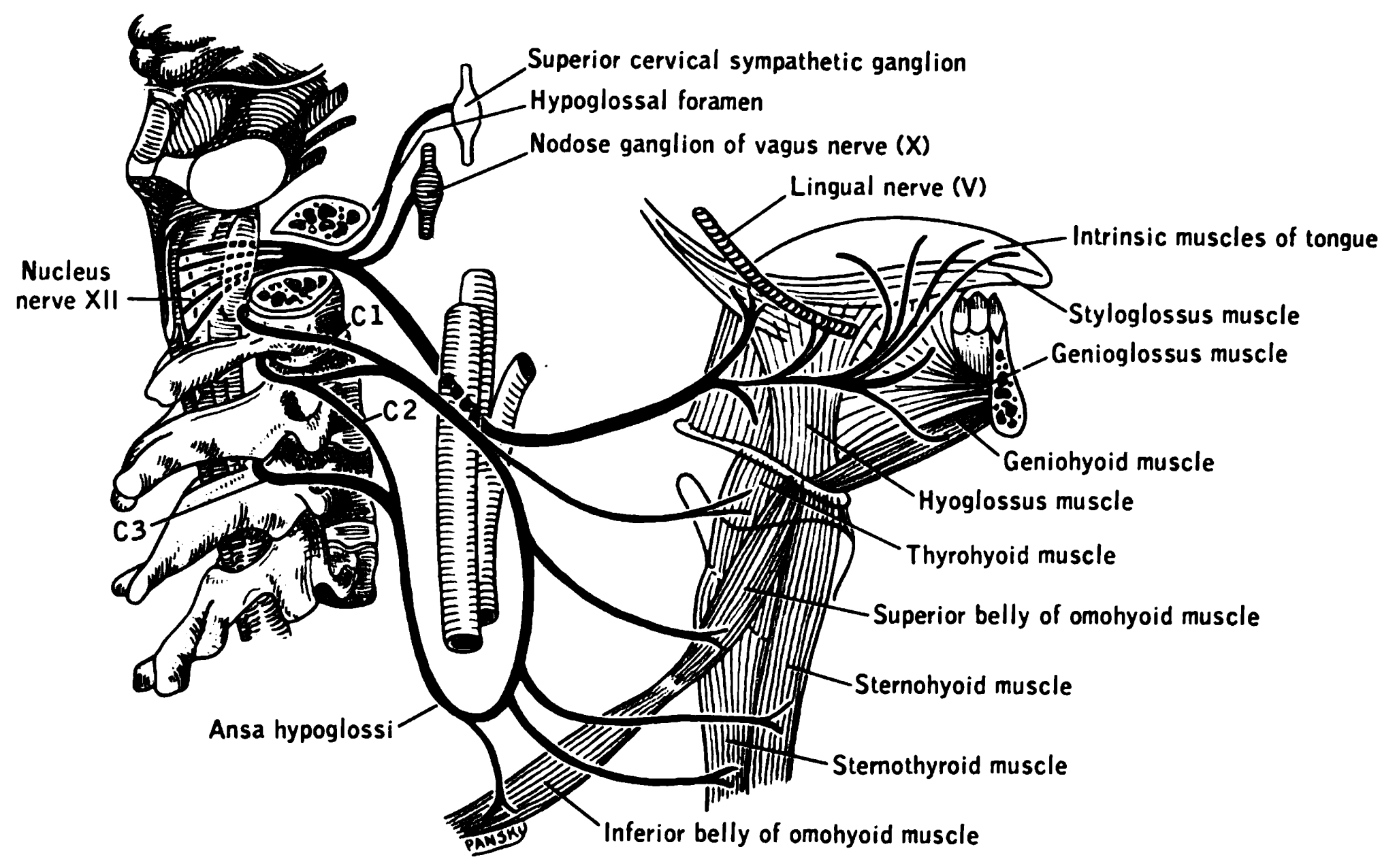
The styloglossus, the shortest and smallest of the three styloid muscles, arises from the anterior and lateral surfaces of the styloid process near its apex, and from the stylomandibular ligament. Passing inferiorly and anteriorly between the internal and external carotid arteries, it divides upon the side of the tongue near its dorsal surface, blending with the fibers of the longitudinalis inferior in front of the hyoglossus; the other, oblique, overlaps the Hyoglossus and decussates with its fibers. Innervation The styloglossus is innervated by the hypoglossal nerve (CN XII) like all muscles of the tongue except palatoglossus which is innervated by the pharyngeal plexus of vagus nerve The pharyngeal plexus is a network of nerve fibers innervating most of the palate and pharynx. (The larynx, which is innervated by the superior and recurrent laryngeal nerves from vagus nerve (CN X), is not included.) It is located on the sur ... (CN X). Action The styloglossus draws up ... [...More Info...] [...Related Items...] OR: [Wikipedia] [Google] [Baidu] |
Mandibular Nerve
In neuroanatomy, the mandibular nerve (V) is the largest of the three divisions of the trigeminal nerve, the fifth cranial nerve (CN V). Unlike the other divisions of the trigeminal nerve ( ophthalmic nerve, maxillary nerve) which contain only afferent fibers, the mandibular nerve contains both afferent and efferent fibers. These nerve fibers innervate structures of the lower jaw and face, such as the tongue, lower lip, and chin. The mandibular nerve also innervates the muscles of mastication. Structure The large sensory root emerges from the lateral part of the trigeminal ganglion and exits the cranial cavity through the foramen ovale. Portio minor, the small motor root of the trigeminal nerve, passes under the trigeminal ganglion and through the foramen ovale to unite with the sensory root just outside the skull. The mandibular nerve immediately passes between tensor veli palatini, which is medial, and lateral pterygoid, which is lateral, and gives off a meningeal ... [...More Info...] [...Related Items...] OR: [Wikipedia] [Google] [Baidu] |
Lingual Branches Of Hypoglossal Nerve
The hypoglossal nerve, also known as the twelfth cranial nerve, cranial nerve XII, or simply CN XII, is a cranial nerve that innervates all the extrinsic and intrinsic muscles of the tongue except for the palatoglossus, which is innervated by the vagus nerve. CN XII is a nerve with a solely motor function. The nerve arises from the hypoglossal nucleus in the medulla as a number of small rootlets, passes through the hypoglossal canal and down through the neck, and eventually passes up again over the tongue muscles it supplies into the tongue. The nerve is involved in controlling tongue movements required for speech and swallowing, including sticking out the tongue and moving it from side to side. Damage to the nerve or the neural pathways which control it can affect the ability of the tongue to move and its appearance, with the most common sources of damage being injury from trauma or surgery, and motor neuron disease. The first recorded description of the nerve is by Heroph ... [...More Info...] [...Related Items...] OR: [Wikipedia] [Google] [Baidu] |
Wisdom Tooth
A third molar, commonly called wisdom tooth, is one of the three molars per quadrant of the human dentition. It is the most posterior of the three. The age at which wisdom teeth come through ( erupt) is variable, but this generally occurs between late teens and early twenties. Most adults have four wisdom teeth, one in each of the four quadrants, but it is possible to have none, fewer, or more, in which case the extras are called supernumerary teeth. Wisdom teeth may get stuck ( impacted) against other teeth if there is not enough space for them to come through normally. Impacted wisdom teeth are still sometimes removed for orthodontic treatment, believing that they move the other teeth and cause crowding, though this is not held anymore as true. Impacted wisdom teeth may suffer from tooth decay if oral hygiene becomes more difficult. Wisdom teeth which are partially erupted through the gum may also cause inflammation and infection in the surrounding gum tissues, termed pericor ... [...More Info...] [...Related Items...] OR: [Wikipedia] [Google] [Baidu] |
Parasympathetic
The parasympathetic nervous system (PSNS) is one of the three divisions of the autonomic nervous system, the others being the sympathetic nervous system and the enteric nervous system. The enteric nervous system is sometimes considered part of the autonomic nervous system, and sometimes considered an independent system. The autonomic nervous system is responsible for regulating the body's unconscious actions. The parasympathetic system is responsible for stimulation of "rest-and-digest" or "feed and breed" activities that occur when the body is at rest, especially after eating, including sexual arousal, salivation, lacrimation (tears), urination, digestion, and defecation. Its action is described as being complementary to that of the sympathetic nervous system, which is responsible for stimulating activities associated with the fight-or-flight response. Nerve fibres of the parasympathetic nervous system arise from the central nervous system. Specific nerves include sever ... [...More Info...] [...Related Items...] OR: [Wikipedia] [Google] [Baidu] |
Chorda Tympani Nerve
The chorda tympani is a branch of the facial nerve that originates from the taste buds in the front of the tongue, runs through the middle ear, and carries taste messages to the brain. It joins the facial nerve (cranial nerve VII) inside the facial canal, at the level where the facial nerve exits the skull via the stylomastoid foramen, but exits through the petrotympanic fissure and descends in the infratemporal fossa. The chorda tympani is part of one of three cranial nerves that are involved in taste. The taste system involves a complicated feedback loop, with each nerve acting to inhibit the signals of other nerves. Structure The chorda tympani exits the cranial cavity through the internal acoustic meatus along with the facial nerve, then it travels through the middle ear, where it runs from posterior to anterior across the tympanic membrane. It passes between the malleus and the incus, on the medial surface of the neck of the malleus. The nerve continues through the pet ... [...More Info...] [...Related Items...] OR: [Wikipedia] [Google] [Baidu] |
General Somatic Afferent
The general somatic afferent fibers (GSA, or somatic sensory fibers) afferent fibers arise from neurons in sensory ganglia and are found in all the spinal nerves, except occasionally the first cervical, and conduct impulses of pain, touch and temperature from the surface of the body through the dorsal roots to the spinal cord and impulses of muscle sense, tendon sense and joint sense from the deeper structures. See also * Afferent nerve A sensory nerve, or afferent nerve, is a general anatomic term for a nerve which contains predominantly somatic afferent nerve fibers. Afferent nerve fibers in a sensory nerve carry sensory information toward the central nervous system (CNS) fro ... References Spinal cord {{Portal bar, Anatomy ... [...More Info...] [...Related Items...] OR: [Wikipedia] [Google] [Baidu] |
Mucous Membrane
A mucous membrane or mucosa is a membrane that lines various cavities in the body of an organism and covers the surface of internal organs. It consists of one or more layers of epithelial cells overlying a layer of loose connective tissue. It is mostly of endodermal origin and is continuous with the skin at body openings such as the eyes, eyelids, ears, inside the nose, inside the mouth, lips, the genital areas, the urethral opening and the anus. Some mucous membranes secrete mucus, a thick protective fluid. The function of the membrane is to stop pathogens and dirt from entering the body and to prevent bodily tissues from becoming dehydrated. Structure The mucosa is composed of one or more layers of epithelial cells that secrete mucus, and an underlying lamina propria of loose connective tissue. The type of cells and type of mucus secreted vary from organ to organ and each can differ along a given tract. Mucous membranes line the digestive, respiratory and reproductive ... [...More Info...] [...Related Items...] OR: [Wikipedia] [Google] [Baidu] |
Submandibular Gland
The paired submandibular glands (historically known as submaxillary glands) are major salivary glands located beneath the floor of the mouth. They each weigh about 15 grams and contribute some 60–67% of unstimulated saliva secretion; on stimulation their contribution decreases in proportion as the parotid secretion rises to 50%. The average length of the normal human submandibular salivary gland is approximately 27mm, while the average width is approximately 14.3mm. Structure Lying superior to the digastric muscles, each submandibular gland is divided into superficial and deep lobes, which are separated by the mylohyoid muscle: * The superficial lobe comprises most of the gland, with the mylohyoid muscle runs under it * The deep lobe is the smaller part Secretions are delivered into the submandibular duct on the deep portion after which they hook around the posterior edge of the mylohyoid muscle and proceed on the superior surface laterally. The excretory ducts are then cro ... [...More Info...] [...Related Items...] OR: [Wikipedia] [Google] [Baidu] |
Hyoglossus
The hyoglossus, thin and quadrilateral, arises from the side of the body and from the whole length of the greater cornu of the hyoid bone, and passes almost vertically upward to enter the side of the tongue, between the styloglossus and the inferior longitudinal muscle of the tongue. It forms a part of the floor of submandibular triangle. Structure The fibers arising from the body of the hyoid bone overlap those from the greater cornu. Structures that are medial/deep to the hyoglossus are the glossopharyngeal nerve (cranial nerve 9), the stylohyoid ligament and the lingual artery and lingual vein. The lingual vein passes medial to the hyoglossus, and the lingual artery passes deep to the hyoglossus. Laterally, in between the hyoglossus muscle and the mylohyoid muscle lay several important structures (from upper to lower): sublingual gland, submandibular duct, lingual nerve, vena comitans of hypoglossal nerve, and the hypoglossal nerve. Note, posteriorly, the lingual ne ... [...More Info...] [...Related Items...] OR: [Wikipedia] [Google] [Baidu] |
Constrictor Pharyngis Superior
The superior pharyngeal constrictor muscle is a muscle in the pharynx. It is the highest located muscle of the three pharyngeal constrictors. The muscle is a quadrilateral muscle, thinner and paler than the inferior pharyngeal constrictor muscle and middle pharyngeal constrictor muscle. The muscle is divided into four parts: A pterygopharyngeal, buccopharyngeal, mylopharyngeal and a glossopharyngeal part. Origin and insertion The four parts of this muscle arise from: - the lower third of the posterior margin of the medial pterygoid plate and its hamulus (Pterygopharyngeal part) - from the pterygomandibular raphe (Buccopharyngeal part) - from the alveolar process of the mandible above the posterior end of the mylohyoid line (Mylopharyngeal part) - and by a few fibers from the side of the tongue (Glossopharyngeal part) The fibers curve backward to be inserted into the median raphe, being also prolonged by means of an aponeurosis to the pharyngeal spine on the basilar part o ... [...More Info...] [...Related Items...] OR: [Wikipedia] [Google] [Baidu] |


