synovial joints on:
[Wikipedia]
[Google]
[Amazon]
A synovial joint, also known as diarthrosis, joins bones or cartilage with a fibrous joint capsule that is continuous with the
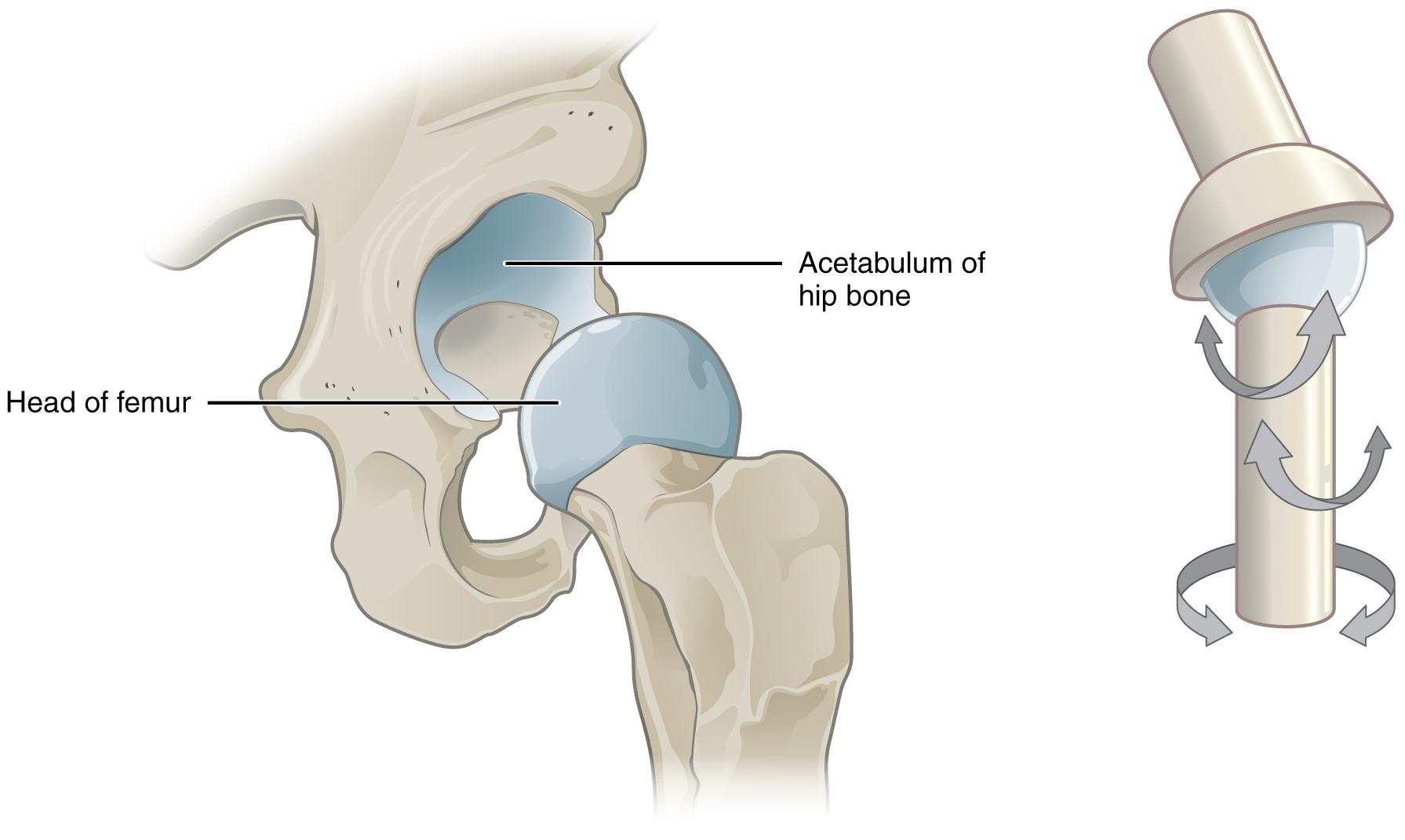
 A multiaxial joint (polyaxial joint or triaxial joint) is a synovial joint that allows for several directions of movement. In the human body, the shoulder and
Tortora & Derrickson () ''Principles of Anatomy & Physiology'' (12th ed.). Wiley & Sons
Rogers, Kara (2010) ''Bone and Muscle: Structure, Force, and Motion'
p.157
/ref> Sharkey, John (2008) ''The Concise Book of Neuromuscular Therapy'
p.33
/ref> Moini (2011) ''Introduction to Pathology for the Physical Therapist Assistant'
pp.231-2
/ref> Bruce Abernethy (2005) ''The Biophysical Foundations Of Human Movement'' pp.23
331
/ref>
periosteum
The periosteum is a membrane that covers the outer surface of all bones, except at the articular surfaces (i.e. the parts within a joint space) of long bones. (At the joints of long bones the bone's outer surface is lined with "articular cartila ...
of the joined bones, constitutes the outer boundary of a synovial cavity, and surrounds the bones' articulating surfaces. This joint unites long bones and permits free bone movement and greater mobility. The synovial cavity/joint is filled with synovial fluid
Synovial fluid, also called synovia, elp 1/sup> is a viscous, non-Newtonian fluid found in the cavities of synovial joints. With its egg white–like consistency, the principal role of synovial fluid is to reduce friction between the articul ...
. The joint capsule is made up of an outer layer of fibrous membrane, which keeps the bones together structurally, and an inner layer, the synovial membrane
Synovial () may refer to:
* Synovial fluid
* Synovial joint
A synovial joint, also known as diarthrosis, joins bones or cartilage with a fibrous joint capsule that is continuous with the periosteum of the joined bones, constitutes the outer bou ...
, which seals in the synovial fluid.
They are the most common and most movable type of joint
A joint or articulation (or articular surface) is the connection made between bones, ossicles, or other hard structures in the body which link an animal's skeletal system into a functional whole.Saladin, Ken. Anatomy & Physiology. 7th ed. McGraw- ...
in the body. As with most other joints, synovial joints achieve movement at the point of contact of the articulating bone
A bone is a rigid organ that constitutes part of the skeleton in most vertebrate animals. Bones protect the various other organs of the body, produce red and white blood cells, store minerals, provide structure and support for the body, ...
s. They originated 400 million years ago in the first jawed vertebrates.
Structure
Synovial joints contain the following structures: * Synovial cavity: all diarthroses have the characteristic space between the bones that is filled withsynovial fluid
Synovial fluid, also called synovia, elp 1/sup> is a viscous, non-Newtonian fluid found in the cavities of synovial joints. With its egg white–like consistency, the principal role of synovial fluid is to reduce friction between the articul ...
.
* Joint capsule: the fibrous capsule, continuous with the periosteum of articulating bones, surrounds the diarthrosis and unites the articulating bones; the joint capsule consists of two layers - (1) the outer fibrous membrane that may contain ligaments and (2) the inner synovial membrane
Synovial () may refer to:
* Synovial fluid
* Synovial joint
A synovial joint, also known as diarthrosis, joins bones or cartilage with a fibrous joint capsule that is continuous with the periosteum of the joined bones, constitutes the outer bou ...
that secretes the lubricating, shock absorbing, and joint-nourishing synovial fluid; the joint capsule is highly innervated, but without blood and lymph vessels, and receives nutrition from the surrounding blood supply via either diffusion
Diffusion is the net movement of anything (for example, atoms, ions, molecules, energy) generally from a region of higher concentration to a region of lower concentration. Diffusion is driven by a gradient in Gibbs free energy or chemical p ...
(slow), or via convection
Convection is single or Multiphase flow, multiphase fluid flow that occurs Spontaneous process, spontaneously through the combined effects of material property heterogeneity and body forces on a fluid, most commonly density and gravity (see buoy ...
(fast, more efficient), induced through exercise.
* Articular cartilage: the bones of a synovial joint are covered by a layer of hyaline cartilage that lines the epiphyses of the joint end of the bone with a smooth, slippery surface that prevents adhesion
Adhesion is the tendency of dissimilar particles or interface (matter), surfaces to cling to one another. (Cohesion (chemistry), Cohesion refers to the tendency of similar or identical particles and surfaces to cling to one another.)
The ...
; articular cartilage functions to absorb shock and reduce friction
Friction is the force resisting the relative motion of solid surfaces, fluid layers, and material elements sliding against each other. Types of friction include dry, fluid, lubricated, skin, and internal -- an incomplete list. The study of t ...
during movement.
Many, but not all, synovial joints also contain additional structures:
* Articular discs or menisci - the fibrocartilage
Fibrocartilage consists of a mixture of white fibrous tissue and cartilaginous tissue in various proportions. It owes its inflexibility and toughness to the former of these constituents, and its elasticity to the latter. It is the only type of ...
pads between opposing surfaces in a joint
* Articular fat pads - adipose tissue
Adipose tissue (also known as body fat or simply fat) is a loose connective tissue composed mostly of adipocytes. It also contains the stromal vascular fraction (SVF) of cells including preadipocytes, fibroblasts, Blood vessel, vascular endothel ...
pads that protect the articular cartilage, as seen in the infrapatellar fat pad in the knee
* Tendons - cords of dense regular connective tissue
Dense regular connective tissue (DRCT) provides connection between different tissues in the human body. The collagen fibers in dense regular connective tissue are bundled in a parallel fashion. DRCT is divided into white fibrous connective tissue ...
composed of parallel bundles of collagen fibers
Type I collagen is the most abundant collagen of the human body, consisting of around 90% of the body's total collagen in vertebrates. Due to this, it is also the most abundant protein type found in all vertebrates. Type I forms large, eosinoph ...
* Accessory ligaments (extracapsular and intracapsular) - the fibers of some fibrous membranes are arranged in parallel bundles of dense regular connective tissue that are highly adapted for resisting strains to prevent extreme movements that may damage the articulation
* Bursae - sac-like structures that are situated strategically to alleviate friction in some joints (shoulder and knee) that are filled with fluid similar to synovial fluid
The bone surrounding the joint on the proximal side is sometimes called the ''plafond'' (French word for ceiling), especially in the talocrural joint. Damage to this structure is referred to as a Gosselin fracture.
Blood supply
The blood supply of a synovial joint is derived from the arteries sharing in the anastomosis around the joint.Types
There are seven types of synovial joints. Some are relatively immobile, therefore more stable. Others have multiple degrees of freedom, but at the expense of greater risk of injury. In ascending order of mobility, they are:Multiaxial joints
 A multiaxial joint (polyaxial joint or triaxial joint) is a synovial joint that allows for several directions of movement. In the human body, the shoulder and
 hip joint
In vertebrate anatomy, the hip, or coxaLatin ''coxa'' was used by Celsus in the sense "hip", but by Pliny the Elder in the sense "hip bone" (Diab, p 77) (: ''coxae'') in medical terminology, refers to either an anatomical region or a joint o ...
s are multiaxial joints. They allow the upper or lower limb to move in an anterior-posterior direction and a medial-lateral direction. In addition, the limb can also be rotated around its long axis. This third movement results in rotation of the limb so that its anterior surface is moved either toward or away from the midline of the body.
Function
The movements possible with synovial joints are: * abduction: movement away from the mid-line of the body *adduction
Motion, the process of movement, is described using specific anatomical terms. Motion includes movement of organs, joints, limbs, and specific sections of the body. The terminology used describes this motion according to its direction relativ ...
: movement toward the mid-line of the body
* extension: straightening limbs at a joint
* flexion
Motion, the process of movement, is described using specific anatomical terminology, anatomical terms. Motion includes movement of Organ (anatomy), organs, joints, Limb (anatomy), limbs, and specific sections of the body. The terminology used de ...
: bending the limbs at a joint
* rotation
Rotation or rotational/rotary motion is the circular movement of an object around a central line, known as an ''axis of rotation''. A plane figure can rotate in either a clockwise or counterclockwise sense around a perpendicular axis intersect ...
: a circular movement around a fixed point
Clinical significance
The ''joint space'' equals the distance between the involved bones of the joint. A ''joint space narrowing'' is a sign of either (or both)osteoarthritis
Osteoarthritis is a type of degenerative joint disease that results from breakdown of articular cartilage, joint cartilage and underlying bone. A form of arthritis, it is believed to be the fourth leading cause of disability in the world, affect ...
and inflammatory degeneration. The normal joint space is at least 2 mm in the hip (at the superior acetabulum
The acetabulum (; : acetabula), also called the cotyloid cavity, is a wikt:concave, concave surface of the pelvis. The femur head, head of the femur meets with the pelvis at the acetabulum, forming the Hip#Articulation, hip joint.
Structure
The ...
), at least 3 mm in the knee
In humans and other primates, the knee joins the thigh with the leg and consists of two joints: one between the femur and tibia (tibiofemoral joint), and one between the femur and patella (patellofemoral joint). It is the largest joint in the hu ...
, and 4–5 mm in the shoulder joint. For the temporomandibular joint
In anatomy, the temporomandibular joints (TMJ) are the two joints connecting the jawbone to the skull. It is a bilateral Synovial joint, synovial articulation between the temporal bone of the skull above and the condylar process of mandible be ...
, a joint space of between 1.5 and 4 mm is regarded as normal. Joint space narrowing is therefore a component of several radiographic classifications of osteoarthritis.
In rheumatoid arthritis
Rheumatoid arthritis (RA) is a long-term autoimmune disorder that primarily affects synovial joint, joints. It typically results in warm, swollen, and painful joints. Pain and stiffness often worsen following rest. Most commonly, the wrist and h ...
, the clinical manifestations are primarily synovial inflammation and joint damage. The fibroblast-like synoviocytes, highly specialized mesenchymal cells found in the synovial membrane
Synovial () may refer to:
* Synovial fluid
* Synovial joint
A synovial joint, also known as diarthrosis, joins bones or cartilage with a fibrous joint capsule that is continuous with the periosteum of the joined bones, constitutes the outer bou ...
, have an active and prominent role in the pathogenic processes in the rheumatic joints. Therapies that target these cells are emerging as promising therapeutic tools, raising hope for future applications in rheumatoid arthritis.
Evolutionary origins
Synovial joints has been found in earliest jawed vertebrates ( gnathostomes) 400 million years ago during theSilurian
The Silurian ( ) is a geologic period and system spanning 23.5 million years from the end of the Ordovician Period, at million years ago ( Mya), to the beginning of the Devonian Period, Mya. The Silurian is the third and shortest period of t ...
and Devonian
The Devonian ( ) is a period (geology), geologic period and system (stratigraphy), system of the Paleozoic era (geology), era during the Phanerozoic eon (geology), eon, spanning 60.3 million years from the end of the preceding Silurian per ...
. This finding overturns an earlier view that these joints first evolved in early tetrapods
A tetrapod (; from Ancient Greek τετρα- ''(tetra-)'' 'four' and πούς ''(poús)'' 'foot') is any four- limbed vertebrate animal of the clade Tetrapoda (). Tetrapods include all extant and extinct amphibians and amniotes, with the lat ...
for terrestrial locomotion
Terrestrial locomotion has evolution, evolved as animals adapted from ecoregion#Marine, aquatic to ecoregion#Terrestrial, terrestrial environments. Animal locomotion, Locomotion on land raises different problems than that in water, with reduced f ...
. Comparative studies find that synovial joints in all major groups of jawed vertebrates, including cartilaginous fishes (sharks, skates, and rays), bony fishes, and tetrapods. They are however absent in jawless vertebrates
Agnatha (; ) or jawless fish is a paraphyletic infraphylum of animals in the subphylum Vertebrata of the phylum Chordata, characterized by the lack of jaws. The group consists of both extant taxon, living (Cyclostomi, cyclostomes such as hagfish ...
such as lampreys and hagfish
Hagfish, of the Class (biology), class Myxini (also known as Hyperotreti) and Order (biology), order Myxiniformes , are eel-shaped Agnatha, jawless fish (occasionally called slime eels). Hagfish are the only known living Animal, animals that h ...
. Cartilaginous fishes have true synovial joints with clear synovial cavities, articular cartilage lined by flattened chondrocytes, and express key developmental signaling molecules including growth differentiation factor-5 (Gdf5) and β-catenin, and require muscle contraction for proper joint cavitation. In contrast, cyclostomes have joints filled with tissue rather than fluid-filled cavities, with proteoglycans uniformly distributed across cartilages. Fossil evidence finds jawless osteostracans had pectoral fin connections filled with canals incompatible with fluid-filled joint cavities, while early jawed placoderms have reciprocally articulating surfaces separated by joint cavities. Synovial joints, it have been suggested, arose due to the high mechanical loads associated with predation and feeding and as a result allowed for the evolution of the complex skeletons of modern jawed vertebrates.
References
p.157
/ref> Sharkey, John (2008) ''The Concise Book of Neuromuscular Therapy'
p.33
/ref> Moini (2011) ''Introduction to Pathology for the Physical Therapist Assistant'
pp.231-2
/ref> Bruce Abernethy (2005) ''The Biophysical Foundations Of Human Movement'' pp.23
331
/ref>
Sources
{{DEFAULTSORT:Synovial Joint Joints