|
Fibrocartilage
Fibrocartilage consists of a mixture of white fibrous tissue and cartilaginous tissue in various proportions. It owes its inflexibility and toughness to the former of these constituents, and its elasticity to the latter. It is the only type of cartilage that contains type I collagen in addition to the normal type II. Structure The extracellular matrix of fibrocartilage is mainly made from type I collagen secreted by chondroblasts. Locations of fibrocartilage in the human body * secondary cartilaginous joints: ** pubic symphysis ** annulus fibrosis of intervertebral discs ** manubriosternal joint * glenoid labrum of shoulder joint * acetabular labrum of hip joint * medial and lateral menisci of the knee joint * location where tendons and ligaments attach to bone * triangular fibrocartilage complex (UTFCC) Function Repair If hyaline cartilage is torn all the way down to the bone, the blood supply from inside the bone is sometimes enough to start some heal ... [...More Info...] [...Related Items...] OR: [Wikipedia] [Google] [Baidu] [Amazon] |
Pubic Symphysis
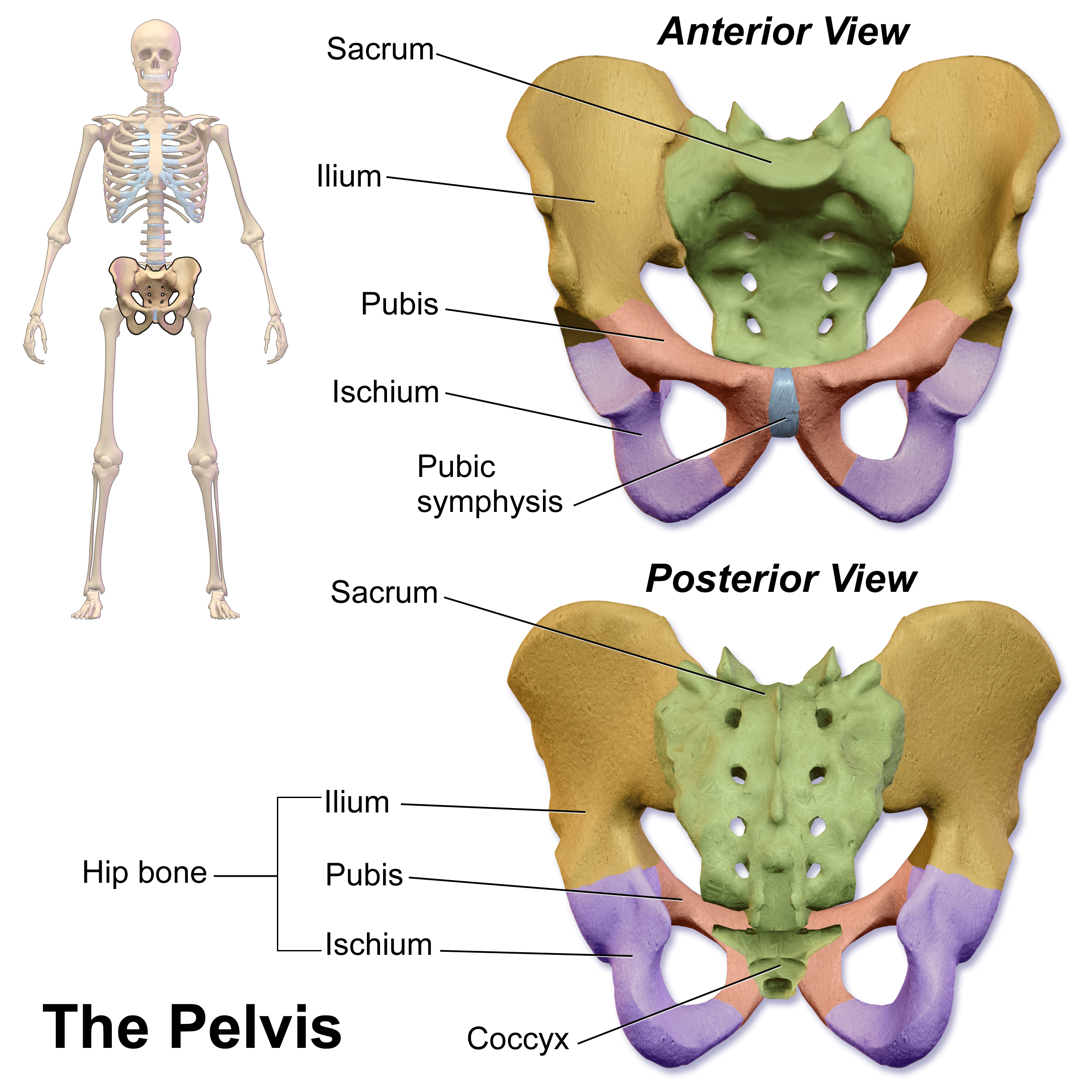
The pubic symphysis (: symphyses) is a secondary cartilaginous joint between the left and right superior rami of the pubis of the hip bones. It is in front of and below the urinary bladder. In males, the suspensory ligament of the penis attaches to the pubic symphysis. In females, the pubic symphysis is attached to the suspensory ligament of the clitoris. In most adults, it can be moved roughly 2 mm and with 1 degree rotation. This increases for women at the time of childbirth. The name comes from the Greek word ''symphysis'', meaning 'growing together'. Structure The pubic symphysis is a nonsynovial amphiarthrodial joint. The width of the pubic symphysis at the front is 3–5 mm greater than its width at the back. This joint is connected by fibrocartilage and may contain a fluid-filled cavity; the center is avascular, possibly due to the nature of the compressive forces passing through this joint, which may lead to harmful vascular disease. The ends of both pubi ... [...More Info...] [...Related Items...] OR: [Wikipedia] [Google] [Baidu] [Amazon] |
Meniscus (anatomy)
A meniscus (: menisci or meniscuses) is a crescent-shaped fibrocartilage, fibrocartilaginous anatomy, anatomical structure that, in contrast to an articular disc, only partly divides a synovial joint#Structure, joint cavity.Platzer (2004), p 208 In humans, they are present in the knee, wrist, acromioclavicular joint, acromioclavicular, sternoclavicular joint, sternoclavicular, and temporomandibular joints; in other animals they may be present in other joints. Generally, the term "meniscus" is used to refer to the cartilage of the knee, either to the lateral meniscus, lateral or medial meniscus. Both are Cartilaginous#Fibrocartilage, cartilaginous tissues that provide structural integrity to the knee when it undergoes tension (mechanics), tension and torsion (mechanics), torsion. The menisci are also known as "semi-lunar" cartilages, referring to their half-moon, crescent shape. The term "meniscus" is from the Ancient Greek word (), meaning "crescent". Structure The menisci of t ... [...More Info...] [...Related Items...] OR: [Wikipedia] [Google] [Baidu] [Amazon] |
List Of Distinct Cell Types In The Adult Human Body
The list of human cell types provides an enumeration and description of the various specialized cells found within the human body, highlighting their distinct functions, characteristics, and contributions to overall physiological processes. Cells may be classified by their physiological function, histology (microscopic anatomy), lineage, or gene expression. Total number of cells The adult human body is estimated to contain about 30 trillion (3×10¹³) human cells, with the number varying between 20 and 100 trillion depending on factors such as sex, age, and weight. Additionally, there are approximately an equal number of bacterial cells. The exact count of human cells has not yet been empirically measured in its entirety and is estimated using different approaches based on smaller samples of empirical observation. It is generally assumed that these cells share features with each other and thus may be organized as belonging to a smaller number of types. Classification ... [...More Info...] [...Related Items...] OR: [Wikipedia] [Google] [Baidu] [Amazon] |
Degenerative Disc Disease
Degenerative disc disease (DDD) is a medical condition typically brought on by the aging process in which there are anatomic changes and possibly a loss of function of one or more intervertebral discs of the vertebral column, spine. DDD can take place with or without symptoms, but is typically identified once symptoms arise. The root cause is thought to be loss of soluble proteins within the fluid contained in the disc with resultant reduction of the oncotic pressure, which in turn causes loss of fluid volume. Normal downward forces cause the affected disc to lose height, and the distance between vertebrae is reduced. The Intervertebral disc, anulus fibrosus, the tough outer layers of a disc, also weakens. This loss of height causes laxity of the longitudinal ligaments, which may allow anterior, posterior, or lateral shifting of the vertebral bodies, causing facet joint malalignment and arthritis; scoliosis; Cervical vertebrae, cervical hyperlordosis; thoracic hyperkyphosis; lumbar ... [...More Info...] [...Related Items...] OR: [Wikipedia] [Google] [Baidu] [Amazon] |
Symphysis
A symphysis (, : symphyses) is a fibrocartilaginous fusion between two bones. It is a type of cartilaginous joint, specifically a secondary cartilaginous joint. # A symphysis is an amphiarthrosis, a slightly movable joint. # A growing together of parts or structures. Unlike synchondroses, symphyses are permanent. Examples The more prominent symphyses are: * the pubic symphysis * sacrococcygeal symphysis * intervertebral disc between two vertebrae * in the sternum, between the manubrium and body * mandibular symphysis In human anatomy, the facial skeleton of the skull the external surface of the mandible is marked in the median line by a faint ridge, indicating the mandibular symphysis (Latin: ''symphysis menti'') or line of junction where the two lateral ha ..., in the jaw Symphysis disorders Pubic symphysis diastasis Pubic symphysis diastasis is an extremely rare complication that occurs in women who are giving birth. Separation of the two pubic bones during deli ... [...More Info...] [...Related Items...] OR: [Wikipedia] [Google] [Baidu] [Amazon] |
Glenoid Labrum
The glenoid labrum (glenoid ligament) is a fibrocartilaginous (but not fibrocartilage, as previously thought) structure attached around the rim of the glenoid cavity on the shoulder blade. The shoulder joint is considered a ball-and-socket joint. However, in bony terms the 'socket' (the Glenoid cavity, glenoid fossa of the scapula) is quite shallow and small, covering at most only a third of the 'ball' (the Humerus, head of the humerus). The socket is deepened by the glenoid labrum, stabilizing the shoulder joint. The labrum is triangular in section; the base is fixed to the circumference of the cavity, while the free edge is thin and sharp. It is continuous above with the tendon of the long head of the biceps brachii, which gives off two muscle fascicle, fascicles to blend with the fibrous tissue of the labrum. Structure Clinical significance Injury Tearing of the labrum can occur from either Major trauma, acute trauma or repetitive shoulder motion such as in the sports of s ... [...More Info...] [...Related Items...] OR: [Wikipedia] [Google] [Baidu] [Amazon] |
Degenerative Disc Disease - High Mag
Degeneracy, degenerate, or degeneration may refer to: Arts and entertainment * ''Degenerate'' (album), a 2010 album by the British band Trigger the Bloodshed * Degenerate art, a term adopted in the 1920s by the Nazi Party in Germany to describe modern art ** Decadent movement The Decadent movement (from the French language, French ''décadence'', ) was a late 19th-century Art movement, artistic and literary movement, literary movement, centered in Western Europe, that followed an aesthetic ideology of excess and artif ..., often associated with degeneracy * Dégénération, a single by Mylène Farmer * ''Degeneration'' (Nordau), an 1892 book by Max Nordau * '' Resident Evil: Degeneration'', a 2008 film * "Degenerate", a song by Blink-182 from the album '' Dude Ranch'' * "Degenerates", a song by A Day to Remember from the album '' You're Welcome'' * "DEGENERATE", a 2024 song by Starset Science, mathematics, and medicine Mathematics * Degeneracy (mathematics), a limi ... [...More Info...] [...Related Items...] OR: [Wikipedia] [Google] [Baidu] [Amazon] |
Hyaline Cartilage
Hyaline cartilage is the glass-like (hyaline) and translucent cartilage found on many joint surfaces. It is also most commonly found in the ribs, nose, larynx, and trachea. Hyaline cartilage is pearl-gray in color, with a firm consistency and has a considerable amount of collagen. It contains no nerves or blood vessels, and its structure is relatively simple. Structure Hyaline cartilage is the most common kind of cartilage in the human body. It is primarily composed of type II collagen and proteoglycans. Hyaline cartilage is located in the trachea, nose, epiphyseal plate, sternum, and ribs. Hyaline cartilage is covered externally by a fibrous membrane known as the perichondrium. The primary cells of cartilage are chondrocytes, which are in a matrix of fibrous tissue, proteoglycans and glycosaminoglycans. As cartilage does not have lymph glands or blood vessels, the movements of solutes, including nutrients, occur via diffusion within the fluid compartments contiguous with ... [...More Info...] [...Related Items...] OR: [Wikipedia] [Google] [Baidu] [Amazon] |
Ligament
A ligament is a type of fibrous connective tissue in the body that connects bones to other bones. It also connects flight feathers to bones, in dinosaurs and birds. All 30,000 species of amniotes (land animals with internal bones) have ligaments. It is also known as ''articular ligament'', ''articular larua'', ''fibrous ligament'', or ''true ligament''. Comparative anatomy Ligaments are similar to tendons and fasciae as they are all made of connective tissue. The differences among them are in the connections that they make: ligaments connect one bone to another bone, tendons connect muscle to bone, and fasciae connect muscles to other muscles. These are all found in the skeletal system of the human body. Ligaments cannot usually be regenerated naturally; however, there are periodontal ligament stem cells located near the periodontal ligament which are involved in the adult regeneration of periodontist ligament. The study of ligaments is known as . Humans Other ligame ... [...More Info...] [...Related Items...] OR: [Wikipedia] [Google] [Baidu] [Amazon] |
Tendon
A tendon or sinew is a tough band of fibrous connective tissue, dense fibrous connective tissue that connects skeletal muscle, muscle to bone. It sends the mechanical forces of muscle contraction to the skeletal system, while withstanding tension (physics), tension. Tendons, like ligaments, are made of collagen. The difference is that ligaments connect bone to bone, while tendons connect muscle to bone. There are about 4,000 tendons in the adult human body. Structure A tendon is made of dense regular connective tissue, whose main cellular components are special fibroblasts called tendon cells (tenocytes). Tendon cells synthesize the tendon's extracellular matrix, which abounds with densely-packed collagen fibers. The collagen fibers run parallel to each other and are grouped into fascicles. Each fascicle is bound by an endotendineum, which is a delicate loose connective tissue containing thin collagen fibrils and elastic fibers. A set of fascicles is bound by an epitenon, whi ... [...More Info...] [...Related Items...] OR: [Wikipedia] [Google] [Baidu] [Amazon] |
Acetabular Labrum
The acetabular labrum (glenoidal labrum of the hip joint or cotyloid ligament in older texts) is a fibrocartilaginous ring which surrounds the circumference of the acetabulum of the hip, deepening the acetabulum. The labrum is attached onto the bony rim and transverse acetabular ligament. It is triangular in cross-section (with the apex represented by the free margin). The labrum contributes to the articular surface of the joint (increasing it by almost 10%). It embraces the femoral head, holding it firmly in the joint socket to stabilise the joint. It thus also seals the joint cavity, facilitating even distribution of synovial fluid Synovial fluid, also called synovia, elp 1/sup> is a viscous, non-Newtonian fluid found in the cavities of synovial joints. With its egg white–like consistency, the principal role of synovial fluid is to reduce friction between the articul ... so that friction is reduced and dissolved nutrients are better distributed. The labrum is about 2 ... [...More Info...] [...Related Items...] OR: [Wikipedia] [Google] [Baidu] [Amazon] |


