microscopic anatomy on:
[Wikipedia]
[Google]
[Amazon]

 Histology,
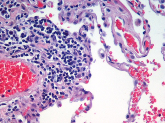
also known as microscopic anatomy or microanatomy, is the branch of
 Chemical fixatives are used to preserve and maintain the structure of tissues and cells; fixation also hardens tissues which aids in cutting the thin sections of tissue needed for observation under the microscope. Fixatives generally preserve tissues (and cells) by irreversibly cross-linking proteins. The most widely used fixative for light microscopy is 10% neutral buffered formalin, or NBF (4%
 For light microscopy,
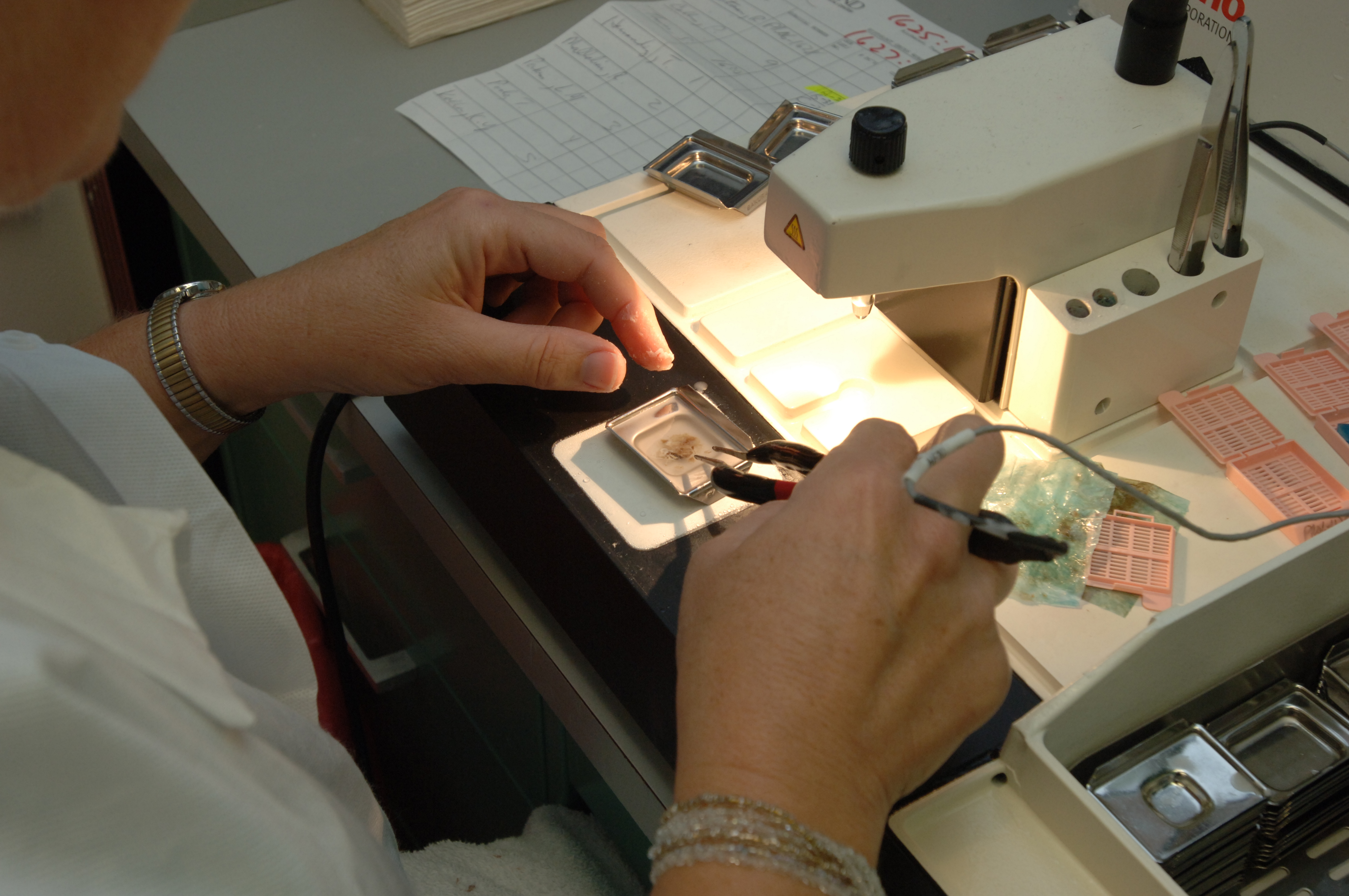
 For light microscopy, a knife mounted in a microtome is used to cut tissue sections (typically between 5-15 micrometers thick) which are mounted on a glass microscope slide. For transmission electron microscopy (TEM), a diamond or glass knife mounted in an ultramicrotome is used to cut between 50 and 150
 In the 17th century the Italian

 Histology,
also known as microscopic anatomy or microanatomy, is the branch of
 biology
Biology is the scientific study of life and living organisms. It is a broad natural science that encompasses a wide range of fields and unifying principles that explain the structure, function, growth, History of life, origin, evolution, and ...
that studies the microscopic anatomy
Anatomy () is the branch of morphology concerned with the study of the internal structure of organisms and their parts. Anatomy is a branch of natural science that deals with the structural organization of living things. It is an old scien ...
of biological tissues. Histology is the microscopic counterpart to gross anatomy
Gross anatomy is the study of anatomy at the visible or macroscopic level. The counterpart to gross anatomy is the field of histology, which studies microscopic anatomy. Gross anatomy of the human body or other animals seeks to understand the ...
, which looks at larger structures visible without a microscope
A microscope () is a laboratory equipment, laboratory instrument used to examine objects that are too small to be seen by the naked eye. Microscopy is the science of investigating small objects and structures using a microscope. Microscopic ...
. Although one may divide microscopic anatomy into ''organology'', the study of organs, ''histology'', the study of tissues, and ''cytology
Cell biology (also cellular biology or cytology) is a branch of biology that studies the structure, function, and behavior of cells. All living organisms are made of cells. A cell is the basic unit of life that is responsible for the living an ...
'', the study of cells, modern usage places all of these topics under the field of histology. In medicine
Medicine is the science and Praxis (process), practice of caring for patients, managing the Medical diagnosis, diagnosis, prognosis, Preventive medicine, prevention, therapy, treatment, Palliative care, palliation of their injury or disease, ...
, histopathology
Histopathology (compound of three Greek words: 'tissue', 'suffering', and '' -logia'' 'study of') is the microscopic examination of tissue in order to study the manifestations of disease. Specifically, in clinical medicine, histopatholog ...
is the branch of histology that includes the microscopic identification and study of diseased tissue. In the field of paleontology
Paleontology, also spelled as palaeontology or palæontology, is the scientific study of the life of the past, mainly but not exclusively through the study of fossils. Paleontologists use fossils as a means to classify organisms, measure ge ...
, the term paleohistology refers to the histology of fossil
A fossil (from Classical Latin , ) is any preserved remains, impression, or trace of any once-living thing from a past geological age. Examples include bones, shells, exoskeletons, stone imprints of animals or microbes, objects preserve ...
organisms.
Biological tissues
Animal tissue classification
There are four basic types of animal tissues:muscle tissue
Muscle is a soft tissue, one of the four basic types of animal tissue. There are three types of muscle tissue in vertebrates: skeletal muscle, cardiac muscle, and smooth muscle. Muscle tissue gives skeletal muscles the ability to contract. ...
, nervous tissue
Nervous tissue, also called neural tissue, is the main tissue component of the nervous system. The nervous system regulates and controls body functions and activity. It consists of two parts: the central nervous system (CNS) comprising the brain ...
, connective tissue
Connective tissue is one of the four primary types of animal tissue, a group of cells that are similar in structure, along with epithelial tissue, muscle tissue, and nervous tissue. It develops mostly from the mesenchyme, derived from the mesod ...
, and epithelial tissue
Epithelium or epithelial tissue is a thin, continuous, protective layer of cells with little extracellular matrix. An example is the epidermis, the outermost layer of the skin. Epithelial ( mesothelial) tissues line the outer surfaces of man ...
. All animal tissues are considered to be subtypes of these four principal tissue types (for example, blood is classified as connective tissue, since the blood cells are suspended in an extracellular matrix
In biology, the extracellular matrix (ECM), also called intercellular matrix (ICM), is a network consisting of extracellular macromolecules and minerals, such as collagen, enzymes, glycoproteins and hydroxyapatite that provide structural and bio ...
, the plasma).
.
Plant tissue classification
For plants, the study of their tissues falls under the field ofplant anatomy
Plant anatomy or phytotomy is the general term for the study of the internal Anatomy, structure of plants. Originally, it included plant morphology, the description of the physical form and external structure of plants, but since the mid-20th centu ...
, with the following four main types:
* Dermal tissue
* Vascular tissue
Vascular tissue is a complex transporting tissue, formed of more than one cell type, found in vascular plants. The primary components of vascular tissue are the xylem and phloem. These two tissues transport fluid and nutrients internally. T ...
* Ground tissue
The ground tissue of plants includes all tissues that are neither dermal nor vascular. It can be divided into three types based on the nature of the cell walls. This tissue system is present between the dermal tissue and forms the main bulk of t ...
* Meristematic tissue
Medical histology
Histopathology
Histopathology (compound of three Greek words: 'tissue', 'suffering', and '' -logia'' 'study of') is the microscopic examination of tissue in order to study the manifestations of disease. Specifically, in clinical medicine, histopatholog ...
is the branch of histology that includes the microscopic identification and study of diseased tissue. It is an important part of anatomical pathology and surgical pathology, as accurate diagnosis of cancer
Cancer is a group of diseases involving Cell growth#Disorders, abnormal cell growth with the potential to Invasion (cancer), invade or Metastasis, spread to other parts of the body. These contrast with benign tumors, which do not spread. Po ...
and other diseases often requires histopathological examination of tissue samples. Trained physicians, frequently licensed pathologist
Pathology is the study of disease. The word ''pathology'' also refers to the study of disease in general, incorporating a wide range of biology research fields and medical practices. However, when used in the context of modern medical treatme ...
s, perform histopathological examination and provide diagnostic information based on their observations.
Occupations
The field of histology that includes the preparation of tissues for microscopic examination is known as histotechnology. Job titles for the trained personnel who prepare histological specimens for examination are numerous and include histotechnicians, histotechnologists, histology technicians and technologists, medical laboratory technicians, andbiomedical scientist
A biomedical scientist is a scientist trained in biology, particularly in the context of medical laboratory sciences or laboratory medicine. These scientists work to gain knowledge on the main principles of how the human body works and to find new ...
s.
Sample preparation
Most histological samples need preparation before microscopic observation; these methods depend on the specimen and method of observation.Fixation
 Chemical fixatives are used to preserve and maintain the structure of tissues and cells; fixation also hardens tissues which aids in cutting the thin sections of tissue needed for observation under the microscope. Fixatives generally preserve tissues (and cells) by irreversibly cross-linking proteins. The most widely used fixative for light microscopy is 10% neutral buffered formalin, or NBF (4%
 formaldehyde
Formaldehyde ( , ) (systematic name methanal) is an organic compound with the chemical formula and structure , more precisely . The compound is a pungent, colourless gas that polymerises spontaneously into paraformaldehyde. It is stored as ...
in phosphate buffered saline).
For electron microscopy, the most commonly used fixative is glutaraldehyde, usually as a 2.5% solution in phosphate buffered saline. Other fixatives used for electron microscopy are osmium tetroxide
Osmium tetroxide (also osmium(VIII) oxide) is the chemical compound with the formula OsO4. The compound is noteworthy for its many uses, despite its toxicity and the rarity of osmium. It also has a number of unusual properties, one being that the ...
or uranyl acetate.
The main action of these aldehyde
In organic chemistry, an aldehyde () (lat. ''al''cohol ''dehyd''rogenatum, dehydrogenated alcohol) is an organic compound containing a functional group with the structure . The functional group itself (without the "R" side chain) can be referred ...
fixatives is to cross-link amino groups in proteins through the formation of methylene bridges (), in the case of formaldehyde, or by C5H10 cross-links in the case of glutaraldehyde. This process, while preserving the structural integrity of the cells and tissue can damage the biological functionality of proteins, particularly enzymes
An enzyme () is a protein that acts as a biological catalyst by accelerating chemical reactions. The molecules upon which enzymes may act are called substrates, and the enzyme converts the substrates into different molecules known as pro ...
.
Formalin fixation leads to degradation of mRNA, miRNA, and DNA as well as denaturation and modification of proteins in tissues. However, extraction and analysis of nucleic acids and proteins from formalin-fixed, paraffin-embedded tissues is possible using appropriate protocols.
Selection and trimming
''Selection'' is the choice of relevant tissue in cases where it is not necessary to put the entire original tissue mass through further processing. The remainder may remain fixed in case it needs to be examined at a later time. ''Trimming'' is the cutting of tissue samples in order to expose the relevant surfaces for later sectioning. It also creates tissue samples of appropriate size to fit into cassettes.Embedding
Tissues are embedded in a harder medium both as a support and to allow the cutting of thin tissue slices. In general, water must first be removed from tissues (dehydration) and replaced with a medium that either solidifies directly, or with an intermediary fluid (clearing) that is miscible with the embedding media.Paraffin wax
 For light microscopy,
 paraffin wax
Paraffin wax (or petroleum wax) is a soft colorless solid derived from petroleum, coal, or oil shale that consists of a mixture of hydrocarbon molecules containing between 20 and 40 carbon atoms. It is solid at room temperature and melting poi ...
is the most frequently used embedding material. Paraffin is immiscible with water, the main constituent of biological tissue, so it must first be removed in a series of dehydration steps. Samples are transferred through a series of progressively more concentrated ethanol
Ethanol (also called ethyl alcohol, grain alcohol, drinking alcohol, or simply alcohol) is an organic compound with the chemical formula . It is an Alcohol (chemistry), alcohol, with its formula also written as , or EtOH, where Et is the ps ...
baths, up to 100% ethanol to remove remaining traces of water. Dehydration is followed by a ''clearing agent'' (typically xylene although other environmental safe substitutes are in use) which removes the alcohol and is miscible with the wax, finally melted paraffin wax is added to replace the xylene and infiltrate the tissue. In most histology, or histopathology laboratories the dehydration, clearing, and wax infiltration are carried out in ''tissue processors'' which automate this process. Once infiltrated in paraffin, tissues are oriented in molds which are filled with wax; once positioned, the wax is cooled, solidifying the block and tissue.
Other materials
Paraffin wax does not always provide a sufficiently hard matrix for cutting very thin sections (which are especially important for electron microscopy). Paraffin wax may also be too soft in relation to the tissue, the heat of the melted wax may alter the tissue in undesirable ways, or the dehydrating or clearing chemicals may harm the tissue. Alternatives to paraffin wax include,epoxy
Epoxy is the family of basic components or Curing (chemistry), cured end products of epoxy Resin, resins. Epoxy resins, also known as polyepoxides, are a class of reactive prepolymers and polymers which contain epoxide groups. The epoxide fun ...
, acrylic, agar
Agar ( or ), or agar-agar, is a jelly-like substance consisting of polysaccharides obtained from the cell walls of some species of red algae, primarily from " ogonori" and " tengusa". As found in nature, agar is a mixture of two components, t ...
, gelatin
Gelatin or gelatine () is a translucent, colorless, flavorless food ingredient, commonly derived from collagen taken from animal body parts. It is brittle when dry and rubbery when moist. It may also be referred to as hydrolyzed collagen, coll ...
, celloidin, and other types of waxes.
In electron microscopy epoxy resins are the most commonly employed embedding media, but acrylic resins are also used, particularly where immunohistochemistry
Immunohistochemistry is a form of immunostaining. It involves the process of selectively identifying antigens in cells and tissue, by exploiting the principle of Antibody, antibodies binding specifically to antigens in biological tissues. Alber ...
is required.
For tissues to be cut in a frozen state, tissues are placed in a water-based embedding medium. Pre-frozen tissues are placed into molds with the liquid embedding material, usually a water-based glycol, OCT, TBS, Cryogen, or resin, which is then frozen to form hardened blocks.
Sectioning
 For light microscopy, a knife mounted in a microtome is used to cut tissue sections (typically between 5-15 micrometers thick) which are mounted on a glass microscope slide. For transmission electron microscopy (TEM), a diamond or glass knife mounted in an ultramicrotome is used to cut between 50 and 150
 nanometer
330px, Different lengths as in respect to the Molecule">molecular scale.
The nanometre (international spelling as used by the International Bureau of Weights and Measures; SI symbol: nm), or nanometer (American spelling
Despite the va ...
thick tissue sections.
A limited number of manufacturers are recognized for their production of microtomes, including vibrating microtomes commonly referred to as vibratomes, primarily for research and clinical studies. Additionally, Leica Biosystems
Leica Biosystems, founded 1872 as Precision Engineering, is a medical devices company that develops and supplies clinical diagnostics to the pathology market. It is also a research, instrument, and medical device company as well as a division of ...
is known for its production of products related to light microscopy in the context of research and clinical studies.
Staining
Biological tissue has little inherent contrast in either the light or electron microscope.Staining
Staining is a technique used to enhance contrast in samples, generally at the Microscope, microscopic level. Stains and dyes are frequently used in histology (microscopic study of biological tissue (biology), tissues), in cytology (microscopic ...
is employed to give both contrast to the tissue as well as highlighting particular features of interest. When the stain is used to target a specific chemical component of the tissue (and not the general structure), the term histochemistry is used.
Light microscopy
Hematoxylin
Haematoxylin American and British English spelling differences#ae and oe, or hematoxylin (), also called natural black 1 or Colour Index International, C.I. 75290, is a chemical compound, compound extracted from wood#Heartwood and sapwood, heart ...
and eosin
Eosin is the name of several fluorescent acidic compounds which bind to and from salts with basic, or eosinophilic, compounds like proteins containing basic amino acid residues such as histidine, arginine and lysine, and stains them dark red ...
(H&E stain
Hematoxylin and eosin stain ( or haematoxylin and eosin stain or hematoxylin–eosin stain; often abbreviated as H&E stain or HE stain) is one of the principal tissue stains used in histology. It is the most widely used stain in medical diag ...
) is one of the most commonly used stains in histology to show the general structure of the tissue. Hematoxylin stains cell nuclei blue; eosin, an acidic
An acid is a molecule or ion capable of either donating a proton (i.e. hydrogen cation, H+), known as a Brønsted–Lowry acid, or forming a covalent bond with an electron pair, known as a Lewis acid.
The first category of acids are the ...
dye, stains the cytoplasm
The cytoplasm describes all the material within a eukaryotic or prokaryotic cell, enclosed by the cell membrane, including the organelles and excluding the nucleus in eukaryotic cells. The material inside the nucleus of a eukaryotic cell a ...
and other tissues in different stains of pink.
In contrast to H&E, which is used as a general stain, there are many techniques that more selectively stain cells, cellular components, and specific substances. A commonly performed histochemical technique that targets a specific chemical is the Perls' Prussian blue reaction, used to demonstrate iron deposits in diseases like hemochromatosis. The Nissl method for Nissl substance and Golgi's method (and related silver stains) are useful in identifying neuron
A neuron (American English), neurone (British English), or nerve cell, is an membrane potential#Cell excitability, excitable cell (biology), cell that fires electric signals called action potentials across a neural network (biology), neural net ...
s are other examples of more specific stains.
Historadiography
In historadiography, a slide (sometimes stained histochemically) is X-rayed. More commonly, autoradiography is used in visualizing the locations to which a radioactive substance has been transported within the body, such as cells inS phase
S phase (Synthesis phase) is the phase of the cell cycle in which DNA is replicated, occurring between G1 phase and G2 phase. Since accurate duplication of the genome is critical to successful cell division, the processes that occur during S ...
(undergoing DNA replication
In molecular biology, DNA replication is the biological process of producing two identical replicas of DNA from one original DNA molecule. DNA replication occurs in all life, living organisms, acting as the most essential part of heredity, biolog ...
) which incorporate tritiated thymidine
Thymidine (nucleoside#List of nucleosides and corresponding nucleobases, symbol dT or dThd), also known as deoxythymidine, deoxyribosylthymine, or thymine deoxyriboside, is a pyrimidine nucleoside, deoxynucleoside. Deoxythymidine is the DNA nuc ...
, or sites to which radiolabeled nucleic acid
Nucleic acids are large biomolecules that are crucial in all cells and viruses. They are composed of nucleotides, which are the monomer components: a pentose, 5-carbon sugar, a phosphate group and a nitrogenous base. The two main classes of nuclei ...
probes bind in in situ hybridization
''In situ'' hybridization (ISH) is a type of Hybridisation (molecular biology), hybridization that uses a labeled complementary DNA, RNA or modified nucleic acid strand (i.e., a Hybridization probe, probe) to localize a specific DNA or RNA seq ...
. For autoradiography on a microscopic level, the slide is typically dipped into liquid nuclear tract emulsion, which dries to form the exposure film. Individual silver grains in the film are visualized with dark field microscopy
Dark-field microscopy, also called dark-ground microscopy, describes microscopy methods, in both light microscopy, light and electron microscopy, which exclude the unscattered beam from the image. Consequently, the field around the specimen (i.e ...
.
Immunohistochemistry
Recently,antibodies
An antibody (Ab) or immunoglobulin (Ig) is a large, Y-shaped protein belonging to the immunoglobulin superfamily which is used by the immune system to identify and neutralize antigens such as bacteria and viruses, including those that caus ...
have been used to specifically visualize proteins, carbohydrates, and lipids. This process is called immunohistochemistry
Immunohistochemistry is a form of immunostaining. It involves the process of selectively identifying antigens in cells and tissue, by exploiting the principle of Antibody, antibodies binding specifically to antigens in biological tissues. Alber ...
, or when the stain is a fluorescent molecule, immunofluorescence
Immunofluorescence (IF) is a light microscopy-based technique that allows detection and localization of a wide variety of target biomolecules within a cell or tissue at a quantitative level. The technique utilizes the binding specificity of anti ...
. This technique has greatly increased the ability to identify categories of cells under a microscope. Other advanced techniques, such as nonradioactive ''in situ'' hybridization, can be combined with immunochemistry to identify specific DNA or RNA molecules with fluorescent probes or tags that can be used for immunofluorescence and enzyme-linked fluorescence amplification (especially alkaline phosphatase and tyramide signal amplification). Fluorescence microscopy and confocal microscopy
Confocal microscopy, most frequently confocal laser scanning microscopy (CLSM) or laser scanning confocal microscopy (LSCM), is an optical imaging technique for increasing optical resolution and contrast (vision), contrast of a micrograph by me ...
are used to detect fluorescent signals with good intracellular detail.
Electron microscopy
For electron microscopyheavy metals
upright=1.2, Crystals of lead.html" ;"title="osmium, a heavy metal nearly twice as dense as lead">osmium, a heavy metal nearly twice as dense as lead
Heavy metals is a controversial and ambiguous term for metallic elements with relatively h ...
are typically used to stain tissue sections. Uranyl acetate and lead citrate are commonly used to impart contrast to tissue in the electron microscope.
Specialized techniques
Cryosectioning
Similar to thefrozen section procedure
The frozen section procedure is a pathological laboratory procedure to perform rapid microscopic analysis of a specimen. It is used most often in oncological surgery. The technical name for this procedure is cryosection. The microtome device tha ...
employed in medicine, cryosectioning is a method to rapidly freeze, cut, and mount sections of tissue for histology. The tissue is usually sectioned on a cryostat or freezing microtome. The frozen sections are mounted on a glass slide and may be stained to enhance the contrast between different tissues. Unfixed frozen sections can be used for studies requiring enzyme localization in tissues and cells. Tissue fixation is required for certain procedures such as antibody-linked immunofluorescence
Immunofluorescence (IF) is a light microscopy-based technique that allows detection and localization of a wide variety of target biomolecules within a cell or tissue at a quantitative level. The technique utilizes the binding specificity of anti ...
staining. Frozen sections are often prepared during surgical removal of tumor
A neoplasm () is a type of abnormal and excessive growth of tissue. The process that occurs to form or produce a neoplasm is called neoplasia. The growth of a neoplasm is uncoordinated with that of the normal surrounding tissue, and persists ...
s to allow rapid identification of tumor margins, as in Mohs surgery, or determination of tumor malignancy, when a tumor is discovered incidentally during surgery.
Ultramicrotomy
Ultramicrotomy is a method of preparing extremely thin sections fortransmission electron microscope
Transmission electron microscopy (TEM) is a microscopy technique in which a beam of electrons is transmitted through a specimen to form an image. The specimen is most often an ultrathin section less than 100 nm thick or a suspension on a gr ...
(TEM) analysis. Tissues are commonly embedded in epoxy
Epoxy is the family of basic components or Curing (chemistry), cured end products of epoxy Resin, resins. Epoxy resins, also known as polyepoxides, are a class of reactive prepolymers and polymers which contain epoxide groups. The epoxide fun ...
or other plastic resin. Very thin sections (less than 0.1 micrometer in thickness) are cut using diamond or glass knives on an ultramicrotome.
Artifacts
Artifacts are structures or features in tissue that interfere with normal histological examination. Artifacts interfere with histology by changing the tissues appearance and hiding structures. Tissue processing artifacts can include pigments formed by fixatives, shrinkage, washing out of cellular components, color changes in different tissues types and alterations of the structures in the tissue. An example is mercury pigment left behind after using Zenker's fixative to fix a section. Formalin fixation can also leave a brown to black pigment under acidic conditions.History
 In the 17th century the Italian
 Marcello Malpighi
Marcello Malpighi (10 March 1628 – 30 November 1694) was an Italians, Italian biologist and physician, who is referred to as the "founder of microscopical anatomy, histology and father of physiology and embryology". Malpighi's name is borne by ...
used microscopes to study tiny biological entities; some regard him as the founder of the fields of histology and microscopic pathology. Malpighi analyzed several parts of the organs of bats, frogs and other animals under the microscope. While studying the structure of the lung, Malpighi noticed its membranous alveoli and the hair-like connections between veins and arteries, which he named capillaries. His discovery established how the oxygen breathed in enters the blood stream and serves the body.
In the 19th century histology was an academic discipline in its own right. The French anatomist Xavier Bichat introduced the concept of tissue in anatomy in 1801, and the term "histology" (), coined to denote the "study of tissues", first appeared in a book by Karl Meyer in 1819. Bichat described twenty-one human tissues, which can be subsumed under the four categories currently accepted by histologists. The usage of illustrations in histology, deemed as useless by Bichat, was promoted by Jean Cruveilhier.
In the early 1830s Purkynĕ invented a microtome with high precision.
During the 19th century many fixation techniques were developed by Adolph Hannover (solutions of chromates and chromic acid
Chromic acid is a chemical compound with the chemical formula . It is also a jargon for a solution formed by the addition of sulfuric acid to aqueous solutions of dichromate. It consists at least in part of chromium trioxide.
The term "chromic ...
), Franz Schulze and Max Schultze ( osmic acid), Alexander Butlerov (formaldehyde
Formaldehyde ( , ) (systematic name methanal) is an organic compound with the chemical formula and structure , more precisely . The compound is a pungent, colourless gas that polymerises spontaneously into paraformaldehyde. It is stored as ...
) and Benedikt Stilling (freezing
Freezing is a phase transition in which a liquid turns into a solid when its temperature is lowered below its freezing point.
For most substances, the melting and freezing points are the same temperature; however, certain substances possess dif ...
).
Mounting techniques were developed by Rudolf Heidenhain (1824–1898), who introduced gum Arabic
Gum arabic (gum acacia, gum sudani, Senegal gum and by other names) () is a tree gum exuded by two species of '' Acacia sensu lato:'' '' Senegalia senegal,'' and '' Vachellia seyal.'' However, the term "gum arabic" does not indicate a partic ...
; Salomon Stricker (1834–1898), who advocated a mixture of wax and oil; and Andrew Pritchard (1804–1884) who, in 1832, used a gum/isinglass
Isinglass ( ) is a form of collagen obtained from the dried swim bladders of fish. The English word origin is from the obsolete Dutch ''huizenblaas'' – ''huizen'' is a kind of sturgeon, and ''blaas'' is a bladder, or German ''Hausenblase'', ...
mixture. In the same year, Canada balsam appeared on the scene, and in 1869 Edwin Klebs (1834–1913) reported that he had for some years embedded his specimens in paraffin.
The 1906 Nobel Prize
The Nobel Prizes ( ; ; ) are awards administered by the Nobel Foundation and granted in accordance with the principle of "for the greatest benefit to humankind". The prizes were first awarded in 1901, marking the fifth anniversary of Alfred N ...
in Physiology or Medicine was awarded to histologists Camillo Golgi
Camillo Golgi (; 7 July 184321 January 1926) was an Italian biologist and pathologist known for his works on the central nervous system. He studied medicine at the University of Pavia (where he later spent most of his professional career) bet ...
and Santiago Ramon y Cajal. They had conflicting interpretations of the neural structure of the brain based on differing interpretations of the same images. Ramón y Cajal won the prize for his correct theory, and Golgi for the silver-staining technique
Technique or techniques may refer to:
Music
* The Techniques, a Jamaican rocksteady vocal group of the 1960s
* Technique (band), a British female synth pop band in the 1990s
* ''Technique'' (album), by New Order, 1989
* ''Techniques'' (album), by ...
that he invented to make it possible.
Future directions
''In vivo'' histology
There is interest in developing techniques for ''in vivo'' histology (predominantly usingMRI
Magnetic resonance imaging (MRI) is a medical imaging technique used in radiology to generate pictures of the anatomy and the physiological processes inside the body. MRI scanners use strong magnetic fields, magnetic field gradients, and rad ...
), which would enable doctors to non-invasively gather information about healthy and diseased tissues in living patients, rather than from fixed tissue samples.
See also
* National Society for Histotechnology * Slice preparationNotes
References
External links
* {{Authority control Histotechnology Staining Histochemistry Anatomy Laboratory healthcare occupations