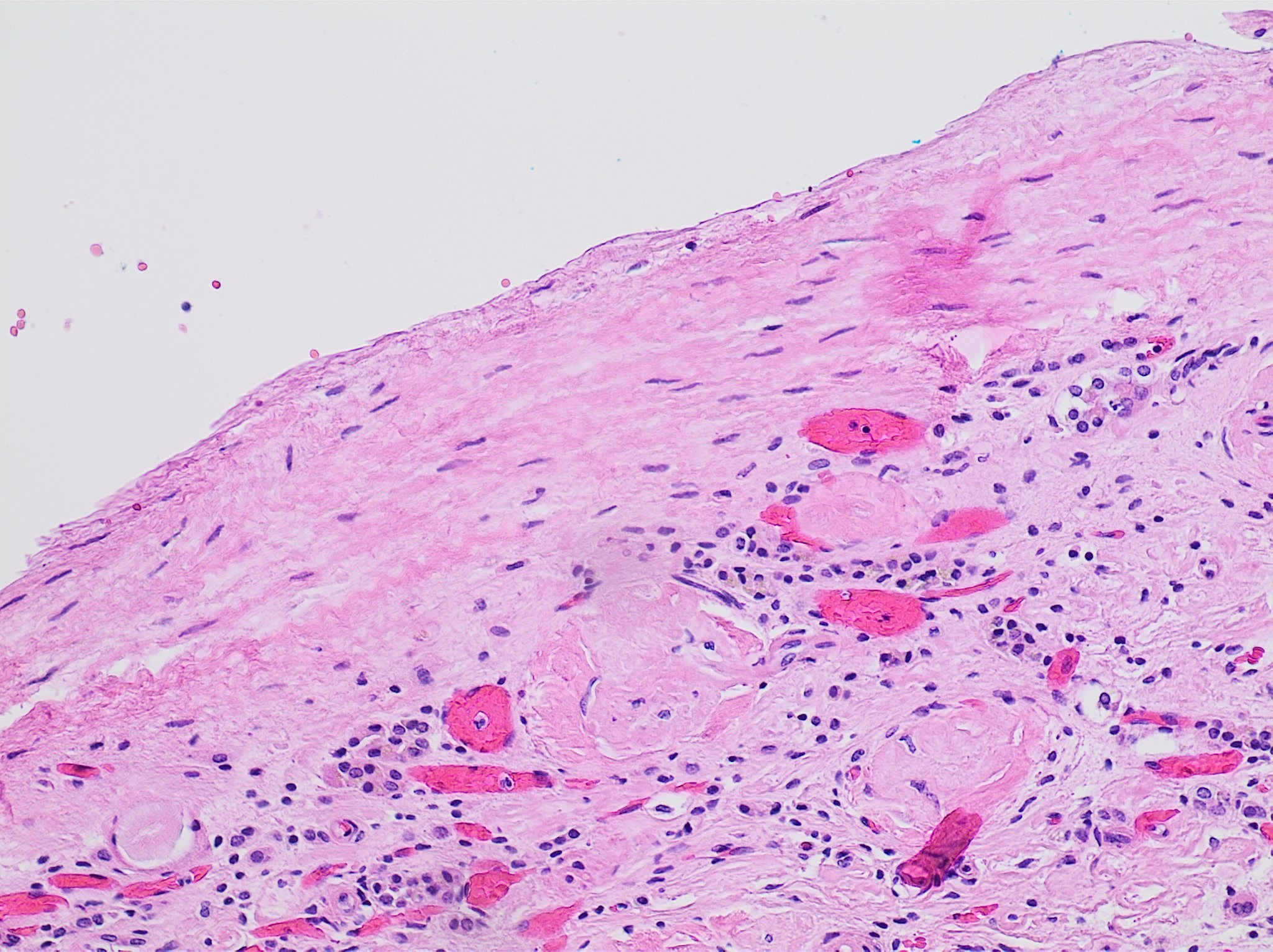
Renal cyst on:
[Wikipedia]
[Google]
[Amazon]
 A renal cyst is a fluid collection in or on the
 This category includes renal cysts with multiple thin septa, a septum thicker than hairline, slightly thick wall, or with calcification, which may be thick. It also includes intrarenal cysts larger than if:
* there is no contrast enhancement (otherwise category III).
* there is high attenuation or there is a maximum 25% of their walls visible outside the kidney (otherwise category II).
Category IIF cysts have a 5–10% risk of being
/ref>
Simple cyst with posterior enhancement.jpg,
 Parapelvic cysts originate from around the kidney at the adjacent renal parenchyma, and plunge into the renal sinus. Peripelvic cysts are contained entirely within the renal sinus, possibly related to dilated lymphatic channels. When viewed on CT in absence of contrast, they can mimic
 A renal cyst is a fluid collection in or on the
 kidney
In humans, the kidneys are two reddish-brown bean-shaped blood-filtering organ (anatomy), organs that are a multilobar, multipapillary form of mammalian kidneys, usually without signs of external lobulation. They are located on the left and rig ...
. There are several types based on the Bosniak classification. The majority are benign, simple cysts that can be monitored and not intervened upon. However, some are cancer
Cancer is a group of diseases involving Cell growth#Disorders, abnormal cell growth with the potential to Invasion (cancer), invade or Metastasis, spread to other parts of the body. These contrast with benign tumors, which do not spread. Po ...
ous or are suspicious for cancer and are commonly removed in a surgical procedure called nephrectomy
A nephrectomy is the surgical removal of a kidney, performed to treat a number of kidney diseases including kidney cancer. It is also done to remove a normal healthy kidney from a living or deceased donor, which is part of a kidney transplant pro ...
.
Numerous renal cysts are seen in the cystic kidney diseases, which include polycystic kidney disease
Polycystic kidney disease (PKD or PCKD, also known as polycystic kidney syndrome) is a genetic disorder in which the renal tubules become structurally abnormal, resulting in the development and growth of multiple cysts within the kidney. These ...
and medullary sponge kidney
Medullary sponge kidney is a congenital disorder of the kidneys characterized by cystic dilatation of the collecting tubules in one or both kidneys. Individuals with medullary sponge kidney are at increased risk for kidney stones and urinary tra ...
.
Classification
Renal cysts are classified by malignant risk using the Bosniak classification system. The system was created by Morton Bosniak (1929–2016), a faculty member at theNew York University
New York University (NYU) is a private university, private research university in New York City, New York, United States. Chartered in 1831 by the New York State Legislature, NYU was founded in 1832 by Albert Gallatin as a Nondenominational ...
Langone Medical Center in New York City
New York, often called New York City (NYC), is the most populous city in the United States, located at the southern tip of New York State on one of the world's largest natural harbors. The city comprises five boroughs, each coextensive w ...
.
The Bosniak classification categorizes renal cysts into five groups.
Category I
:Benign simple cyst with thin wall withoutsepta
SEPTA, the Southeastern Pennsylvania Transportation Authority, is a regional public transportation authority that operates bus, rapid transit, commuter rail, light rail, and electric trolleybus services for nearly four million people througho ...
, calcifications, or solid components, and has a density of 0–20 Hounsfield units
The Hounsfield scale ( ), named after Sir Godfrey Hounsfield, is a quantitative scale for describing radiodensity. It is frequently used in CT scans, where its value is also termed CT number.
Definition
The Hounsfield unit (HU) scale is a linear t ...
(HU) (about equal to that of water). In such cases, a CT scan without intravenous contrast is enough for classification. Still, if a contrast CT
Contrast CT, or contrast-enhanced computed tomography (CECT), is CT scan, X-ray computed tomography (CT) using radiocontrast. Radiocontrasts for X-ray CT are generally Iodinated contrast, iodine-based types. This is useful to highlight structure ...
is performed, a category I cyst should not show significant enhancement, which can be regarded as an increase of less than 10 HU.
Category II
:Benign cyst with a few thin septa, which may contain fine calcifications or a small segment of mildly thickened calcification. This includes homogenous, high-attenuation (60–70 Hounsfield units) lesions less than 3 cm with sharp margins but without enhancement. Hyperdense cysts must be exophytic with at least 75 percent of its wall outside the kidney to allow for appropriate assessment of margins, otherwise they are categorized as IIF.Category IIF
 This category includes renal cysts with multiple thin septa, a septum thicker than hairline, slightly thick wall, or with calcification, which may be thick. It also includes intrarenal cysts larger than if:
* there is no contrast enhancement (otherwise category III).
* there is high attenuation or there is a maximum 25% of their walls visible outside the kidney (otherwise category II).
Category IIF cysts have a 5–10% risk of being
 kidney cancer
Kidney cancer, also known as renal cancer, is a group of cancers that starts in the kidney. Symptoms may include blood in the urine, a lump in the abdomen, or back pain. Fever, weight loss, and tiredness may also occur. Complications can include ...
, and therefore follow-up is recommended. However, there is no consensus recommendation on the appropriate interval of follow up.
Category III
:Indeterminate cystic masses with thickened, irregular or smooth walls or septa with measurable enhancement. Approximately 40 to 60% of these lesions are ultimately found to be malignant, most commonly in the forms of cystic renal cell carcinoma and its multiloculated variant. The remaining lesions are benign and include hemorrhagic cysts, chronic infected cysts, and multiloculated cystic nephromas.Category IV
:Malignant cystic masses with all the characteristics of category III lesions but also with enhancing soft tissue components independent of but adjacent to the septa. Approximately 85 to 100% of these lesions are malignant. The presence of measurable contrast enhancement of the lesion is the most important characteristic in distinguishing between high-risk cysts (classifications III and IV) from the typically benign, low-risk Bosniak I, II, and IIF cysts. Such contrast enhancement should be at least 10 to 15 Hounsfield units higher when compared with unenhanced images.Diagnosis
The complex cyst can be further evaluated withdoppler ultrasonography
Doppler ultrasonography is medical ultrasonography that employs the Doppler effect to perform imaging of the movement of tissues and body fluids (usually blood), and their relative velocity to the probe. By calculating the frequency shift of a ...
, and for Bosniak classification and follow-up of complex cysts, either contrast-enhanced ultrasound
Contrast-enhanced ultrasound (CEUS) is the application of ultrasound contrast medium to traditional medical sonography. Ultrasound contrast agents rely on the different ways in which sound waves are reflected from interfaces between substances. T ...
(CEUS) or contrast CT
Contrast CT, or contrast-enhanced computed tomography (CECT), is CT scan, X-ray computed tomography (CT) using radiocontrast. Radiocontrasts for X-ray CT are generally Iodinated contrast, iodine-based types. This is useful to highlight structure ...
is used.(CC-BY 4.0)/ref>

Renal ultrasonography
Renal ultrasonography (Renal US) is the examination of one or both kidneys using medical ultrasound.
Ultrasonography of the kidneys is essential in the diagnosis and management of kidney-related diseases. The kidneys are easily examined, and most ...
of a simple renal cyst with posterior enhancement.
File:Advanced polycystic kidney disease with multiple cysts.jpg, Advanced polycystic kidney disease
Polycystic kidney disease (PKD or PCKD, also known as polycystic kidney syndrome) is a genetic disorder in which the renal tubules become structurally abnormal, resulting in the development and growth of multiple cysts within the kidney. These ...
with multiple cysts.
Renal cyst ultrasound.jpg, Renal cyst as seen on abdominal ultrasound
Abdominal ultrasonography (also called abdominal ultrasound imaging or abdominal sonography) is a form of medical ultrasonography (medicine, medical application of ultrasound technology) to visualise abdomen, abdominal anatomy, anatomical structu ...
Renal cyst ultrasound 2.jpg, Renal cyst as seen on abdominal ultrasound
Renal cyst ultrasound 3.jpg, Renal cyst as seen on abdominal ultrasound
Small simple renal cyst.jpg, A very small (8 mm) simple renal cyst.
Complex cysts at the lower pole of right kidney showing septations and sediment within.jpg, Bosniak II cyst at the lower pole of right kidney with septations within.
Treatment
This system is more directly focused on the most appropriate management. These alternatives are broadly to ignore the cyst, schedule follow-up or perform a surgical excision of it. When a cyst shows discrepancy in severity across categories, it is the most worrisome feature that is used in deciding about management. There is no established rule regarding the follow-up frequency, but one possibility is after 6 months, which can later be doubled if unchanged.Peripelvic versus parapelvic cysts
 Parapelvic cysts originate from around the kidney at the adjacent renal parenchyma, and plunge into the renal sinus. Peripelvic cysts are contained entirely within the renal sinus, possibly related to dilated lymphatic channels. When viewed on CT in absence of contrast, they can mimic
 hydronephrosis
Hydronephrosis is the hydrostatic dilation of the renal pelvis and Renal calyx, calyces as a result of obstruction to urine flow downstream. Alternatively, hydroureter describes the dilation of the ureter, and hydronephroureter describes the dila ...
. If symptomatic, they can be laparoscopically decorticated - removal of the outer layer or cortex.
Epidemiology
Up to 27 percent of individuals older than 50 years may have simple renal cysts that cause no symptoms.See also
*Renal tumor
Kidney tumours are tumours, or growths, on or in the kidney. These growths can be benign or malignant (kidney cancer).
Presentation
Kidney tumours may be discovered on medical imaging incidentally (i.e. an incidentaloma), or may be present in pati ...
References
External links
{{Authority control Kidney diseases