|
Renal Parenchyma
upright=1.6, Lung parenchyma showing damage due to large subpleural bullae. Parenchyma () is the bulk of functional substance in an animal organ such as the brain or lungs, or a structure such as a tumour. In zoology, it is the tissue that fills the interior of flatworms. In botany, it is some layers in the cross-section of the leaf. Etymology The term ''parenchyma'' is Neo-Latin from the Ancient Greek word meaning 'visceral flesh', and from meaning 'to pour in' from 'beside' + 'in' + 'to pour'. Originally, Erasistratus and other anatomists used it for certain human tissues. Later, it was also applied to plant tissues by Nehemiah Grew. Structure The parenchyma is the ''functional'' parts of an organ, or of a structure such as a tumour in the body. This is in contrast to the stroma, which refers to the ''structural'' tissue of organs or of structures, namely, the connective tissues. Brain The brain parenchyma refers to the functional tissue in the brain ... [...More Info...] [...Related Items...] OR: [Wikipedia] [Google] [Baidu] |
Brain Cell
Brain cells make up the functional tissue of the brain. The rest of the brain tissue is the structural stroma that includes connective tissue such as the meninges, blood vessels, and ducts. The two main types of cells in the brain are neurons, also known as nerve cells, and glial cells, also known as neuroglia. There are many types of neuron, and several types of glial cell. Neurons are the excitable cells of the brain that function by communicating with other neurons and interneurons (via synapses), in neural circuits and larger brain networks. The two main neuronal classes in the cerebral cortex are excitatory projection neurons (around 70-80%) and inhibitory interneurons (around 20–30%). Neurons are often grouped into a cluster known as a nucleus where they usually have roughly similar connections and functions. Nuclei are connected to other nuclei by tracts of white matter. Glia are the supporting cells of the neurons and have many functions of which not all are clear ... [...More Info...] [...Related Items...] OR: [Wikipedia] [Google] [Baidu] |
Neoplasm
A neoplasm () is a type of abnormal and excessive growth of tissue. The process that occurs to form or produce a neoplasm is called neoplasia. The growth of a neoplasm is uncoordinated with that of the normal surrounding tissue, and persists in growing abnormally, even if the original trigger is removed. This abnormal growth usually forms a mass, which may be called a tumour or tumor.'' ICD-10 classifies neoplasms into four main groups: benign neoplasms, in situ neoplasms, malignant neoplasms, and neoplasms of uncertain or unknown behavior. Malignant neoplasms are also simply known as cancers and are the focus of oncology. Prior to the abnormal growth of tissue, such as neoplasia, cells often undergo an abnormal pattern of growth, such as metaplasia or dysplasia. However, metaplasia or dysplasia does not always progress to neoplasia and can occur in other conditions as well. The word neoplasm is from Ancient Greek 'new' and 'formation, creation'. Types A neoplasm ... [...More Info...] [...Related Items...] OR: [Wikipedia] [Google] [Baidu] |
Renal Pyramid
The renal medulla (Latin: ''medulla renis'' 'marrow of the kidney') is the innermost part of the kidney. The renal medulla is split up into a number of sections, known as the renal pyramids. Blood enters into the kidney via the renal artery, which then splits up to form the segmental arteries which then branch to form interlobar arteries. The interlobar arteries each in turn branch into arcuate arteries, which in turn branch to form interlobular arteries, and these finally reach the glomerulus (kidney), glomeruli. At the glomerulus the blood reaches a highly disfavourable pressure gradient and a large exchange surface area, which forces the serum (blood), serum portion of the blood out of the vessel and into the renal tubules. Flow continues through the renal tubules, including the proximal tubule, the loop of Henle, through the distal tubule and finally leaves the kidney by means of the collecting duct, leading to the renal pelvis, the dilated portion of the ureter. The renal med ... [...More Info...] [...Related Items...] OR: [Wikipedia] [Google] [Baidu] |
Renal Lobe
The renal lobe is a portion of a kidney consisting of a renal pyramid and the renal cortex above it. In humans, on average there are 7 to 18 renal lobes. It is visible without a microscope, though it is easier to see in humans than in other animals. It is composed of many renal lobules, which are not visible without a microscope. See also * Renal capsule * Renal medulla The renal medulla (Latin: ''medulla renis'' 'marrow of the kidney') is the innermost part of the kidney. The renal medulla is split up into a number of sections, known as the renal pyramids. Blood enters into the kidney via the renal artery, which ... References External links * Kidney anatomy {{Portal bar, Anatomy ... [...More Info...] [...Related Items...] OR: [Wikipedia] [Google] [Baidu] |
Renal Medulla
The renal medulla (Latin: ''medulla renis'' 'marrow of the kidney') is the innermost part of the kidney. The renal medulla is split up into a number of sections, known as the renal pyramids. Blood enters into the kidney via the renal artery, which then splits up to form the segmental arteries which then branch to form interlobar arteries. The interlobar arteries each in turn branch into arcuate arteries, which in turn branch to form interlobular arteries, and these finally reach the glomerulus (kidney), glomeruli. At the glomerulus the blood reaches a highly disfavourable pressure gradient and a large exchange surface area, which forces the serum (blood), serum portion of the blood out of the vessel and into the renal tubules. Flow continues through the renal tubules, including the proximal tubule, the loop of Henle, through the distal tubule and finally leaves the kidney by means of the collecting duct, leading to the renal pelvis, the dilated portion of the ureter. The renal med ... [...More Info...] [...Related Items...] OR: [Wikipedia] [Google] [Baidu] |
Renal Cortex
The renal cortex is the outer portion of the kidney between the renal capsule and the renal medulla. In the adult, it forms a continuous smooth outer zone with a number of projections ( cortical columns) that extend down between the pyramids. It contains the renal corpuscles and the renal tubules except for parts of the loop of Henle which descend into the renal medulla. It also contains blood vessels and cortical collecting ducts. The renal cortex is the part of the kidney where ultrafiltration occurs. Erythropoietin is produced in the renal cortex. Additional images File:Njuren.gif, Kidney File:Kidney-Cortex.JPG, Microscopic cross section of the renal cortex File:Kidney_cd10_ihc.jpg, CD10 immunohistochemical staining of normal kidney In humans, the kidneys are two reddish-brown bean-shaped blood-filtering organ (anatomy), organs that are a multilobar, multipapillary form of mammalian kidneys, usually without signs of external lobulation. They are located on the le ... [...More Info...] [...Related Items...] OR: [Wikipedia] [Google] [Baidu] |
Kidney
In humans, the kidneys are two reddish-brown bean-shaped blood-filtering organ (anatomy), organs that are a multilobar, multipapillary form of mammalian kidneys, usually without signs of external lobulation. They are located on the left and right in the retroperitoneal space, and in adult humans are about in length. They receive blood from the paired renal artery, renal arteries; blood exits into the paired renal veins. Each kidney is attached to a ureter, a tube that carries excreted urine to the urinary bladder, bladder. The kidney participates in the control of the volume of various body fluids, fluid osmolality, Acid-base homeostasis, acid-base balance, various electrolyte concentrations, and removal of toxins. Filtration occurs in the glomerulus (kidney), glomerulus: one-fifth of the blood volume that enters the kidneys is filtered. Examples of substances reabsorbed are solute-free water, sodium, bicarbonate, glucose, and amino acids. Examples of substances secreted are hy ... [...More Info...] [...Related Items...] OR: [Wikipedia] [Google] [Baidu] |
Hepatocyte
A hepatocyte is a cell of the main parenchymal tissue of the liver. Hepatocytes make up 80% of the liver's mass. These cells are involved in: * Protein synthesis * Protein storage * Transformation of carbohydrates * Synthesis of cholesterol, bile salts and phospholipids * Detoxification, modification, and excretion of exogenous and endogenous substances * Initiation of formation and secretion of bile Structure The typical hepatocyte is cubical with sides of 20-30 μm, (in comparison, a human hair has a diameter of 17 to 180 μm).The diameter of human hair ranges from 17 to 181 μm. The typical volume of a hepatocyte is 3.4 x 10⁻⁹ cm3. Smooth endoplasmic reticulum is abundant in hepatocytes, in contrast to most other cell types. Microanatomy Hepatocytes display an eosinophilic cytoplasm, reflecting numerous mitochondria, and basophilic stippling due to large amounts of rough endoplasmic reticulum and free ribosomes. Brown lipofuscin granules are also observed (wit ... [...More Info...] [...Related Items...] OR: [Wikipedia] [Google] [Baidu] |
Liver
The liver is a major metabolic organ (anatomy), organ exclusively found in vertebrates, which performs many essential biological Function (biology), functions such as detoxification of the organism, and the Protein biosynthesis, synthesis of various proteins and various other Biochemistry, biochemicals necessary for digestion and growth. In humans, it is located in the quadrants and regions of abdomen, right upper quadrant of the abdomen, below the thoracic diaphragm, diaphragm and mostly shielded by the lower right rib cage. Its other metabolic roles include carbohydrate metabolism, the production of a number of hormones, conversion and storage of nutrients such as glucose and glycogen, and the decomposition of red blood cells. Anatomical and medical terminology often use the prefix List of medical roots, suffixes and prefixes#H, ''hepat-'' from ἡπατο-, from the Greek language, Greek word for liver, such as hepatology, and hepatitis The liver is also an accessory digestive ... [...More Info...] [...Related Items...] OR: [Wikipedia] [Google] [Baidu] |
Pulmonary Alveolus
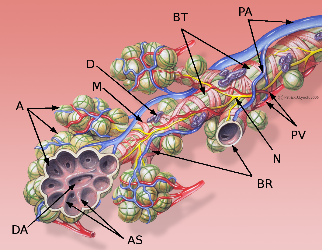
A pulmonary alveolus (; ), also called an air sac or air space, is one of millions of hollow, distensible cup-shaped cavities in the lungs where pulmonary gas exchange takes place. Oxygen is exchanged for carbon dioxide at the blood–air barrier between the alveolar air and the pulmonary capillary. Alveoli make up the functional tissue of the mammalian lungs known as the lung parenchyma, which takes up 90 percent of the total lung volume. Alveoli are first located in the respiratory bronchioles that mark the beginning of the respiratory zone. They are located sparsely in these bronchioles, line the walls of the alveolar ducts, and are more numerous in the blind-ended alveolar sacs. The acini are the basic units of respiration, with gas exchange taking place in all the alveoli present. The alveolar membrane is the gas exchange surface, surrounded by a network of capillaries. Oxygen is diffused across the membrane into the capillaries and carbon dioxide is released fr ... [...More Info...] [...Related Items...] OR: [Wikipedia] [Google] [Baidu] |
Gas Exchange
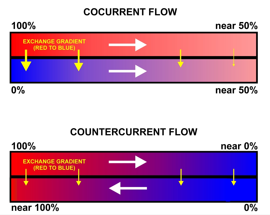
Gas exchange is the physical process by which gases move passively by diffusion across a surface. For example, this surface might be the air/water interface of a water body, the surface of a gas bubble in a liquid, a gas-permeable membrane, or a biological membrane that forms the boundary between an organism and its extracellular environment. Gases are constantly consumed and produced by cellular and metabolic reactions in most living things, so an efficient system for gas exchange between, ultimately, the interior of the cell(s) and the external environment is required. Small, particularly unicellular organisms, such as bacteria and protozoa, have a high surface-area to volume ratio. In these creatures the gas exchange membrane is typically the cell membrane. Some small multicellular organisms, such as flatworms, are also able to perform sufficient gas exchange across the skin or cuticle that surrounds their bodies. However, in most larger organisms, which have small surface-a ... [...More Info...] [...Related Items...] OR: [Wikipedia] [Google] [Baidu] |


