|
Tympanic Membrane Retraction
Tympanic membrane retraction describes a condition in which a part of the eardrum lies deeper within the ear than its normal position. The eardrum comprises two parts: the pars tensa, which is the main part of the eardrum, and the pars flaccida, which is a smaller part of the eardrum located above the pars tensa. Either or both of these parts may become retracted. The retracted segment of eardrum is often known as a retraction pocket. The terms ''atelectasis'' or sometimes ''adhesive otitis media'' can be used to describe retraction of a large area of the pars tensa. Tympanic membrane retraction is fairly common and has been observed in one quarter of a population of British school children. Retraction of both eardrums is less common than having a retraction in just one ear. It is more common in children with cleft palate. Tympanic membrane retraction also occurs in adults. Attempts have been made to categorise the extent of tympanic membrane retraction though the validit ... [...More Info...] [...Related Items...] OR: [Wikipedia] [Google] [Baidu] |
Pars Tensa
In the anatomy of humans and various other tetrapods, the eardrum, also called the tympanic membrane or myringa, is a thin, cone-shaped membrane that separates the external ear from the middle ear. Its function is to transmit changes in pressure of sound from the air to the ossicles inside the middle ear, and thence to the oval window in the fluid-filled cochlea. The ear thereby converts and amplifies vibration in the air to vibration in cochlear fluid. The malleus bone bridges the gap between the eardrum and the other ossicles. Rupture or perforation of the eardrum can lead to conductive hearing loss. Collapse or retraction of the eardrum can cause conductive hearing loss or cholesteatoma. Structure Orientation and relations The tympanic membrane is oriented obliquely in the anteroposterior, mediolateral, and superoinferior planes. Consequently, its superoposterior end lies lateral to its anteroinferior end. Anatomically, it relates superiorly to the middle cranial fossa, p ... [...More Info...] [...Related Items...] OR: [Wikipedia] [Google] [Baidu] |
Pars Flaccida
In human anatomy, the pars flaccida of tympanic membrane or Shrapnell's membrane (also known as Rivinus' ligament) is the small, triangular, flaccid portion of the tympanic membrane, or eardrum. It lies above the malleolar folds attached directly to the petrous bone at the notch of Rivinus. On the inner surface of the tympanic membrane, the chorda tympani Chorda tympani is a branch of the facial nerve that carries gustatory (taste) sensory innervation from the front of the tongue and parasympathetic ( secretomotor) innervation to the submandibular and sublingual salivary glands. Chorda tymp ... crosses this area. The name ''Shrapnell's membrane'' refers to Henry Jones Shrapnell, and the name ''Rivinus' ligament'' to Augustus Quirinus Rivinus. References Auditory system {{anatomy-stub ... [...More Info...] [...Related Items...] OR: [Wikipedia] [Google] [Baidu] |
Tympanic Membrane
In the anatomy of humans and various other tetrapods, the eardrum, also called the tympanic membrane or myringa, is a thin, cone-shaped membrane that separates the external ear from the middle ear. Its function is to transmit changes in pressure of sound from the air to the ossicles inside the middle ear, and thence to the oval window in the fluid-filled cochlea. The ear thereby converts and amplifies vibration in the air to vibration in cochlear fluid. The malleus bone bridges the gap between the eardrum and the other ossicles. Rupture or perforation of the eardrum can lead to conductive hearing loss. Collapse or retraction of the eardrum can cause conductive hearing loss or cholesteatoma. Structure Orientation and relations The tympanic membrane is oriented obliquely in the anteroposterior, mediolateral, and superoinferior planes. Consequently, its superoposterior end lies lateral to its anteroinferior end. Anatomically, it relates superiorly to the middle cranial fos ... [...More Info...] [...Related Items...] OR: [Wikipedia] [Google] [Baidu] |
Cleft Palate
A cleft lip contains an opening in the upper lip that may extend into the nose. The opening may be on one side, both sides, or in the middle. A cleft palate occurs when the palate (the roof of the mouth) contains an opening into the nose. The term orofacial cleft refers to either condition or to both occurring together. These disorders can result in feeding problems, speech problems, hearing problems, and frequent ear infections. Less than half the time the condition is associated with other disorders. Cleft lip and palate are the result of tissues of the face not joining properly during development. As such, they are a type of birth defect. The cause is unknown in most cases. Risk factors include smoking during pregnancy, diabetes, obesity, an older mother, and certain medications (such as some used to treat seizures). Cleft lip and cleft palate can often be diagnosed during pregnancy with an ultrasound exam. A cleft lip or palate can be successfully treated with surge ... [...More Info...] [...Related Items...] OR: [Wikipedia] [Google] [Baidu] |
Ossicles
The ossicles (also called auditory ossicles) are three irregular bones in the middle ear of humans and other mammals, and are among the smallest bones in the human body. Although the term "ossicle" literally means "tiny bone" (from Latin ''ossiculum'') and may refer to any small bone throughout the body, it typically refers specifically to the malleus, incus and stapes ("hammer, anvil, and stirrup") of the middle ear. The auditory ossicles serve as a kinematic chain to transmit and amplify ( intensify) sound vibrations collected from the air by the ear drum to the fluid-filled labyrinth ( cochlea). The absence or pathology of the auditory ossicles would constitute a moderate-to-severe conductive hearing loss. Structure The ossicles are, in order from the eardrum to the inner ear (from superficial to deep): the malleus, incus, and stapes, terms that in Latin are translated as "the hammer, anvil, and stirrup". * The malleus () articulates with the incus through the ... [...More Info...] [...Related Items...] OR: [Wikipedia] [Google] [Baidu] |
Cholesteatoma
Cholesteatoma is a destructive and expanding growth consisting of keratinizing squamous epithelium in the middle ear and/or mastoid process. Cholesteatomas are not cancerous as the name may suggest, but can cause significant problems because of their erosive and expansile properties. This can result in the destruction of the bones of the middle ear (ossicles), as well as growth through the base of skull, base of the skull into the brain. They often become infected and can result in chronically draining ears. Treatment almost always consists of surgical removal. Signs and symptoms Other more common conditions (e.g. otitis externa) may also present with these symptoms, but cholesteatoma is much more serious and should not be overlooked. If a patient presents to a doctor with ear discharge and hearing loss, the doctor should consider cholesteatoma until the disease is definitely excluded. Other less common symptoms (all less than 15%) of cholesteatoma may include pain, balance disorde ... [...More Info...] [...Related Items...] OR: [Wikipedia] [Google] [Baidu] |
Eustachian Tube
The Eustachian tube (), also called the auditory tube or pharyngotympanic tube, is a tube that links the nasopharynx to the middle ear, of which it is also a part. In adult humans, the Eustachian tube is approximately long and in diameter. It is named after the sixteenth-century Italian anatomist Bartolomeo Eustachi. In humans and other tetrapods, both the middle ear and the ear canal are normally filled with air. Unlike the air of the ear canal, however, the air of the middle ear is not in direct contact with the atmosphere outside the body; thus, a pressure difference can develop between the atmospheric pressure of the ear canal and the middle ear. Normally, the Eustachian tube is collapsed, but it gapes open with swallowing and with positive pressure, allowing the middle ear's pressure to adjust to the atmospheric pressure. When taking off in an aircraft, the ambient air pressure goes from higher (on the ground) to lower (in the sky). The air in the middle ear expands as ... [...More Info...] [...Related Items...] OR: [Wikipedia] [Google] [Baidu] |
Patulous Eustachian Tube
Patulous Eustachian tube is the name of a physical disorder where the Eustachian tube, which is normally closed, instead stays intermittently open. When this occurs, the person experiences autophony, the hearing of self-generated sounds. These sounds, such as one's own breathing, voice, and heartbeat, vibrate directly onto the ear drum and can create a "bucket on the head" effect, making it difficult for the patient to attend to environmental sounds. Patulous Eustachian tube is a form of Eustachian tube dysfunction, which is said to be present in about 1 percent of the general population. Signs and symptoms With patulous Eustachian tube, variations in upper airway pressure associated with respiration are transmitted to the middle ear through the Eustachian tube. This causes an unpleasant fullness feeling in the middle ear and alters the auditory perception. Complaints seem to include muffled hearing and autophony. In addition, patulous Eustachian tube generally feels dry with ... [...More Info...] [...Related Items...] OR: [Wikipedia] [Google] [Baidu] |
Tympanostomy Tube
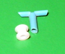
Tympanostomy tube, also known as a grommet, myringotomy tube, or pressure equalizing tube, is a small tube inserted into the eardrum via a surgical procedure called myringotomy to keep the middle ear aerated for a prolonged period of time, typically to prevent accumulation of fluid in the middle ear. The tube itself is made in a variety of designs, most often shaped like a grommet for short-term use, or with long flanges and sometimes resembling a T-shape for long-term use. Materials used to manufacture the tubes are often made from fluoroplastic or silicone, which have largely replaced the use of metal tubes made from stainless steel, titanium, or gold. Medical uses Inserting tympanostomy tubes is one of the most common pediatric surgical procedures in the United States, with 9% of children having had tubes placed sometime in their lives. Tympanostomy tubes are typically placed in one or both eardrums to help children suffering from recurrent acute otitis media (ear infec ... [...More Info...] [...Related Items...] OR: [Wikipedia] [Google] [Baidu] |
Keratin
Keratin () is one of a family of structural fibrous proteins also known as ''scleroproteins''. It is the key structural material making up Scale (anatomy), scales, hair, Nail (anatomy), nails, feathers, horn (anatomy), horns, claws, Hoof, hooves, and the outer layer of skin in vertebrates. Keratin also protects epithelial cells from damage or stress. Keratin is extremely insoluble in water and organic solvents. Keratin monomers assemble into bundles to form intermediate filaments, which are tough and form strong mineralization (biology), unmineralized epidermal appendages found in reptiles, birds, amphibians, and mammals. Excessive keratinization participate in fortification of certain tissues such as in horns of cattle and rhinos, and armadillos' osteoderm. The only other biology, biological matter known to approximate the toughness of keratinized tissue is chitin. Keratin comes in two types: the primitive, softer forms found in all vertebrates and the harder, derived forms fou ... [...More Info...] [...Related Items...] OR: [Wikipedia] [Google] [Baidu] |
Ossicle
The ossicles (also called auditory ossicles) are three irregular bones in the middle ear of humans and other mammals, and are among the smallest bones in the human body. Although the term "ossicle" literally means "tiny bone" (from Latin ''ossiculum'') and may refer to any small bone throughout the body, it typically refers specifically to the malleus, incus and stapes ("hammer, anvil, and stirrup") of the middle ear. The auditory ossicles serve as a kinematic chain to transmit and amplify (intensity (physics), intensify) sound vibrations collected from the air by the ear drum to the fluid-filled labyrinth (inner ear), labyrinth (cochlea). The absence or pathology of the auditory ossicles would constitute a moderate-to-severe conductive hearing loss. Structure The ossicles are, in order from the eardrum to the inner ear (from superficial to deep): the malleus, incus, and stapes, terms that in Latin are translated as "the hammer, anvil, and stirrup". * The malleus () articulat ... [...More Info...] [...Related Items...] OR: [Wikipedia] [Google] [Baidu] |
Valsalva Maneuver
The Valsalva maneuver is performed by a forceful attempt of exhalation against a closed airway, usually done by closing one's mouth and pinching one's nose shut while expelling air, as if blowing up a balloon. Variations of the maneuver can be used either in medicine, medical examination as a test of cardiac function and autonomic nervous system, autonomic nervous control of the heart (because the maneuver raises the pressure in the lungs), or to clear the ears and paranasal sinuses, sinuses (that is, to equalize pressure between them) when ambient pressure changes, as in scuba diving, hyperbaric oxygen therapy, or air travel. A modified version is done by expiring against a closed glottis. This will elicit the cardiovascular responses described below but will not force air into the Eustachian tubes. History The technique is named after Antonio Maria Valsalva, a 17th-century physician and anatomist from Bologna whose principal scientific interest was the human ear. He descri ... [...More Info...] [...Related Items...] OR: [Wikipedia] [Google] [Baidu] |


