|
Eustachian Tube
The Eustachian tube (), also called the auditory tube or pharyngotympanic tube, is a tube that links the nasopharynx to the middle ear, of which it is also a part. In adult humans, the Eustachian tube is approximately long and in diameter. It is named after the sixteenth-century Italian anatomist Bartolomeo Eustachi. In humans and other tetrapods, both the middle ear and the ear canal are normally filled with air. Unlike the air of the ear canal, however, the air of the middle ear is not in direct contact with the atmosphere outside the body; thus, a pressure difference can develop between the atmospheric pressure of the ear canal and the middle ear. Normally, the Eustachian tube is collapsed, but it gapes open with swallowing and with positive pressure, allowing the middle ear's pressure to adjust to the atmospheric pressure. When taking off in an aircraft, the ambient air pressure goes from higher (on the ground) to lower (in the sky). The air in the middle ear expands as ... [...More Info...] [...Related Items...] OR: [Wikipedia] [Google] [Baidu] |
Middle Ear
The middle ear is the portion of the ear medial to the eardrum, and distal to the oval window of the cochlea (of the inner ear). The mammalian middle ear contains three ossicles (malleus, incus, and stapes), which transfer the vibrations of the eardrum into waves in the fluid and membranes of the inner ear. The hollow space of the middle ear is also known as the tympanic cavity and is surrounded by the tympanic part of the temporal bone. The auditory tube (also known as the Eustachian tube or the pharyngotympanic tube) joins the tympanic cavity with the nasal cavity ( nasopharynx), allowing pressure to equalize between the middle ear and throat. The primary function of the middle ear is to efficiently transfer acoustic energy from compression waves in air to fluid–membrane waves within the cochlea. Structure Ossicles The middle ear contains three tiny bones known as the ossicles: '' malleus'', ''incus'', and '' stapes''. The ossicles were given their Latin names f ... [...More Info...] [...Related Items...] OR: [Wikipedia] [Google] [Baidu] |
Air Cell Of Temporal Bone
An atmosphere () is a layer of gases that envelop an astronomical object, held in place by the gravity of the object. A planet retains an atmosphere when the gravity is great and the temperature of the atmosphere is low. A stellar atmosphere is the outer region of a star, which includes the layers above the opaque photosphere; stars of low temperature might have outer atmospheres containing compound molecules. The atmosphere of Earth is composed of nitrogen (78%), oxygen (21%), argon (0.9%), carbon dioxide (0.04%) and trace gases. Most organisms use oxygen for respiration; lightning and bacteria perform nitrogen fixation which produces ammonia that is used to make nucleotides and amino acids; plants, algae, and cyanobacteria use carbon dioxide for photosynthesis. The layered composition of the atmosphere minimises the harmful effects of sunlight, ultraviolet radiation, solar wind, and cosmic rays and thus protects the organisms from genetic damage. The current composition o ... [...More Info...] [...Related Items...] OR: [Wikipedia] [Google] [Baidu] |
Transverse Section
A transverse plane is a plane that is rotated 90° from two other planes. Anatomy The transverse plane is an anatomical plane that is perpendicular to the sagittal plane and the dorsal plane. It is also called the axial plane or horizontal plane, especially in human anatomy, but horizontal plane can be misleading with other animals. The plane splits the body into a cranial (head) side and caudal (tail) side, so in humans the plane will be horizontal (dividing the body into Anatomical terms of location#Superior and inferior, superior and inferior sections) but in quadrupeds it will be vertical. Human anatomy Clinically relevant anatomical planes * Transverse ''thoracic plane'' * ''Xiphosternal plane'' (or xiphosternal junction) * ''Transpyloric plane'' * ''Subcostal plane'' * ''Umbilical plane'' (or transumbilical plane) * ''Supracristal plane'' * ''Intertubercular plane'' (or transtubercular plane) * ''Interspinous plane'' Associated structures * The transverse ''thoracic ... [...More Info...] [...Related Items...] OR: [Wikipedia] [Google] [Baidu] |
Torus Tubarius
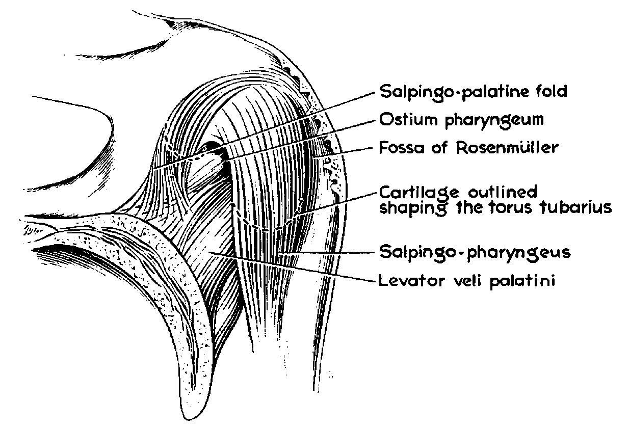
The torus tubarius (or torus of the auditory tube) is an elevation of the mucous membrane of the Pharynx#Nasopharynx, nasal part of the pharynx formed by the underlying base of the Eustachian tube, cartilaginous portion of the Eustachian tube, Eustachian tube (auditory tube). The torus tubarius is situated behind the pharyngeal orifice of the auditory tube. The torus tubarius is very close to the tubal tonsil, which is sometimes also referred to as the ''tonsil of (the) torus tubarius''. Two folds run anteriorly and posteriorly to the torus tubarius: the salpingopalatine fold (anteriorly), and the salpingopharyngeal fold (posteriorly). See also * Tubarial salivary gland References External links * * {{Authority control Ear ... [...More Info...] [...Related Items...] OR: [Wikipedia] [Google] [Baidu] |
Pharynx
The pharynx (: pharynges) is the part of the throat behind the human mouth, mouth and nasal cavity, and above the esophagus and trachea (the tubes going down to the stomach and the lungs respectively). It is found in vertebrates and invertebrates, though its structure varies across species. The pharynx carries food to the esophagus and air to the larynx. The flap of cartilage called the epiglottis stops food from entering the larynx. In humans, the pharynx is part of the Digestion, digestive system and the conducting zone of the respiratory system. (The conducting zone—which also includes the nostrils of the Human nose, nose, the larynx, trachea, bronchus, bronchi, and bronchioles—filters, warms, and moistens air and conducts it into the lungs). The human pharynx is conventionally divided into three sections: the nasopharynx, oropharynx, and laryngopharynx (hypopharynx). In humans, two sets of pharyngeal muscles form the pharynx and determine the shape of its lumen (anatomy), ... [...More Info...] [...Related Items...] OR: [Wikipedia] [Google] [Baidu] |
Mucous Membrane
A mucous membrane or mucosa is a membrane that lines various cavities in the body of an organism and covers the surface of internal organs. It consists of one or more layers of epithelial cells overlying a layer of loose connective tissue. It is mostly of endodermal origin and is continuous with the skin at body openings such as the eyes, eyelids, ears, inside the nose, inside the mouth, lips, the genital areas, the urethral opening and the anus. Some mucous membranes secrete mucus, a thick protective fluid. The function of the membrane is to stop pathogens and dirt from entering the body and to prevent bodily tissues from becoming dehydrated. Structure The mucosa is composed of one or more layers of epithelial cells that secrete mucus, and an underlying lamina propria of loose connective tissue. The type of cells and type of mucus secreted vary from organ to organ and each can differ along a given tract. Mucous membranes line the digestive, respiratory and rep ... [...More Info...] [...Related Items...] OR: [Wikipedia] [Google] [Baidu] |
Elastic Fibrocartilage
Cartilage is a resilient and smooth type of connective tissue. Semi-transparent and non-porous, it is usually covered by a tough and fibrous membrane called perichondrium. In tetrapods, it covers and protects the ends of long bones at the joints as articular cartilage, and is a structural component of many body parts including the rib cage, the neck and the bronchial tubes, and the intervertebral discs. In other taxa, such as chondrichthyans and cyclostomes, it constitutes a much greater proportion of the skeleton. It is not as hard and rigid as bone, but it is much stiffer and much less flexible than muscle. The matrix of cartilage is made up of glycosaminoglycans, proteoglycans, collagen fibers and, sometimes, elastin. It usually grows quicker than bone. Because of its rigidity, cartilage often serves the purpose of holding tubes open in the body. Examples include the rings of the trachea, such as the cricoid cartilage and carina. Cartilage is composed of specialized cells ... [...More Info...] [...Related Items...] OR: [Wikipedia] [Google] [Baidu] |
University Of Michigan Medical School
The University of Michigan Medicine (branded as Michigan Medicine) is the academic medical center of the University of Michigan, a public research university in Ann Arbor, Michigan. It consists of the university's Medical School, affiliated hospitals, and affiliated healthcare centers. History In 1869 the University of Michigan opened the first hospital in the country owned and operated by a university, in a house in Ann Arbor originally built as a professor's residence. In 1876 a new hospital building was opened adjacent to the old one. At the insistence of the Michigan Legislature, the new building had two separate departments, one for medicine and the other for homeopathy. In 1891 the hospital moved to a set of new buildings away from the university campus, on Catherine Street. The homeopathy department closed in 1921. In 1925 the university opened a hospital building at a cost of $3.85 million. It was designed by Albert Kahn (architect), Albert Kahn and built by T ... [...More Info...] [...Related Items...] OR: [Wikipedia] [Google] [Baidu] |
Temporal Bone
The temporal bone is a paired bone situated at the sides and base of the skull, lateral to the temporal lobe of the cerebral cortex. The temporal bones are overlaid by the sides of the head known as the temples where four of the cranial bones fuse. Each temple is covered by a temporal muscle. The temporal bones house the structures of the ears. The lower seven cranial nerves and the major vessels to and from the brain traverse the temporal bone. Structure The temporal bone consists of four parts—the squamous, mastoid, petrous and tympanic parts. The squamous part is the largest and most superiorly positioned relative to the rest of the bone. The zygomatic process is a long, arched process projecting from the lower region of the squamous part and it articulates with the zygomatic bone. Posteroinferior to the squamous is the mastoid part. Fused with the squamous and mastoid parts and between the sphenoid and occipital bones lies the petrous part, which is shaped li ... [...More Info...] [...Related Items...] OR: [Wikipedia] [Google] [Baidu] |
Petrous Part Of The Temporal Bone
The petrous part of the temporal bone is pyramid-shaped and is wedged in at the base of the skull between the sphenoid and occipital bones. Directed medially, forward, and a little upward, it presents a base, an apex, three surfaces, and three angles, and houses in its interior the components of the inner ear. The petrous portion is among the most basal elements of the skull and forms part of the endocranium. Petrous comes from the Latin word ''petrosus'', meaning "stone-like, hard". It is one of the densest bones in the body. In other mammals, it is a separate bone, the petrosal bone. The petrous bone is important for studies of ancient DNA from skeletal remains, as it tends to contain extremely well-preserved DNA. Base The base is fused with the internal surfaces of the squamous, tympanic, and mastoid parts. Apex The apex, which is rough and uneven, is received into the angular interval between the posterior border of the great wing of the sphenoid bone and the basi ... [...More Info...] [...Related Items...] OR: [Wikipedia] [Google] [Baidu] |
Squamous Part Of Temporal Bone
The squamous part of temporal bone, or temporal squama, forms the front and upper part of the temporal bone, and is scale-like, thin, and translucent. Surfaces Its outer surface is smooth and convex; it affords attachment to the temporal muscle, and forms part of the temporal fossa; on its hinder part is a vertical groove for the middle temporal artery. A curved line, the ''temporal line'', or ''supramastoid crest'', runs backward and upward across its posterior part; it serves for the attachment of the temporal fascia, and limits the origin of the temporalis muscle. The boundary between the squamous part and the mastoid portion of the bone, as indicated by traces of the original suture, lies about 1 cm. below this line. Projecting from the lower part of the squamous part is a long, arched process, the ''zygomatic process''. This process is at first directed lateralward, its two surfaces looking upward and downward; it then appears as if twisted inward upon itself, and runs ... [...More Info...] [...Related Items...] OR: [Wikipedia] [Google] [Baidu] |
Tympanic Cavity
The tympanic cavity is a small cavity surrounding the bones of the middle ear. Within it sit the ossicles, three small bones that transmit vibrations used in the detection of sound. Structure On its lateral surface, it abuts the external auditory meatus (ear canal) from which it is separated by the tympanic membrane (eardrum). Walls The tympanic cavity is bounded by: * Facing the inner ear, the medial wall (or ''labyrinthic wall'', ''labyrinthine wall'') is vertical, and has the oval window and round window, the promontory, and the prominence of the facial canal. * Facing the outer ear, the lateral wall (or ''membranous wall''), is formed mainly by the tympanic membrane, partly by the ring of bone into which this membrane is inserted. This ring of bone is incomplete at its upper part, forming a notch (notch of Rivinus), close to which are three small apertures: the "iter chordæ posterius", the petrotympanic fissure, and the "iter chordæ anterius". The iter chordæ pos ... [...More Info...] [...Related Items...] OR: [Wikipedia] [Google] [Baidu] |


