|
Trophoblastic Neoplasm
Gestational trophoblastic neoplasia (GTN) is group of rare diseases related to pregnancy and included in gestational trophoblastic disease (GTD) in which abnormal trophoblast cells grow in the uterus. GTN can be classified into benign and malignant lesions. Benign lesions include placental site nodule and hydatidiform moles while malignant lesions have four subtypes including invasive mole, gestational choriocarcinoma, placental site trophoblastic tumor (PSTT) and epithelioid trophoblastic tumor (ETT). The choriocarcinoma has 2 significant subtypes including gestational and non-gestational and they are differentiated by their different biological feature and prognosis. Signs and symptoms of GTN will appear vary from person to person and depending upon the type of the disease. They may include uterine bleeding not related to menstruation, pain or pressure in pelvis, large uterus and high blood pressure during pregnancy. The cause of this disease is unknown but the identification ... [...More Info...] [...Related Items...] OR: [Wikipedia] [Google] [Baidu] |
Micrograph
A micrograph or photomicrograph is a photograph or digital image taken through a microscope or similar device to show a magnified image of an object. This is opposed to a macrograph or photomacrograph, an image which is also taken on a microscope but is only slightly magnified, usually less than 10 times. Micrography is the practice or art of using microscopes to make photographs. A micrograph contains extensive details of microstructure. A wealth of information can be obtained from a simple micrograph like behavior of the material under different conditions, the phases found in the system, failure analysis, grain size estimation, elemental analysis and so on. Micrographs are widely used in all fields of microscopy. Types Photomicrograph A light micrograph or photomicrograph is a micrograph prepared using an optical microscope, a process referred to as ''photomicroscopy''. At a basic level, photomicroscopy may be performed simply by connecting a camera to a microscope ... [...More Info...] [...Related Items...] OR: [Wikipedia] [Google] [Baidu] |
Anemia
Anemia or anaemia (British English) is a blood disorder in which the blood has a reduced ability to carry oxygen due to a lower than normal number of red blood cells, or a reduction in the amount of hemoglobin. When anemia comes on slowly, the symptoms are often vague, such as tiredness, weakness, shortness of breath, headaches, and a reduced ability to exercise. When anemia is acute, symptoms may include confusion, feeling like one is going to pass out, loss of consciousness, and increased thirst. Anemia must be significant before a person becomes noticeably pale. Symptoms of anemia depend on how quickly hemoglobin decreases. Additional symptoms may occur depending on the underlying cause. Preoperative anemia can increase the risk of needing a blood transfusion following surgery. Anemia can be temporary or long term and can range from mild to severe. Anemia can be caused by blood loss, decreased red blood cell production, and increased red blood cell breakdown. Causes ... [...More Info...] [...Related Items...] OR: [Wikipedia] [Google] [Baidu] |
Syncytiotrophoblast
Syncytiotrophoblast (from the Greek 'syn'- "together"; 'cytio'- "of cells"; 'tropho'- "nutrition"; 'blast'- "bud") is the epithelial covering of the highly vascular embryonic placental villi, which invades the wall of the uterus to establish nutrient circulation between the embryo and the mother. It is a multi-nucleate, terminally differentiated syncytium, extending to 13cm. Function It is the outer layer of the trophoblasts and actively invades the uterine wall, during implantation, rupturing maternal capillaries and thus establishing an interface between maternal blood and embryonic extracellular fluid, facilitating passive exchange of material between the mother and the embryo. The syncytial property is important since the mother's immune system includes white blood cells that are able to migrate into tissues by "squeezing" in between cells. If they were to reach the fetal side of the placenta many foreign proteins would be recognised, triggering an immune reaction. ... [...More Info...] [...Related Items...] OR: [Wikipedia] [Google] [Baidu] |
Cytotrophoblast
"Cytotrophoblast" is the name given to both the inner layer of the trophoblast (also called layer of Langhans) or the cells that live there. It is interior to the syncytiotrophoblast and external to the wall of the blastocyst in a developing embryo. The cytotrophoblast is considered to be the trophoblastic stem cell because the layer surrounding the blastocyst remains while daughter cells differentiate and proliferate to function in multiple roles. There are two lineages that cytotrophoblastic cells may differentiate through: fusion and invasive. The fusion lineage yields syncytiotrophoblast and the invasive lineage yields interstitial cytotrophoblast cells. Cytotrophoblastic cells play an important role in the implantation of an embryo in the uterus. Fusion lineage The formation of all syncytiotrophoblast is from the fusion of two or more cytotrophoblasts via this fusion pathway. This pathway is important because the syncytiotrophoblast plays an important role in fetal-mater ... [...More Info...] [...Related Items...] OR: [Wikipedia] [Google] [Baidu] |
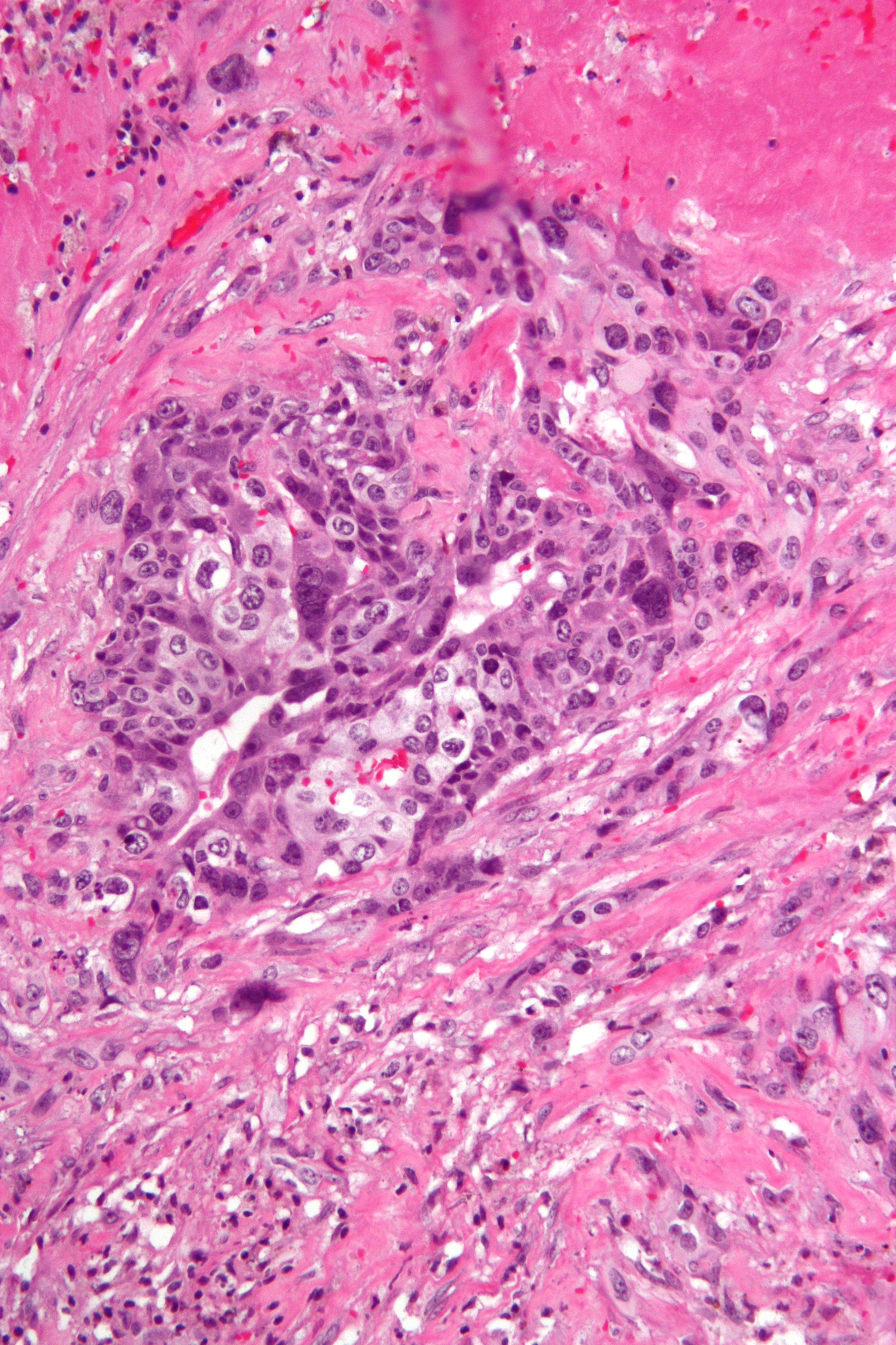
Choriocarcinoma -2- High Mag
Choriocarcinoma is a malignant, trophoblastic cancer, usually of the placenta. It is characterized by early hematogenous spread to the lungs. It belongs to the malignant end of the spectrum in gestational trophoblastic disease (GTD). It is also classified as a germ cell tumor and may arise in the testis or ovary. Signs and symptoms * increased quantitative chorionic gonadotropin (the "pregnancy hormone") levels * vaginal bleeding * shortness of breath * hemoptysis (coughing up blood) * chest pain * chest X-ray shows multiple infiltrates of various shapes in both lungs * presents in males as a testicular cancer, sometimes with skin hyperpigmentation (from excess chorionic gonadotropin cross reacting with the alpha MSH receptor), gynecomastia, and weight loss (from excess chorionic gonadotropin cross reacting with the LH, FSH, and TSH receptor) in males * can present with decreased thyroid-stimulating hormone (TSH) due to hyperthyroidism. Cause Choriocarcinoma of the placenta dur ... [...More Info...] [...Related Items...] OR: [Wikipedia] [Google] [Baidu] |
Broad Ligament Of The Uterus
The broad ligament of the uterus is the wide fold of peritoneum that connects the sides of the uterus to the walls and floor of the pelvis. Structure Subdivisions Contents The contents of the broad ligament include the following: * Reproductive ** uterine tubes (or Fallopian tube) ** ovary (some sources consider the ovary to be on the broad ligament, but not in it.) * vessels ** ovarian artery (in the suspensory ligament) ** uterine artery (in reality, travels in the cardinal ligament) * ligaments ** ovarian ligament ** round ligament of uterus ** suspensory ligament of the ovary (Some sources consider it a part of the broad ligament, while other sources just consider it a "termination" of the ligament.) Relations The peritoneum surrounds the uterus like a flat sheet that folds over its fundus, covering it anteriorly and posteriorly; on the sides of the uterus, this sheet of peritoneum comes in direct contact with itself, forming the double layer of peritoneum known as ... [...More Info...] [...Related Items...] OR: [Wikipedia] [Google] [Baidu] |
Adnexa Of Uterus
The uterine appendages (or adnexa of uterus) are the structures most closely related structurally and functionally to the uterus. Terminology They can be defined in slightly different ways: * Some sources define the adnexa as the fallopian tubes and ovaries. * Others include the supporting tissues". * Another source defines the appendages as the "regions of the true pelvis posterior to the broad ligaments". * One dictionary includes the fallopian tubes, ovaries, and ligaments (without specifying precisely which ligaments are included). Clinical significance The term "adnexitis" is sometimes used to describe an inflammation of the uterine appendages (adnexa). In this context, it replaces the terms oophoritis and salpingitis. The term adnexal mass is sometimes used when the location of a uterine mass is not yet more precisely known. 63% of ectopic pregnancies present with an adnexal mass. Depending on the size of the mass, it could be a medical emergency. Term "Adnexectomy" ... [...More Info...] [...Related Items...] OR: [Wikipedia] [Google] [Baidu] |
FIGO Classification Of Uterine Bleeding
The International Federation of Gynecology and ObstetricsFIGO is an international organization that links about 125 international professional societies of Obstetricians and Gynecologists. In 2011 FIGO recognized two systems designed to aid research, education, and clinical care of women with abnormal uterine bleeding (AUB) in the reproductive years. This page is a summary of the systems and their use in contemporary gynecology. Background Abnormal uterine bleeding (AUB) in the reproductive years, unrelated to pregnancy, is rarely life-threatening, but is frequently life altering. The symptoms frequently interfere with quality of life and those girls and women affected by chronic AUB spend significant amounts of personal resources on menstrual products and medications. Such women are 30 per cent less productive at work, and, consequently, suffer a similar reduction in income. For low resource countries, the combination of poor nutrition, lack of access to simple therapy with iron ... [...More Info...] [...Related Items...] OR: [Wikipedia] [Google] [Baidu] |
Cerebrospinal Fluid
Cerebrospinal fluid (CSF) is a clear, colorless body fluid found within the tissue that surrounds the brain and spinal cord of all vertebrates. CSF is produced by specialised ependymal cells in the choroid plexus of the ventricles of the brain, and absorbed in the arachnoid granulations. There is about 125 mL of CSF at any one time, and about 500 mL is generated every day. CSF acts as a shock absorber, cushion or buffer, providing basic mechanical and immunological protection to the brain inside the skull. CSF also serves a vital function in the cerebral autoregulation of cerebral blood flow. CSF occupies the subarachnoid space (between the arachnoid mater and the pia mater) and the ventricular system around and inside the brain and spinal cord. It fills the ventricles of the brain, cisterns, and sulci, as well as the central canal of the spinal cord. There is also a connection from the subarachnoid space to the bony labyrinth of the inner ear via the ... [...More Info...] [...Related Items...] OR: [Wikipedia] [Google] [Baidu] |
Miscarriage
Miscarriage, also known in medical terms as a spontaneous abortion and pregnancy loss, is the death of an embryo or fetus before it is able to survive independently. Miscarriage before 6 weeks of gestation is defined by ESHRE as biochemical loss. Once ultrasound or histological evidence shows that a pregnancy has existed, the used term is clinical miscarriage, which can be ''early'' before 12 weeks and ''late'' between 12-21 weeks. Fetal death after 20 weeks of gestation is also known as a stillbirth. The most common symptom of a miscarriage is vaginal bleeding with or without pain. Sadness, anxiety, and guilt may occur afterwards. Tissue and clot-like material may leave the uterus and pass through and out of the vagina. Recurrent miscarriage (also referred to medically as Recurrent Spontaneous Abortion or RSA) may also be considered a form of infertility. Risk factors for miscarriage include being an older parent, previous miscarriage, exposure to tobacco smoke, ... [...More Info...] [...Related Items...] OR: [Wikipedia] [Google] [Baidu] |
H&E Stain
Hematoxylin and eosin stain ( or haematoxylin and eosin stain or hematoxylin-eosin stain; often abbreviated as H&E stain or HE stain) is one of the principal tissue stains used in histology. It is the most widely used stain in medical diagnosis and is often the gold standard. For example, when a pathologist looks at a biopsy of a suspected cancer, the histological section is likely to be stained with H&E. H&E is the combination of two histological stains: hematoxylin and eosin. The hematoxylin stains cell nuclei a purplish blue, and eosin stains the extracellular matrix and cytoplasm pink, with other structures taking on different shades, hues, and combinations of these colors. Hence a pathologist can easily differentiate between the nuclear and cytoplasmic parts of a cell, and additionally, the overall patterns of coloration from the stain show the general layout and distribution of cells and provides a general overview of a tissue sample's structure. Thus, pattern recognit ... [...More Info...] [...Related Items...] OR: [Wikipedia] [Google] [Baidu] |
Human Chorionic Gonadotropin
Human chorionic gonadotropin (hCG) is a hormone for the maternal recognition of pregnancy produced by trophoblast cells that are surrounding a growing embryo (syncytiotrophoblast initially), which eventually forms the placenta after implantation. The presence of hCG is detected in some pregnancy tests (HCG pregnancy strip tests). Some cancerous tumors produce this hormone; therefore, elevated levels measured when the patient is not pregnant may lead to a cancer diagnosis and, if high enough, paraneoplastic syndromes, however, it is not known whether this production is a contributing cause, or an effect of carcinogenesis. The pituitary analog of hCG, known as luteinizing hormone (LH), is produced in the pituitary gland of males and females of all ages. Various endogenous forms of hCG exist. The measurement of these diverse forms is used in the diagnosis of pregnancy and a variety of disease states. Preparations of hCG from various sources have also been used therapeutically, ... [...More Info...] [...Related Items...] OR: [Wikipedia] [Google] [Baidu] |


