|
Tuberosity
In the skeleton of humans and other animals, a tubercle, tuberosity or apophysis is a protrusion or eminence that serves as an attachment for skeletal muscles. The muscles attach by tendons, where the enthesis is the connective tissue between the tendon and bone. A ''tuberosity'' is generally a larger tubercle. Main tubercles Humerus The humerus has two tubercles, the greater tubercle and the lesser tubercle. These are situated at the proximal end of the bone, that is the end that connects with the scapula. The greater/lesser tubercule is located from the top of the acromion laterally and inferiorly. The radius has two, the radial tuberosity and Lister's tubercle. Ribs On a rib, tubercle is an eminence on the back surface, at the junction between the neck and the body of the rib. It consists of an articular and a non-articular area. The lower and more medial articular area is a small oval surface for articulation with the transverse process of the lower of the two vertebrae wh ... [...More Info...] [...Related Items...] OR: [Wikipedia] [Google] [Baidu] |
Radial Tuberosity
Beneath the neck of the radius, on the medial side, is an eminence, the radial tuberosity; its surface is divided into: * a ''posterior, rough portion'', for the insertion of the tendon of the biceps brachii. * an ''anterior, smooth portion'', on which a bursa ( grc-gre, Προῦσα, Proûsa, Latin: Prusa, ota, بورسه, Arabic:بورصة) is a city in northwestern Turkey and the administrative center of Bursa Province. The fourth-most populous city in Turkey and second-most populous in t ... is interposed between the tendon and the bone. Ligaments that support the elbow joint also attach to the radial tuberosity. References External links * * () Additional images File:Sobo 1909 124.png, Radial tuberosity shown. File:Radius.jpg, Anterior View. Radial tuberosity. File:Radius2.jpg, Posterior View. Radial tuberosity. File:Radius_post.jpg, Radial tuberosity below neck. File:Radius_ant.jpg, Radial tuberosity below neck. File:Left_radius_-_close-up_-_animation ... [...More Info...] [...Related Items...] OR: [Wikipedia] [Google] [Baidu] |
Humerus
The humerus (; ) is a long bone in the arm that runs from the shoulder to the elbow. It connects the scapula and the two bones of the lower arm, the radius and ulna, and consists of three sections. The humeral upper extremity consists of a rounded head, a narrow neck, and two short processes (tubercles, sometimes called tuberosities). The body is cylindrical in its upper portion, and more prismatic below. The lower extremity consists of 2 epicondyles, 2 processes (trochlea & capitulum), and 3 fossae (radial fossa, coronoid fossa, and olecranon fossa). As well as its true anatomical neck, the constriction below the greater and lesser tubercles of the humerus is referred to as its surgical neck due to its tendency to fracture, thus often becoming the focus of surgeons. Etymology The word "humerus" is derived from la, humerus, umerus meaning upper arm, shoulder, and is linguistically related to Gothic ''ams'' shoulder and Greek ''ōmos''. Structure Upper extremity The upper or pr ... [...More Info...] [...Related Items...] OR: [Wikipedia] [Google] [Baidu] |
Tibia
The tibia (; ), also known as the shinbone or shankbone, is the larger, stronger, and anterior (frontal) of the two bones in the leg below the knee in vertebrates (the other being the fibula, behind and to the outside of the tibia); it connects the knee with the ankle. The tibia is found on the medial side of the leg next to the fibula and closer to the median plane. The tibia is connected to the fibula by the interosseous membrane of leg, forming a type of fibrous joint called a syndesmosis with very little movement. The tibia is named for the flute '' tibia''. It is the second largest bone in the human body, after the femur. The leg bones are the strongest long bones as they support the rest of the body. Structure In human anatomy, the tibia is the second largest bone next to the femur. As in other vertebrates the tibia is one of two bones in the lower leg, the other being the fibula, and is a component of the knee and ankle joints. The ossification or formation of the bo ... [...More Info...] [...Related Items...] OR: [Wikipedia] [Google] [Baidu] |
Tibial Tuberosity
The tuberosity of the tibia or tibial tuberosity or tibial tubercle is an elevation on the proximal, anterior aspect of the tibia, just below where the anterior surfaces of the lateral and medial tibial condyles end. Structure The tuberosity of the tibia gives attachment to the patellar ligament, which attaches to the patella from where the suprapatellar ligament forms the distal tendon of the quadriceps femoris muscles. The quadriceps muscles consist of the rectus femoris, vastus lateralis, vastus medialis, and vastus intermedius. These quadriceps muscles are innervated by the femoral nerve. KneeHipPain (1998) The tibial tuberosity thus forms the terminal part of the large structure that acts as a lever to extend the knee-joint and prevents the knee from collapsing when the foot strikes the ground. The t ... [...More Info...] [...Related Items...] OR: [Wikipedia] [Google] [Baidu] |
Knee
In humans and other primates, the knee joins the thigh with the human leg, leg and consists of two joints: one between the femur and tibia (tibiofemoral joint), and one between the femur and patella (patellofemoral joint). It is the largest joint in the human body. The knee is a modified hinge joint, which permits flexion and extension (kinesiology), extension as well as slight internal and external rotation. The knee is vulnerable to injury and to the development of osteoarthritis. It is often termed a ''compound joint'' having tibiofemoral and patellofemoral components. (The fibular collateral ligament is often considered with tibiofemoral components.) Structure The knee is a modified hinge joint, a type of synovial joint, which is composed of three functional compartments: the patellofemoral articulation, consisting of the patella, or "kneecap", and the patellar groove on the front of the femur through which it slides; and the medial and lateral tibiofemoral articulation ... [...More Info...] [...Related Items...] OR: [Wikipedia] [Google] [Baidu] |
Fifth Metatarsal
The fifth metatarsal bone is a long bone in the foot, and is palpable along the distal outer edges of the feet. It is the second smallest of the five metatarsal bones. The fifth metatarsal is analogous to the fifth metacarpal bone in the hand. As with the four other metatarsal bones it can be divided into three parts; a base, body and head. The base is the part closest to the ankle and the head is closest to the toes. The narrowed part in the middle is referred to as the body (or shaft) of the bone. The bone is somewhat flat giving it two surfaces; the plantar (towards the sole of the foot) and the dorsal side (the area facing upwards while standing). These surfaces are rough for the attachment of ligaments. The bone is curved longitudinally, so as to be concave below, slightly convex above. The base articulates behind, by a triangular surface cut obliquely in a transverse direction, with the cuboid; and medially, with the fourth metatarsal. The fifth metatarsal has a rough emin ... [...More Info...] [...Related Items...] OR: [Wikipedia] [Google] [Baidu] |
Third Trochanter
In human anatomy, the third trochanter is a bony projection occasionally present on the proximal femur near the superior border of the gluteal tuberosity. When present, it is oblong, rounded, or conical in shape and sometimes continuous with the gluteal ridge. It generally occurs bilaterally without significant side to side dimorphism. A structure of minor importance in humans, the incidence of the third trochanter varies from 17 to 72% between ethnic groups and it is frequently reported as more common in females than in males. Structures analogous to the third trochanter are present in other mammals, including some primates. It is called the third trochanter in reference to the greater and lesser trochanters that are always present on the femur. Function Its function is to provide an attachment for the ascending tendon of the gluteus maximus muscle. It may function as (1) a reinforcement mechanism for the proximal femoral diaphysis in response to increased ground reaction ... [...More Info...] [...Related Items...] OR: [Wikipedia] [Google] [Baidu] |
Femur
The femur (; ), or thigh bone, is the proximal bone of the hindlimb in tetrapod vertebrates. The head of the femur articulates with the acetabulum in the pelvic bone forming the hip joint, while the distal part of the femur articulates with the tibia (shinbone) and patella (kneecap), forming the knee joint. By most measures the two (left and right) femurs are the strongest bones of the body, and in humans, the largest and thickest. Structure The femur is the only bone in the upper leg. The two femurs converge medially toward the knees, where they articulate with the proximal ends of the tibiae. The angle of convergence of the femora is a major factor in determining the femoral-tibial angle. Human females have thicker pelvic bones, causing their femora to converge more than in males. In the condition ''genu valgum'' (knock knee) the femurs converge so much that the knees touch one another. The opposite extreme is ''genu varum'' (bow-leggedness). In the general pop ... [...More Info...] [...Related Items...] OR: [Wikipedia] [Google] [Baidu] |
Gerdy's Tubercle
Gerdy's tubercle is a lateral tubercle of the tibia, located where the iliotibial tract inserts. It was named after French surgeon Pierre Nicolas Gerdy (1797–1856). Gerdy's tubercle is a smooth facet on the lateral aspect of the upper part of the tibia, just below the knee joint and adjacent to the proximal tibio-fibular joint, where the iliotibial tract runs down the outside part of the thigh. It is the point of insertion for the Iliotibial band of the lateral thigh. It is used as a site for the insertion of a periosteal needle by which intramedullary fluids may be infused in neonates. It can be fractured along with the tibial tuberosity. It has been used as a source for bone grafts. The peroneal nerve The common fibular nerve (also known as the common peroneal nerve, external popliteal nerve, or lateral popliteal nerve) is a nerve in the lower leg that provides sensation over the posterolateral part of the leg and the knee joint. It divides at ... runs near to it. Referen ... [...More Info...] [...Related Items...] OR: [Wikipedia] [Google] [Baidu] |
Scapula
The scapula (plural scapulae or scapulas), also known as the shoulder blade, is the bone that connects the humerus (upper arm bone) with the clavicle (collar bone). Like their connected bones, the scapulae are paired, with each scapula on either side of the body being roughly a mirror image of the other. The name derives from the Classical Latin word for trowel or small shovel, which it was thought to resemble. In compound terms, the prefix omo- is used for the shoulder blade in medical terminology. This prefix is derived from ὦμος (ōmos), the Ancient Greek word for shoulder, and is cognate with the Latin , which in Latin signifies either the shoulder or the upper arm bone. The scapula forms the back of the shoulder girdle. In humans, it is a flat bone, roughly triangular in shape, placed on a posterolateral aspect of the thoracic cage. Structure The scapula is a thick, flat bone lying on the thoracic wall that provides an attachment for three groups of muscles: i ... [...More Info...] [...Related Items...] OR: [Wikipedia] [Google] [Baidu] |
Tubercle
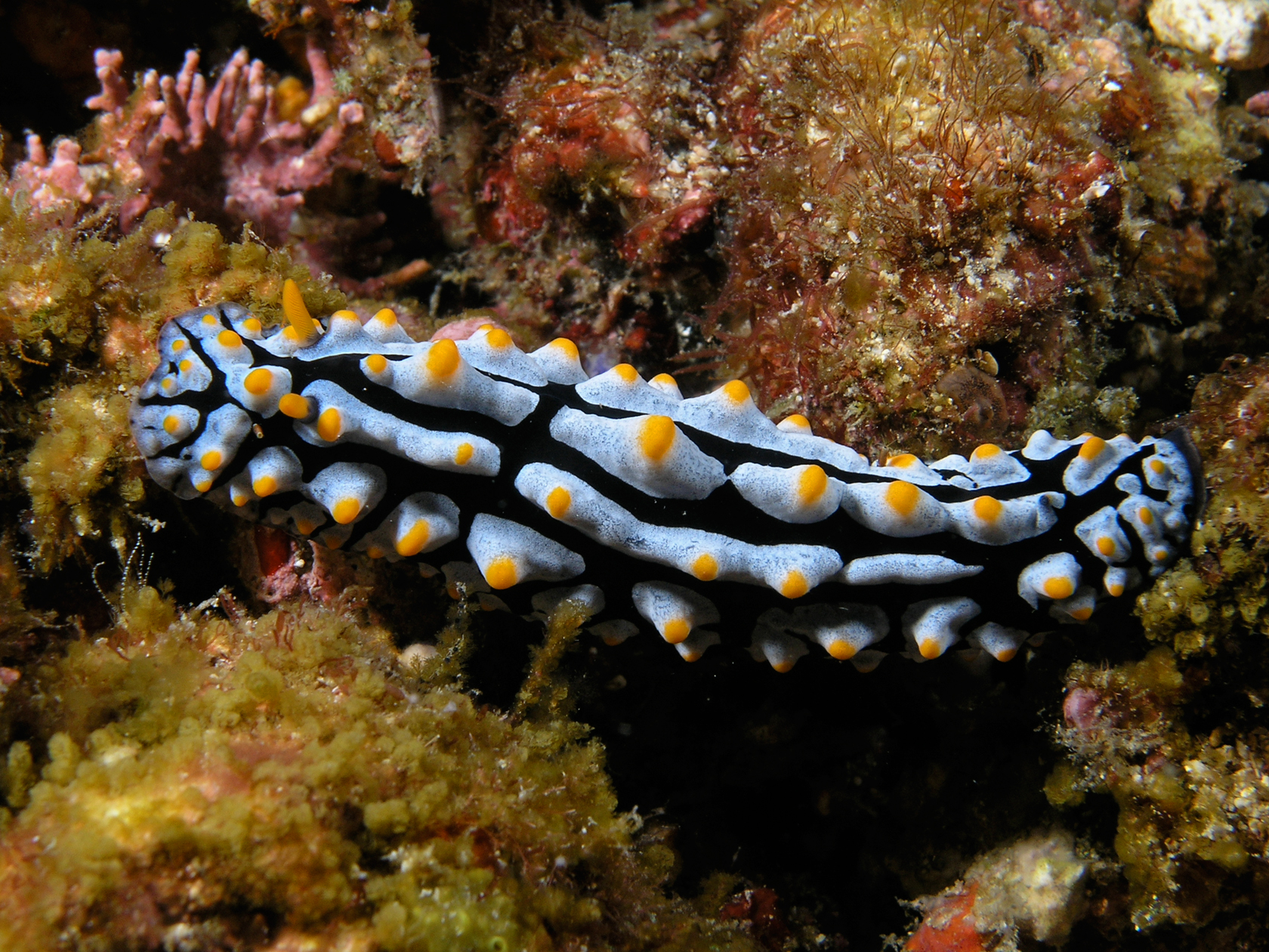
In anatomy, a tubercle (literally 'small tuber', Latin for 'lump') is any round nodule, small eminence, or warty outgrowth found on external or internal organs of a plant or an animal. In plants A tubercle is generally a wart-like projection, but it has slightly different meaning depending on which family of plants or animals it is used to refer to. In the case of certain orchids and cacti, it denotes a round nodule, small eminence, or warty outgrowth found on the lip. They are also known as podaria (singular ''podarium''). When referring to some members of the pea family, it is used to refer to the wart-like excrescences that are found on the roots. In fungi In mycology, a tubercle is used to refer to a mass of hyphae from which a mushroom is made. In animals When it is used in relation to certain dorid nudibranchs such as '' Peltodoris nobilis'', it means the nodules on the dorsum of the animal. The tubercles in nudibranchs can present themselves in different ways: ... [...More Info...] [...Related Items...] OR: [Wikipedia] [Google] [Baidu] |
Bone
A bone is a rigid organ that constitutes part of the skeleton in most vertebrate animals. Bones protect the various other organs of the body, produce red and white blood cells, store minerals, provide structure and support for the body, and enable mobility. Bones come in a variety of shapes and sizes and have complex internal and external structures. They are lightweight yet strong and hard and serve multiple functions. Bone tissue (osseous tissue), which is also called bone in the uncountable sense of that word, is hard tissue, a type of specialized connective tissue. It has a honeycomb-like matrix internally, which helps to give the bone rigidity. Bone tissue is made up of different types of bone cells. Osteoblasts and osteocytes are involved in the formation and mineralization of bone; osteoclasts are involved in the resorption of bone tissue. Modified (flattened) osteoblasts become the lining cells that form a protective layer on the bone surface. The mineralize ... [...More Info...] [...Related Items...] OR: [Wikipedia] [Google] [Baidu] |


