|
Transsphenoidal Surgery
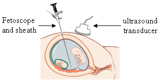
Transsphenoidal surgery is a type of surgery in which an endoscope or surgical instruments are inserted into part of the brain by going through the nose and the sphenoid bone (a butterfly-shaped bone forming the anterior inferior portion of the brain case) into the sphenoidal sinus cavity. Transsphenoidal surgery is used to remove tumors of the pituitary gland. (Such tumours, although within the skull, are outside the brain itself). History The transsphenoidal approach was first attempted by Hermann Schloffer in 1907. Use of the procedure grew in the 1950s and 1960s with the introduction of intraoperative fluoroscopy and operating microscope. See also * Pituitary adenoma * Endoscopic endonasal surgery, particularly Surgical approaches to the anterior skull base References External links Transsphenoidal surgeryentry in the public domain NCI Dictionary of Cancer Terms. Endoscopic transsphenoidal pituitary adenoma resection (IPG32) National Institute for Clinical Excellence ... [...More Info...] [...Related Items...] OR: [Wikipedia] [Google] [Baidu] |
Surgery
Surgery is a medical specialty that uses manual and instrumental techniques to diagnose or treat pathological conditions (e.g., trauma, disease, injury, malignancy), to alter bodily functions (e.g., malabsorption created by bariatric surgery such as gastric bypass), to reconstruct or alter aesthetics and appearance (cosmetic surgery), or to remove unwanted tissue (biology), tissues (body fat, glands, scars or skin tags) or foreign bodies. The act of performing surgery may be called a surgical procedure or surgical operation, or simply "surgery" or "operation". In this context, the verb "operate" means to perform surgery. The adjective surgical means pertaining to surgery; e.g. surgical instruments, operating theater, surgical facility or surgical nurse. Most surgical procedures are performed by a pair of operators: a surgeon who is the main operator performing the surgery, and a surgical assistant who provides in-procedure manual assistance during surgery. Modern surgical opera ... [...More Info...] [...Related Items...] OR: [Wikipedia] [Google] [Baidu] |
Endoscope
An endoscope is an inspection instrument composed of image sensor, optical lens, light source and mechanical device, which is used to look deep into the body by way of openings such as the mouth or anus. A typical endoscope applies several modern technologies including optics, Human factors and ergonomics, ergonomics, precision mechanics, electronics, and software engineering. With an endoscope, it is possible to observe lesions that cannot be detected by X-ray, making it useful in medical diagnosis. An endoscope uses tubes only a few millimeters thick to transfer illumination in one direction and high-resolution video in the other, allowing minimally invasive surgeries. It is used to examine the internal organs like the throat or esophagus. Specialized instruments are named after their target organ. Examples include the cystoscope (bladder), Nephroscopy, nephroscope (kidney), Bronchoscopy, bronchoscope (bronchus), arthroscope (joints) and colonoscope (colon), and Laparoscopy, lapar ... [...More Info...] [...Related Items...] OR: [Wikipedia] [Google] [Baidu] |
Surgical Instrument
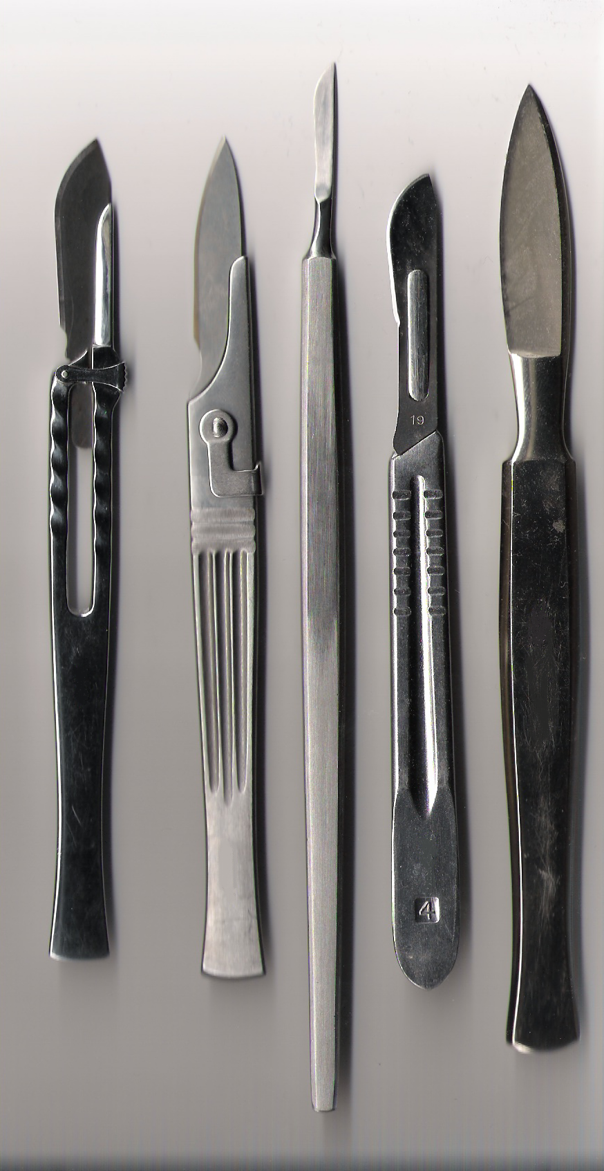
A surgical instrument is a medical device for performing specific actions or carrying out desired effects during a surgery or operation, such as modifying biological tissue, or to provide access for viewing it. Over time, many different kinds of surgical instruments and tools have been invented. Some surgical instruments are designed for general use in all sorts of surgeries, while others are designed for only certain specialties or specific procedures. Classification of surgical instruments helps surgeons to understand the functions and purposes of the instruments. With the goal of optimizing surgical results and performing more difficult operations, more instruments continue to be invented in the modern era. History Many different kinds of surgical instruments and tools have been invented and some have been repurposed as medical knowledge and surgical practices have developed. As surgery practice diversified, some tools are advanced for higher accuracy and stability while so ... [...More Info...] [...Related Items...] OR: [Wikipedia] [Google] [Baidu] |
Sphenoid Bone
The sphenoid bone is an unpaired bone of the neurocranium. It is situated in the middle of the skull towards the front, in front of the basilar part of occipital bone, basilar part of the occipital bone. The sphenoid bone is one of the seven bones that articulate to form the orbit (anatomy), orbit. Its shape somewhat resembles that of a butterfly, bat or wasp with its wings extended. The name presumably originates from this shape, since () means in Ancient Greek. Structure It is divided into the following parts: * a median portion, known as the body of sphenoid bone, containing the sella turcica, which houses the pituitary gland as well as the paired paranasal sinuses, the sphenoidal sinuses * two Greater wing of sphenoid bone, greater wings on the lateral side of the body and two Lesser wing of sphenoid bone, lesser wings from the anterior side. * Pterygoid processes of the sphenoides, directed downwards from the junction of the body and the greater wings. Two sphenoidal co ... [...More Info...] [...Related Items...] OR: [Wikipedia] [Google] [Baidu] |
Brain Case
In human anatomy, the neurocranium, also known as the braincase, brainpan, brain-pan, or brainbox, is the upper and back part of the skull, which forms a protective case around the brain. In the human skull, the neurocranium includes the calvaria or skullcap. The remainder of the skull is the facial skeleton. In comparative anatomy, neurocranium is sometimes used synonymously with endocranium or chondrocranium. Structure The neurocranium is divided into two portions: * the membranous part, consisting of flat bones, which surround the brain; and * the cartilaginous part, or chondrocranium, which forms bones of the base of the skull. In humans, the neurocranium is usually considered to include the following eight bones: * 1 ethmoid bone * 1 frontal bone * 1 occipital bone * 2 parietal bones * 1 sphenoid bone * 2 temporal bones The ossicles (three on each side) are usually not included as bones of the neurocranium. There may variably also be extra sutural bones present. Belo ... [...More Info...] [...Related Items...] OR: [Wikipedia] [Google] [Baidu] |
Sphenoidal Sinus
The sphenoid sinus is a paired paranasal sinus in the Body of sphenoid bone, body of the sphenoid bone. It is one pair of the four paired paranasal sinuses.Illustrated Anatomy of the Head and Neck, Fehrenbach and Herring, Elsevier, 2012, page 64 The two sphenoid sinuses are separated from each other by a septum. Each sphenoid sinus communicates with the nasal cavity via the opening of sphenoidal sinus. The two sphenoid sinuses vary in size and shape, and are usually asymmetrical. Structure On average, a sphenoid sinus measures 2.2 cm vertical height, 2 cm in transverse breadth; and 2.2 cm antero-posterior depth. Each spehoid sinus is in the body of sphenoid bone, just under the sella turcica. The sphenoid sinuses are separated from each other medially by the septum of sphenoidal sinuses, which is usually asymmetrical. An opening of sphenoidal sinus forms a passage between each sphenoidal sinus and the nasal cavity. Posteriorly, an opening of sphenoidal sinus opens ... [...More Info...] [...Related Items...] OR: [Wikipedia] [Google] [Baidu] |
Tumor
A neoplasm () is a type of abnormal and excessive growth of tissue. The process that occurs to form or produce a neoplasm is called neoplasia. The growth of a neoplasm is uncoordinated with that of the normal surrounding tissue, and persists in growing abnormally, even if the original trigger is removed. This abnormal growth usually forms a mass, which may be called a tumour or tumor.'' ICD-10 classifies neoplasms into four main groups: benign neoplasms, in situ neoplasms, malignant neoplasms, and neoplasms of uncertain or unknown behavior. Malignant neoplasms are also simply known as cancers and are the focus of oncology. Prior to the abnormal growth of tissue, such as neoplasia, cells often undergo an abnormal pattern of growth, such as metaplasia or dysplasia. However, metaplasia or dysplasia does not always progress to neoplasia and can occur in other conditions as well. The word neoplasm is from Ancient Greek 'new' and 'formation, creation'. Types A neopla ... [...More Info...] [...Related Items...] OR: [Wikipedia] [Google] [Baidu] |
Pituitary Gland
The pituitary gland or hypophysis is an endocrine gland in vertebrates. In humans, the pituitary gland is located at the base of the human brain, brain, protruding off the bottom of the hypothalamus. The pituitary gland and the hypothalamus control much of the body's endocrine system. It is seated in part of the sella turcica a fossa (anatomy), depression in the sphenoid bone, known as the hypophyseal fossa. The human pituitary gland is ovoid, oval shaped, about 1 cm in diameter, in weight on average, and about the size of a kidney bean. Digital version. There are two main lobes of the pituitary, an anterior pituitary, anterior lobe, and a posterior pituitary, posterior lobe joined and separated by a small intermediate lobe. The anterior lobe (adenohypophysis) is the glandular part that produces and secretes several hormones. The posterior lobe (neurohypophysis) secretes neurohypophysial hormones produced in the hypothalamus. Both lobes have different origins and they are both co ... [...More Info...] [...Related Items...] OR: [Wikipedia] [Google] [Baidu] |
Hermann Schloffer
Hermann Schloffer (May 13, 1868 in Graz - January 21, 1937) was an Austrian surgeon. He studied medicine at the University of Freiburg and University of Graz, where in 1892 he earned his medical doctorate. He spent several years in Prague as a surgical assistant and associate professor, and in 1903-1911 was a surgeon and professor at the University of Innsbruck. Afterwards he was a professor at Charles University in Prague. On March 16, 1907 Schloffer performed the first transsphenoidal surgery for removal of a pituitary adenoma at the University of Innsbruck. Unfortunately, the patient died several weeks afterwards from a residual tumor. His name is lent to the eponymous "Schloffer tumor", described as an uncommon pseudo-tumor of the abdominal wall that usually appears several years after abdominal surgery. In 1916 Schloffer became the first to remove a spleen for idiopathic thrombocytopenic purpura (ITP). His student Paul Kaznelson (1898-1959) hypothesized - in analogy wit ... [...More Info...] [...Related Items...] OR: [Wikipedia] [Google] [Baidu] |
Fluoroscopy
Fluoroscopy (), informally referred to as "fluoro", is an imaging technique that uses X-rays to obtain real-time moving images of the interior of an object. In its primary application of medical imaging, a fluoroscope () allows a surgeon to see the internal anatomy, structure and physiology, function of a patient, so that the pumping action of the heart or the motion of swallowing, for example, can be watched. This is useful for both medical diagnosis, diagnosis and therapy and occurs in general radiology, interventional radiology, and image-guided surgery. In its simplest form, a fluoroscope consists of an X-ray generator, X-ray source and a fluorescence, fluorescent screen, between which a patient is placed. However, since the 1950s most fluoroscopes have included X-ray image intensifiers and cameras as well, to improve the image's visibility and make it available on a remote display screen. For many decades, fluoroscopy tended to produce live pictures that were not recorded, bu ... [...More Info...] [...Related Items...] OR: [Wikipedia] [Google] [Baidu] |
Operating Microscope
An operating microscope or surgical microscope is an optical microscope specifically designed to be used in a surgical setting, typically to perform microsurgery. Design features of an operating microscope are: magnification typically in the range from 4x-40x, components that are easy to sterilize or disinfect in order to ensure cross-infection control. There is often a prism that allows splitting of the light beam in order that assistants may also visualize the procedure or to allow photography or video to be taken of the operating field. Typically an operating microscope might cost several thousand dollars for a basic model, more advanced models may be much more expensive. Additionally, specialized microsurgical instruments may be required to make full use of the improved vision the microscope affords. It can take time to master use of an operating microscope. Fields of medicine that make significant use of the operating microscope include plastic surgery, dentistry (esp ... [...More Info...] [...Related Items...] OR: [Wikipedia] [Google] [Baidu] |
Pituitary Adenoma
Pituitary adenomas are tumors that occur in the pituitary gland. Most pituitary tumors are benign, approximately 35% are invasive and just 0.1% to 0.2% are carcinomas.Pituitary Tumors Treatment (PDQ®)–Health Professional Version NIH National Cancer Institute Pituitary adenomas represent from 10% to 25% of all intracranial neoplasia, neoplasms, with an estimated prevalence, prevalence rate in the general population of approximately 17%. Non-invasive and non-secreting pituitary adenomas are considered to be Benign tumors, benign in the literal as well as the clinical sense, though a 2011 meta-analysis of available research showed that research to either support or refute this assumption was scant and of questionable qua ... [...More Info...] [...Related Items...] OR: [Wikipedia] [Google] [Baidu] |


