|
Fluoroscopy
Fluoroscopy (), informally referred to as "fluoro", is an imaging technique that uses X-rays to obtain real-time moving images of the interior of an object. In its primary application of medical imaging, a fluoroscope () allows a surgeon to see the internal anatomy, structure and physiology, function of a patient, so that the pumping action of the heart or the motion of swallowing, for example, can be watched. This is useful for both medical diagnosis, diagnosis and therapy and occurs in general radiology, interventional radiology, and image-guided surgery. In its simplest form, a fluoroscope consists of an X-ray generator, X-ray source and a fluorescence, fluorescent screen, between which a patient is placed. However, since the 1950s most fluoroscopes have included X-ray image intensifiers and cameras as well, to improve the image's visibility and make it available on a remote display screen. For many decades, fluoroscopy tended to produce live pictures that were not recorded, bu ... [...More Info...] [...Related Items...] OR: [Wikipedia] [Google] [Baidu] |
Interventional Radiology
Interventional radiology (IR) is a medical specialty that performs various minimally-invasive procedures using medical imaging guidance, such as Fluoroscopy, x-ray fluoroscopy, CT scan, computed tomography, magnetic resonance imaging, or ultrasound. IR performs both diagnostic and therapeutic procedures Minimally invasive procedure, through very small incisions or body orifices. Diagnostic IR procedures are those intended to help make a diagnosis or guide further medical treatment, and include image-guided biopsy of a tumor or injection of an Radiocontrast agent, imaging contrast agent into a hollow structure, such as a blood vessel or a Bile duct, duct. By contrast, Therapy, therapeutic IR procedures provide direct treatment—they include catheter-based medicine delivery, medical device placement (e.g., stents), and angioplasty of narrowed structures. The main benefits of IR techniques are that they can reach the deep structures of the body through a body orifice or tiny incisi ... [...More Info...] [...Related Items...] OR: [Wikipedia] [Google] [Baidu] |
Radiography
Radiography is an imaging technology, imaging technique using X-rays, gamma rays, or similar ionizing radiation and non-ionizing radiation to view the internal form of an object. Applications of radiography include medical ("diagnostic" radiography and "therapeutic radiography") and industrial radiography. Similar techniques are used in airport security, (where "body scanners" generally use backscatter X-ray). To create an image in conventional radiography, a beam of X-rays is produced by an X-ray generator and it is projected towards the object. A certain amount of the X-rays or other radiation are absorbed by the object, dependent on the object's density and structural composition. The X-rays that pass through the object are captured behind the object by a X-ray detector, detector (either photographic film or a digital detector). The generation of flat two-dimensional images by this technique is called Projection radiography, projectional radiography. In computed tomography (C ... [...More Info...] [...Related Items...] OR: [Wikipedia] [Google] [Baidu] |
X-ray Image Intensifier
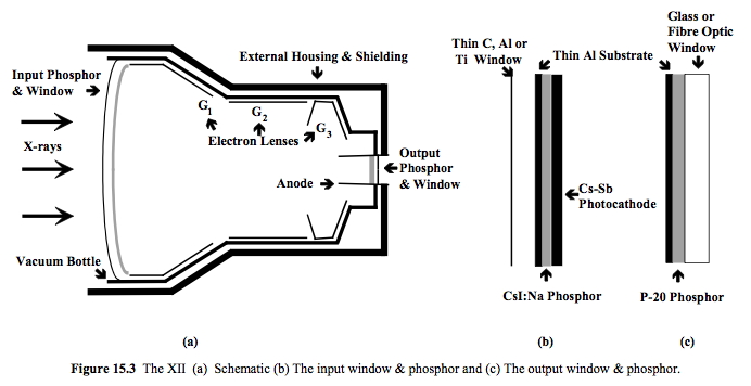
An X-ray image intensifier (XRII) is an image intensifier that converts X-rays into visible light at higher intensity (physics), intensity than the more traditional fluorescence, fluorescent screens can. Such intensifiers are used in X-ray imaging systems (such as fluoroscopy, fluoroscopes) to allow low-intensity X-rays to be converted to a conveniently brightness, bright visible light output. The device contains a low absorbency/scatter input window, typically aluminum, input fluorescent screen, photocathode, electron optics, output fluorescent screen and output window. These parts are all mounted in a high vacuum environment within glass or, more recently, metal/ceramic. By its amplifier, intensifying effect, It allows the viewer to more easily see the structure of the object being imaged than fluorescent screens alone, whose images are dim. The XRII requires lower absorbed doses due to more efficient conversion of X-ray quanta to visible light. This device was originally introdu ... [...More Info...] [...Related Items...] OR: [Wikipedia] [Google] [Baidu] |
X-ray Generator
An X-ray machine is a device that uses X-rays for a variety of applications including medicine, X-ray fluorescence, electronic assembly inspection, and measurement of material thickness in manufacturing operations. In medical applications, X-ray machines are used by radiographers to acquire x-ray images of the internal structures (e.g., bones) of living organisms, and also in sterilization. Structure An X-ray generator generally contains an X-ray tube to produce the X-rays. Possibly, radioisotopes can also be used to generate X-rays. An X-ray tube is a simple vacuum tube that contains a cathode, which directs a stream of electrons into a vacuum, and an anode, which collects the electrons and is made of tungsten to evacuate the heat generated by the collision. When the electrons collide with the target, about 1% of the resulting energy is emitted as X-rays, with the remaining 99% released as heat. Due to the high energy of the electrons that reach relativistic speeds, the t ... [...More Info...] [...Related Items...] OR: [Wikipedia] [Google] [Baidu] |
X-ray Computed Tomography
An X-ray (also known in many languages as Röntgen radiation) is a form of high-energy electromagnetic radiation with a wavelength shorter than those of ultraviolet rays and longer than those of gamma rays. Roughly, X-rays have a wavelength ranging from 10 nanometers to 10 picometers, corresponding to frequencies in the range of 30 petahertz to 30 exahertz ( to ) and photon energies in the range of 100 eV to 100 keV, respectively. X-rays were discovered in 1895 by the German scientist Wilhelm Conrad Röntgen, who named it ''X-radiation'' to signify an unknown type of radiation.Novelline, Robert (1997). ''Squire's Fundamentals of Radiology''. Harvard University Press. 5th edition. . X-rays can penetrate many solid substances such as construction materials and living tissue, so X-ray radiography is widely used in medical diagnostics (e.g., checking for broken bones) and materials science (e.g., identification of some chemical elements and ... [...More Info...] [...Related Items...] OR: [Wikipedia] [Google] [Baidu] |
Radiology
Radiology ( ) is the medical specialty that uses medical imaging to diagnose diseases and guide treatment within the bodies of humans and other animals. It began with radiography (which is why its name has a root referring to radiation), but today it includes all imaging modalities. This includes technologies that use no ionizing electromagnetic radiation, such as medical ultrasound, ultrasonography and magnetic resonance imaging (MRI), as well as others that do use radiation, such as x-ray computed tomography, computed tomography (CT), fluoroscopy, and nuclear medicine including positron emission tomography (PET). Interventional radiology is the performance of usually invasiveness of surgical procedures, minimally invasive medical procedures with the guidance of imaging technologies such as those mentioned above. The modern practice of radiology involves a team of several different healthcare professionals. A radiologist, who is a medical doctor with specialized post-graduate tr ... [...More Info...] [...Related Items...] OR: [Wikipedia] [Google] [Baidu] |
Digital Imaging
Digital imaging or digital image acquisition is the creation of a digital representation of the visual characteristics of an object, such as a physical scene or the interior structure of an object. The term is often assumed to imply or include the processing, compression, storage, printing and display of such images. A key advantage of a digital image, versus an analog image such as a film photograph, is the ability to digitally propagate copies of the original subject indefinitely without any loss of image quality. Digital imaging can be classified by the type of electromagnetic radiation or other waves whose variable attenuation, as they pass through or reflect off objects, conveys the information that constitutes the image. In all classes of digital imaging, the information is converted by image sensors into digital signals that are processed by a computer and made output as a visible-light image. For example, the medium of visible light allows digital photog ... [...More Info...] [...Related Items...] OR: [Wikipedia] [Google] [Baidu] |
Anatomy
Anatomy () is the branch of morphology concerned with the study of the internal structure of organisms and their parts. Anatomy is a branch of natural science that deals with the structural organization of living things. It is an old science, having its beginnings in prehistoric times. Anatomy is inherently tied to developmental biology, embryology, comparative anatomy, evolutionary biology, and phylogeny, as these are the processes by which anatomy is generated, both over immediate and long-term timescales. Anatomy and physiology, which study the structure and function of organisms and their parts respectively, make a natural pair of related disciplines, and are often studied together. Human anatomy is one of the essential basic sciences that are applied in medicine, and is often studied alongside physiology. Anatomy is a complex and dynamic field that is constantly evolving as discoveries are made. In recent years, there has been a significant increase in the use of ... [...More Info...] [...Related Items...] OR: [Wikipedia] [Google] [Baidu] |
Medical Imaging
Medical imaging is the technique and process of imaging the interior of a body for clinical analysis and medical intervention, as well as visual representation of the function of some organs or tissues (physiology). Medical imaging seeks to reveal internal structures hidden by the skin and bones, as well as to diagnose and treat disease. Medical imaging also establishes a database of normal anatomy and physiology to make it possible to identify abnormalities. Although imaging of removed organ (anatomy), organs and Tissue (biology), tissues can be performed for medical reasons, such procedures are usually considered part of pathology instead of medical imaging. Measurement and recording techniques that are not primarily designed to produce images, such as electroencephalography (EEG), magnetoencephalography (MEG), electrocardiography (ECG), and others, represent other technologies that produce data susceptible to representation as a parameter graph versus time or maps that contain ... [...More Info...] [...Related Items...] OR: [Wikipedia] [Google] [Baidu] |
Transparency And Translucency
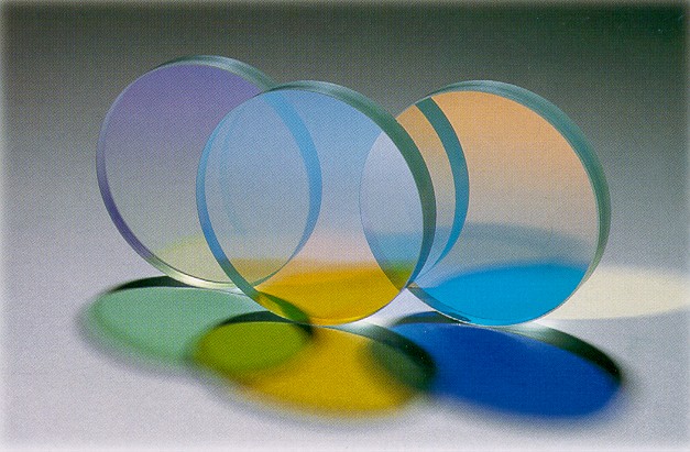
In the field of optics, transparency (also called pellucidity or diaphaneity) is the physical property of allowing light to pass through the material without appreciable scattering of light. On a macroscopic scale (one in which the dimensions are much larger than the wavelengths of the photons in question), the photons can be said to follow Snell's law. Translucency (also called translucence or translucidity) is the physical property of allowing light to pass through the material (with or without scattering of light). It allows light to pass through but the light does not necessarily follow Snell's law on the macroscopic scale; the photons may be scattered at either of the two interfaces, or internally, where there is a change in the index of refraction. In other words, a translucent material is made up of components with different indices of refraction. A transparent material is made up of components with a uniform index of refraction. Transparent materials appear clear, with t ... [...More Info...] [...Related Items...] OR: [Wikipedia] [Google] [Baidu] |
Radiodensity
Radiodensity (or radiopacity) is opacity to the radio wave and X-ray portion of the electromagnetic spectrum: that is, the relative inability of those kinds of electromagnetic radiation to pass through a particular material. Radiolucency or hypodensity indicates greater passage (greater transradiancy) to X-ray photonsNovelline, Robert. ''Squire's Fundamentals of Radiology''. Harvard University Press. 5th edition. 1997. . and is the analogue of transparency and translucency with visible light. Materials that inhibit the passage of electromagnetic radiation are called radiodense or radiopaque, while those that allow radiation to pass more freely are referred to as radiolucent. Radiopaque volumes of material have white appearance on radiographs, compared with the relatively darker appearance of radiolucent volumes. For example, on typical radiographs, bones look white or light gray (radiopaque), whereas muscle and skin look black or dark gray, being mostly invisible (radiolucent). Th ... [...More Info...] [...Related Items...] OR: [Wikipedia] [Google] [Baidu] |


