|
Radial Groove
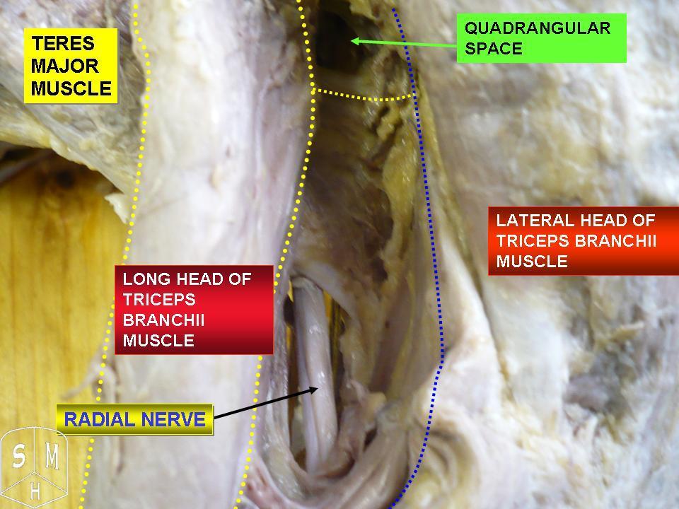
The radial groove (also known as the musculospiral groove, radial sulcus, or spiral groove) is a broad but shallow oblique depression for the radial nerve and deep brachial artery. It is located on the center of the lateral border of the humerus bone. It is situated alongside the posterior margin of the deltoid tuberosity In human anatomy, the deltoid tuberosity is a rough, triangular area on the anterolateral (front-side) surface of the middle of the humerus. It is a site of attachment of deltoid muscle. Structure Variation The deltoid tuberosity has been r ..., ending at its inferior margin. Although it provides protection to the radial nerve, it is often involved in compressions on the nerve (due to external pressure due to surgery) that can cause radial nerve palsy. See also * Intertubercular groove * Triceps brachii muscle Additional images File:Gray413_color.png, Cross-section through the middle of upper arm. File:Gray525.png, The brachial artery. File:Gray8 ... [...More Info...] [...Related Items...] OR: [Wikipedia] [Google] [Baidu] |
Radial Nerve
The radial nerve is a nerve in the human body that supplies the posterior portion of the upper limb. It innervates the medial and lateral heads of the triceps brachii muscle of the arm, as well as all 12 muscles in the Posterior compartment of the forearm, posterior osteofascial compartment of the forearm and the associated joints and overlying skin. It originates from the brachial plexus, carrying fibers from the posterior roots of spinal nerves C5, C6, C7, C8 and T1. The radial nerve and its branches provide Motor neuron, motor innervation to the dorsal arm muscles (the triceps brachii and the anconeus) and the extrinsic extensors of the wrists and hands; it also provides cutaneous Nerve supply to the skin, sensory innervation to most of the back of the hand, except for the back of the little finger and adjacent half of the ring finger (which are innervated by the ulnar nerve). The radial nerve divides into a deep branch, which becomes the posterior interosseous nerve, and a su ... [...More Info...] [...Related Items...] OR: [Wikipedia] [Google] [Baidu] |
Profunda Brachii
The deep artery of arm (also known as deep brachial artery) is a large artery of the arm which arises from the brachial artery. It descends in the arm before ending by anastomosing with the radial recurrent artery. Structure Origin The deep artery of arm arises from the posterolateral aspect of the brachial artery, just below the lower border of the teres major. Course It follows closely the radial nerve, running at first backward between the long and medial heads of the triceps brachii, then along the groove for the radial nerve (the radial sulcus), where it is covered by the lateral head of the triceps brachii, to the lateral side of the arm; there it pierces the lateral intermuscular septum, and, descending between the brachioradialis and the brachialis to the front of the lateral epicondyle of the humerus, ends by anastomosing with the radial recurrent artery. Branches and anastomoses It gives branches to the deltoid muscle (which, however, primarily is supplied b ... [...More Info...] [...Related Items...] OR: [Wikipedia] [Google] [Baidu] |
Humerus
The humerus (; : humeri) is a long bone in the arm that runs from the shoulder to the elbow. It connects the scapula and the two bones of the lower arm, the radius (bone), radius and ulna, and consists of three sections. The humeral upper extremity of humerus, upper extremity consists of a rounded head, a narrow neck, and two short processes (tubercles, sometimes called tuberosities). The body of humerus, body is cylindrical in its upper portion, and more prism (geometry), prismatic below. The lower extremity of humerus, lower extremity consists of 2 epicondyles, 2 processes (trochlea of the humerus, trochlea and capitulum of the humerus, capitulum), and 3 fossae (radial fossa, coronoid fossa, and olecranon fossa). As well as its true anatomical neck, the constriction below the greater and lesser tubercles of the humerus is referred to as its Surgical neck of the humerus, surgical neck due to its tendency to fracture, thus often becoming the focus of surgeons. Etymology The word ... [...More Info...] [...Related Items...] OR: [Wikipedia] [Google] [Baidu] |
Deltoid Tuberosity
In human anatomy, the deltoid tuberosity is a rough, triangular area on the anterolateral (front-side) surface of the middle of the humerus. It is a site of attachment of deltoid muscle. Structure Variation The deltoid tuberosity has been reported as very prominent in less than 10% of people. Development The deltoid tuberosity develops through endochondral ossification in a two-phase process. The initiating signal is tendon-dependent, whilst the growth phase is muscle-dependent. Clinical significance The deltoid tuberosity is at risk of avulsion fracture. These fractures may be managed conservatively with rest. Other animals In mammals, the humerus displays a wide morphological variation. The size and orientation of its functionally important features, including the deltoid tubercle, greater tubercle, and medial epicondyle, are pivotal to an animal's style of locomotion and habitat. In cursorial (running) animals such as the pronghorn, the deltoid tubercle is locate ... [...More Info...] [...Related Items...] OR: [Wikipedia] [Google] [Baidu] |
Radial Nerve Palsy
Radial nerve dysfunction is a problem associated with the radial nerve resulting from injury consisting of acute trauma to the radial nerve. The damage has sensory consequences, as it interferes with the radial nerve's innervation of the skin of the posterior forearm, lateral three digits, and the dorsal surface of the lateral side of the palm. The damage also has motor consequences, as it interferes with the radial nerve's innervation of the muscles associated with the extension at the elbow, wrist, and fingers, as well the supination of the forearm. This type of injury can be difficult to localize, but relatively common, as many ordinary occurrences can lead to the injury and resulting mononeuropathy. One out of every ten patients with radial nerve dysfunction do so because of a fractured humerus. Signs and symptoms People experiencing radial nerve dysfunction may also experience any of the following symptoms: * Lost ability or discomfort in extending the elbow * Lost ability or ... [...More Info...] [...Related Items...] OR: [Wikipedia] [Google] [Baidu] |
Intertubercular Groove
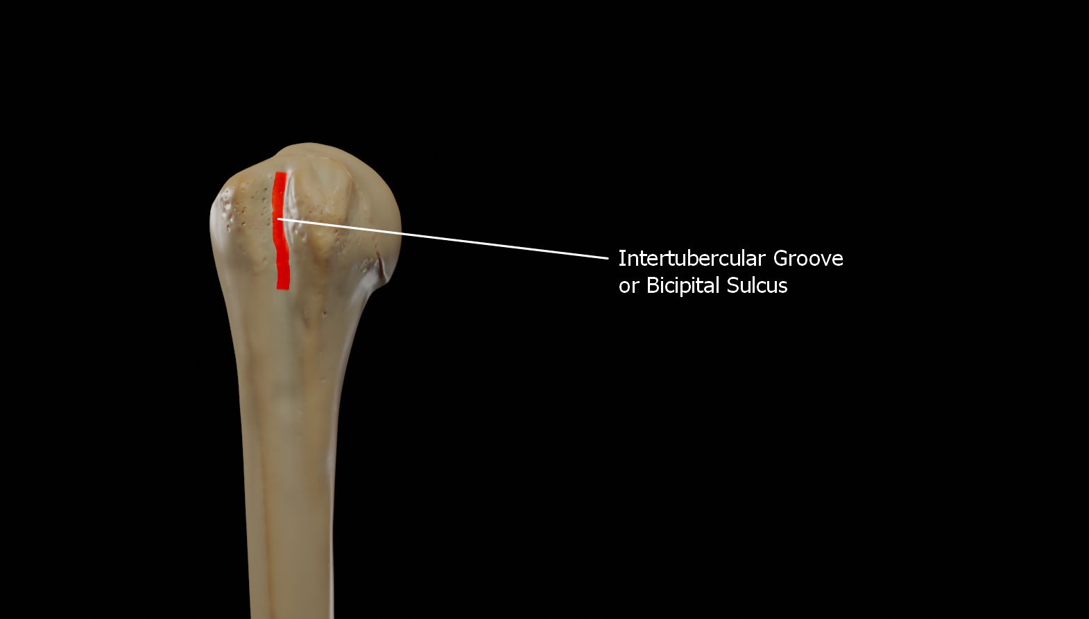
The bicipital groove (intertubercular groove, sulcus intertubercularis) is a deep groove on the humerus that separates the greater tubercle from the lesser tubercle. It allows for the long tendon of the biceps brachii muscle to pass. Structure The bicipital groove separates the greater tubercle from the lesser tubercle. It is usually around 8 cm long and 1 cm wide in adults. The groove lodges the long tendon of the biceps brachii muscle, positioned between the tendon of the pectoralis major muscle on the lateral lip and the tendon of the teres major muscle on the medial lip. It also transmits a branch of the anterior humeral circumflex artery to the shoulder joint. The insertion of the latissimus dorsi muscle is found along the floor of the bicipital groove. The teres major muscle inserts on the medial lip of the groove. It runs obliquely downward, and ends near the junction of the upper with the middle third of the bone. It is the lateral wall of the axilla. Function The ... [...More Info...] [...Related Items...] OR: [Wikipedia] [Google] [Baidu] |
Triceps Brachii Muscle
The triceps, or triceps brachii (Latin for "three-headed muscle of the arm"), is a large muscle on the back of the upper limb of many vertebrates. It consists of three parts: the medial, lateral, and long head. All three heads cross the elbow joint. However, the long head also crosses the shoulder joint. The triceps muscle contracts when the elbow is straightened and expands when the elbow is bent. The long head gets a further contraction when the arm is behind the torso due to how it crosses the shoulder joint. It is the muscle principally responsible for extension of the elbow joint (straightening of the arm). Structure * The long head arises from the infraglenoid tubercle of the scapula. It extends distally anterior to the teres minor and posterior to the teres major. * The medial head arises proximally in the humerus, just inferior to the groove of the radial nerve; from the dorsal (back) surface of the humerus; from the medial intermuscular septum; and its dista ... [...More Info...] [...Related Items...] OR: [Wikipedia] [Google] [Baidu] |


