|
Pilon Fracture
A pilon fracture, is a fracture of the distal part of the tibia, involving its articular surface at the ankle joint. Pilon fractures are caused by rotational or axial forces, mostly as a result of falls from a height or motor vehicle accidents. Pilon fractures are rare, comprising 3 to 10 percent of all fractures of the tibia and 1 percent of all lower extremity fractures, but they involve a large part of the weight-bearing surface of the tibia in the ankle joint. Because of this, they may be difficult to fixate and are historically associated with high rates of complications and poor outcome. ''Pilon'' is the French word for "pestle" and was introduced into orthopedic literature in 1911 by pioneer French radiologist Étienne Destot. Classification Pilon fractures are categorized by two main X-ray schemes, Ruedi-Allgower classification system. and Müller AO Classification of fractures. Treatment The treatment of pilon fractures depends on the extent of the injury. This i ... [...More Info...] [...Related Items...] OR: [Wikipedia] [Google] [Baidu] |
X-ray
An X-ray, or, much less commonly, X-radiation, is a penetrating form of high-energy electromagnetic radiation. Most X-rays have a wavelength ranging from 10 picometers to 10 nanometers, corresponding to frequencies in the range 30 petahertz to 30 exahertz ( to ) and energies in the range 145 eV to 124 keV. X-ray wavelengths are shorter than those of UV rays and typically longer than those of gamma rays. In many languages, X-radiation is referred to as Röntgen radiation, after the German scientist Wilhelm Conrad Röntgen, who discovered it on November 8, 1895. He named it ''X-radiation'' to signify an unknown type of radiation.Novelline, Robert (1997). ''Squire's Fundamentals of Radiology''. Harvard University Press. 5th edition. . Spellings of ''X-ray(s)'' in English include the variants ''x-ray(s)'', ''xray(s)'', and ''X ray(s)''. The most familiar use of X-rays is checking for fractures (broken bones), but X-rays are also used in other ways. ... [...More Info...] [...Related Items...] OR: [Wikipedia] [Google] [Baidu] |
Müller AO Classification Of Fractures
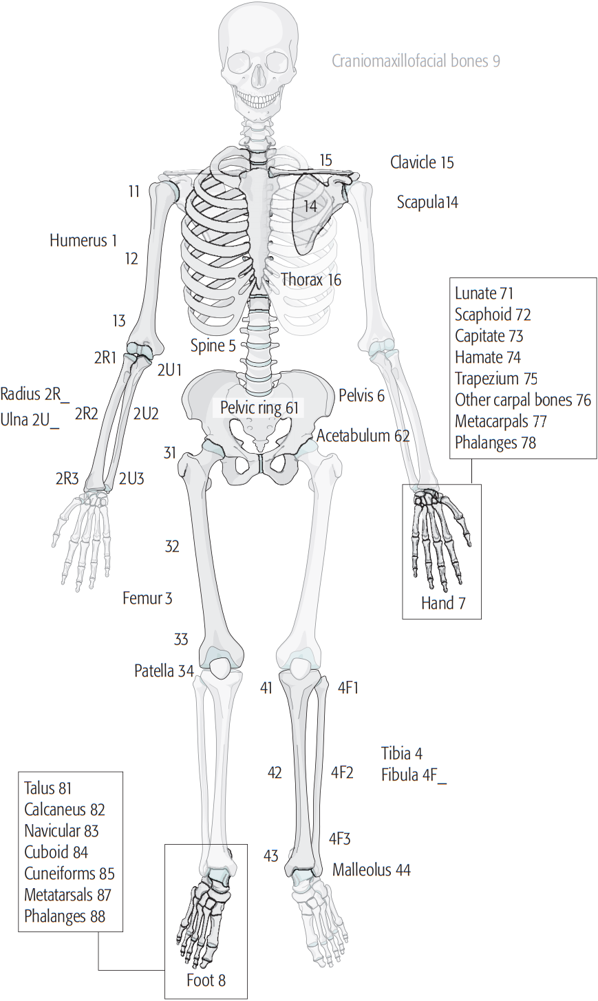
The Müller AO Classification of fractures is a system for classifying bone fractures initially published in 1987 by the AO Foundation The AO Foundation is a nonprofit organization dedicated to improving the care of patients with musculoskeletal injuries or pathologies and their sequelae through research, development, and education of surgeons and operating room personnel. The AO ... as a method of categorizing injuries according to therognosis of the patient's anatomical and functional outcome. "AO" is an initialism for the German "Arbeitsgemeinschaft für Osteosynthesefragen", the predecessor of the AO Foundation. It is one of the few complete fracture classification systems to remain in use today after validation. Comprehensive classification of the long bones The English language version of the system allows consistent in detail description of a fracture in defined terminology by creating a 5-element alphanumeric code: Localisation First, each fracture is given 2 numbers ... [...More Info...] [...Related Items...] OR: [Wikipedia] [Google] [Baidu] |
Ankle Fracture
An ankle fracture is a break of one or more of the bones that make up the ankle joint. Symptoms may include pain, swelling, bruising, and an inability to walk on the injured leg. Complications may include an associated high ankle sprain, compartment syndrome, stiffness, malunion, and post-traumatic arthritis. Ankle fractures may result from excessive stress on the joint such as from rolling an ankle or from blunt trauma. Types of ankle fractures include lateral malleolus, medial malleolus, posterior malleolus, bimalleolar, and trimalleolar fractures. The Ottawa ankle rule can help determine the need for X-rays. Special X-ray views called stress views help determine whether an ankle fracture is unstable. Treatment depends on the fracture type. Ankle stability largely dictates non-operative vs. operative treatment. Non-operative treatment includes splinting or casting while operative treatment includes fixing the fracture with metal implants through an open reduction internal ... [...More Info...] [...Related Items...] OR: [Wikipedia] [Google] [Baidu] |
Vacuum Assisted Closure Wound Therapy
Negative-pressure wound therapy (NPWT), also known as a vacuum assisted closure (VAC), is a therapeutic technique using a suction pump, tubing and a dressing to remove excess exudate and promote healing in acute or chronic wounds and second- and third-degree burns. The therapy involves the controlled application of subatmospheric pressure to the local wound environment using a sealed wound dressing connected to a vacuum pump. The use of this technique in wound management started in the 1990s and this technique is often recommended for treatment of a range of wounds including dehisced surgical wounds, closed surgical wounds, open abdominal wounds, open fractures, pressure injuries or pressure ulcers, diabetic foot ulcers, venous insufficiency ulcers, some types of skin grafts, burns, sternal wounds. It may also be considered after a clean surgery in a person who is obese. NPWT is performed by applying a vacuum through a special sealed dressing. The continued vacuum draws ou ... [...More Info...] [...Related Items...] OR: [Wikipedia] [Google] [Baidu] |

