|
Pseudoathetosis
Pseudoathetosis is abnormal writhing movements, usually of the fingers, caused by a failure of joint position sense (proprioception) and indicates disruption of the proprioceptive pathway, from nerve to parietal cortex. Presentation Analogous to Romberg's sign, the abnormal posturing is most pronounced when the eyes are closed as visual inputs are unavailable to guide corrective movements. Paradoxically, eye closure may decrease the amount of movement as the visual cues probably trigger corrective movements which return the limb to the desired "baseline" allowing a new phase of involuntary drift before a subsequent corrective phase occurs. Variants Hemipseudaoathetosis refers to pseudoathetosis on one side of the body, usually the upper limb and is most commonly caused by a lesion affecting the cuneate tract or cuneate nucleus in the cervical spine or lower brainstem (medulla) respectively. Diagnosis Differential diagnosis It may be mistaken for choreoathetosis; but these abnormal ... [...More Info...] [...Related Items...] OR: [Wikipedia] [Google] [Baidu] |
Finger
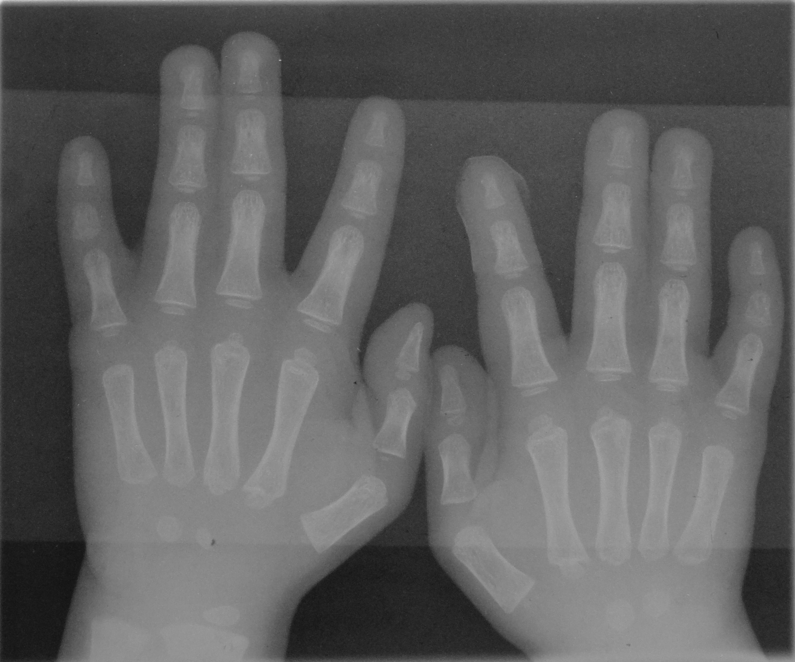
A finger is a prominent digit (anatomy), digit on the forelimbs of most tetrapod vertebrate animals, especially those with prehensile extremities (i.e. hands) such as humans and other primates. Most tetrapods have five digits (dactyly, pentadactyly),#Cha1998, Chambers 1998 p. 603#OxfIll, Oxford Illustrated pp. 311, 380 and short digits (i.e. significantly shorter than the metacarpal/metatarsals) are typically referred to as toes, while those that are notably elongated are called fingers. In humans, the fingers are flexibly joint, articulated and opposable, serving as an important organ of somatosensory, tactile sensation and fine motor skill, fine movements, which are crucial to the dexterity of the hands and the ability to grasp and object manipulation, manipulate objects. Land vertebrate fingers As terrestrial vertebrates were evolution, evolved from lobe-finned fish, their forelimbs are phylogeny, phylogenetically equivalent to the pectoral fins of fish. Within the taxon, ... [...More Info...] [...Related Items...] OR: [Wikipedia] [Google] [Baidu] |
Proprioception
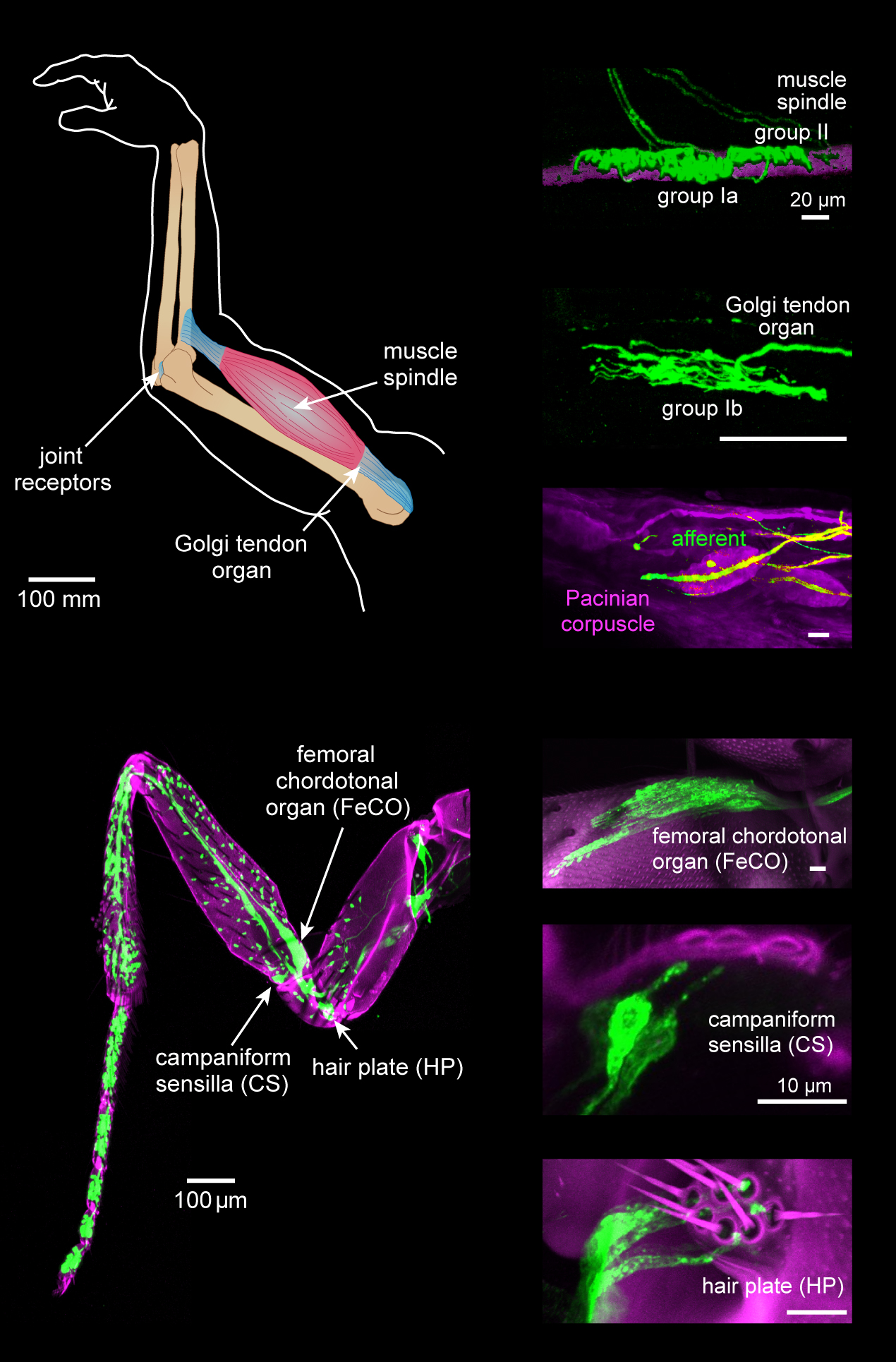
Proprioception ( ) is the sense of self-movement, force, and body position. Proprioception is mediated by proprioceptors, a type of sensory receptor, located within muscles, tendons, and joints. Most animals possess multiple subtypes of proprioceptors, which detect distinct kinesthetic parameters, such as joint position, movement, and load. Although all mobile animals possess proprioceptors, the structure of the sensory organs can vary across species. Proprioceptive signals are transmitted to the central nervous system, where they are integrated with information from other Sensory nervous system, sensory systems, such as Visual perception, the visual system and the vestibular system, to create an overall representation of body position, movement, and acceleration. In many animals, sensory feedback from proprioceptors is essential for stabilizing body posture and coordinating body movement. System overview In vertebrates, limb movement and velocity (muscle length and the rate ... [...More Info...] [...Related Items...] OR: [Wikipedia] [Google] [Baidu] |
Nerve
A nerve is an enclosed, cable-like bundle of nerve fibers (called axons). Nerves have historically been considered the basic units of the peripheral nervous system. A nerve provides a common pathway for the Electrochemistry, electrochemical nerve impulses called action potentials that are transmitted along each of the axons to peripheral organs or, in the case of sensory nerves, from the periphery back to the central nervous system. Each axon is an extension of an individual neuron, along with other supportive cells such as some Schwann cells that coat the axons in myelin. Each axon is surrounded by a layer of connective tissue called the endoneurium. The axons are bundled together into groups called Nerve fascicle, fascicles, and each fascicle is wrapped in a layer of connective tissue called the perineurium. The entire nerve is wrapped in a layer of connective tissue called the epineurium. Nerve cells (often called neurons) are further classified as either Sensory neuron, sens ... [...More Info...] [...Related Items...] OR: [Wikipedia] [Google] [Baidu] |
Parietal Cortex
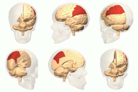
The parietal lobe is one of the four major lobes of the cerebral cortex in the brain of mammals. The parietal lobe is positioned above the temporal lobe and behind the frontal lobe and central sulcus. The parietal lobe integrates sensory information among various modalities, including spatial sense and navigation (proprioception), the main sensory receptive area for the sense of touch in the somatosensory cortex which is just posterior to the central sulcus in the postcentral gyrus, and the dorsal stream of the visual system. The major sensory inputs from the skin (touch, temperature, and pain receptors), relay through the thalamus to the parietal lobe. Several areas of the parietal lobe are important in language processing. The somatosensory cortex can be illustrated as a distorted figure – the cortical homunculus (Latin: "little man") in which the body parts are rendered according to how much of the somatosensory cortex is devoted to them. The superior parietal lobule an ... [...More Info...] [...Related Items...] OR: [Wikipedia] [Google] [Baidu] |
Romberg's Sign
Romberg's test, Romberg's sign, or the Romberg maneuver is a test used in an exam of neurological function for balance. The exam is based on the premise that a person requires at least two of the three following senses to maintain balance while standing: * proprioception (the ability to know one's body position in space) * vestibular function (the ability to know one's head position in space) * vision (which can be used to monitor and adjust for changes in body position). A patient who has a problem with proprioception can still maintain balance by using vestibular function and vision. In the Romberg test, the standing patient is asked to close their eyes. An increased loss of balance is interpreted as a positive Romberg's test. The Romberg test is a test of the body's sense of positioning (proprioception), which requires healthy functioning of the dorsal columns of the spinal cord. The Romberg test is used to investigate the cause of loss of motor coordination (ataxia). A ... [...More Info...] [...Related Items...] OR: [Wikipedia] [Google] [Baidu] |
Cuneate Tract , a tract from the spinal cord into the brainstem
{{disambiguation ...
Cuneate means "wedge-shaped", and can apply to: * Cuneate leaf, a leaf shape * Cuneate nucleus, a part of the brainstem * Cuneate fasciculus Cuneate means "wedge-shaped", and can apply to: * Cuneate leaf, a leaf shape * Cuneate nucleus The dorsal column nuclei are a pair of nuclei in the dorsal columns of the dorsal column–medial lemniscus pathway (DCML) in the brainstem. The ... [...More Info...] [...Related Items...] OR: [Wikipedia] [Google] [Baidu] |
Cuneate Nucleus
The dorsal column nuclei are a pair of nuclei in the dorsal columns of the dorsal column–medial lemniscus pathway (DCML) in the brainstem. The name refers collectively to the cuneate nucleus and gracile nucleus, which are situated at the lower end of the medulla oblongata. Both nuclei contain second-order neurons of the DCML, which convey fine touch and proprioceptive information from the body to the brain via the thalamus. Structure Nerve pathways The dorsal column nuclei each have an associated nerve tract in the spinal cord, the gracile fasciculus and the cuneate fasciculus, together forming the dorsal columns. Both dorsal column nuclei contain synapses from afferent nerve fibers that have travelled in the spinal cord. They then send on second-order neurons of the dorsal column–medial lemniscal pathway. Neurons of the dorsal column nuclei eventually reach the midbrain and the thalamus. They send axons that form the internal arcuate fibers. These cross over ... [...More Info...] [...Related Items...] OR: [Wikipedia] [Google] [Baidu] |
Cervical Spine
In tetrapods, cervical vertebrae (: vertebra) are the vertebrae of the neck, immediately below the skull. Truncal vertebrae (divided into thoracic and lumbar vertebrae in mammals) lie caudal (toward the tail) of cervical vertebrae. In sauropsid species, the cervical vertebrae bear cervical ribs. In lizards and saurischian dinosaurs, the cervical ribs are large; in birds, they are small and completely fused to the vertebrae. The vertebral transverse processes of mammals are homologous to the cervical ribs of other amniotes. Most mammals have seven cervical vertebrae, with the only three known exceptions being the manatee with six, the two-toed sloth with five or six, and the three-toed sloth with nine. In humans, cervical vertebrae are the smallest of the true vertebrae and can be readily distinguished from those of the thoracic or lumbar regions by the presence of a transverse foramen, an opening in each transverse process, through which the vertebral artery, verte ... [...More Info...] [...Related Items...] OR: [Wikipedia] [Google] [Baidu] |
Brainstem
The brainstem (or brain stem) is the posterior stalk-like part of the brain that connects the cerebrum with the spinal cord. In the human brain the brainstem is composed of the midbrain, the pons, and the medulla oblongata. The midbrain is continuous with the thalamus of the diencephalon through the tentorial notch, and sometimes the diencephalon is included in the brainstem. The brainstem is very small, making up around only 2.6 percent of the brain's total weight. It has the critical roles of regulating heart and respiratory system, respiratory function, helping to control heart rate and breathing rate. It also provides the main motor and sensory nerve supply to the face and neck via the cranial nerves. Ten pairs of cranial nerves come from the brainstem. Other roles include the regulation of the central nervous system and the body's sleep cycle. It is also of prime importance in the conveyance of motor and sensory pathways from the rest of the brain to the body, and from the b ... [...More Info...] [...Related Items...] OR: [Wikipedia] [Google] [Baidu] |
Medulla Oblongata
The medulla oblongata or simply medulla is a long stem-like structure which makes up the lower part of the brainstem. It is anterior and partially inferior to the cerebellum. It is a cone-shaped neuronal mass responsible for autonomic (involuntary) functions, ranging from vomiting to sneezing. The medulla contains the cardiovascular center, the respiratory center, vomiting and vasomotor centers, responsible for the autonomic functions of breathing, heart rate and blood pressure as well as the sleep–wake cycle. "Medulla" is from Latin, ‘pith or marrow’. And "oblongata" is from Latin, ‘lengthened or longish or elongated'. During embryonic development, the medulla oblongata develops from the myelencephalon. The myelencephalon is a secondary brain vesicle which forms during the maturation of the rhombencephalon, also referred to as the hindbrain. The bulb is an archaic term for the medulla oblongata. In modern clinical usage, the word bulbar (as in bulbar palsy) is r ... [...More Info...] [...Related Items...] OR: [Wikipedia] [Google] [Baidu] |
Choreoathetosis
Choreoathetosis is the occurrence of involuntary movements in a combination of chorea (irregular migrating contractions) and athetosis (twisting and writhing). It is caused by many different diseases and agents. It is a symptom of several diseases, including GLUT1 deficiency syndrome, Lesch–Nyhan syndrome, phenylketonuria, and Huntington disease and can be a feature of kernicterus (rapidly increasing unconjugated bilirubin that cross the blood-brain-barrier in infants). Choreoathetosis is also a common presentation of dyskinesia as a side effect of levodopa-carbidopa in the treatment of Parkinson disease. The use of crack cocaine or amphetamines Substituted amphetamines, or simply amphetamines, are a chemical class, class of compounds based upon the amphetamine structure; it includes all derivative (chemistry), derivative compounds which are formed by replacing, or substitution reacti ... can result in conditions nicknamed ''crack dancing'', or ''tweaking'' respectively ... [...More Info...] [...Related Items...] OR: [Wikipedia] [Google] [Baidu] |
Proprioceptive
Proprioception ( ) is the sense of self-movement, force, and body position. Proprioception is mediated by proprioceptors, a type of sensory receptor, located within muscles, tendons, and joints. Most animals possess multiple subtypes of proprioceptors, which detect distinct kinesthetic parameters, such as joint position, movement, and load. Although all mobile animals possess proprioceptors, the structure of the sensory organs can vary across species. Proprioceptive signals are transmitted to the central nervous system, where they are integrated with information from other sensory systems, such as the visual system and the vestibular system, to create an overall representation of body position, movement, and acceleration. In many animals, sensory feedback from proprioceptors is essential for stabilizing body posture and coordinating body movement. System overview In vertebrates, limb movement and velocity (muscle length and the rate of change) are encoded by one group of se ... [...More Info...] [...Related Items...] OR: [Wikipedia] [Google] [Baidu] |


