|
Optic Nerve Glioma
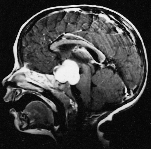
Optic nerve glioma (or optic glioma), a form of glioma which affects the optic nerve, is often one of the central nervous system manifestations of neurofibromatosis 1. Optic gliomas are usually pilocytic tumors, and can involve the optic nerve or optic chiasm. Optic gliomas are usually associated with neurofibromatosis type 1 in 30% of people with the condition. Optic nerve gliomas have low mortality but extremely high prevalence of vision loss & eye-bulging exophthalmos) in children. As of 2014, approximately 1000 cases had been reported. Diagnosis Optic nerve gliomas are diagnosed using magnetic resonance imaging (MRI) and CT scans. The tumor adopts a fusiform appearance, appearing wider in the middle and tapered at the ends. Enlargement of the optic nerve along with a downward kink in the mid-orbit is usually observed. While CT scans allow for optic nerve evaluation, MRI allows for intracranial evaluation to observe if the tumor has extended to other regions such as the opti ... [...More Info...] [...Related Items...] OR: [Wikipedia] [Google] [Baidu] |
Glioma
A glioma is a type of tumor that starts in the glial cells of the brain or the spine. Gliomas comprise about 30 percent of all brain tumors and central nervous system tumours, and 80 percent of all malignant brain tumours. Signs and symptoms Symptoms of gliomas depend on which part of the central nervous system is affected. A brain glioma can cause headaches, vomiting, seizures, and cranial nerve disorders as a result of increased intracranial pressure. A glioma of the optic nerve can cause visual loss. Spinal cord gliomas can cause pain, weakness, or numbness in the extremities. Gliomas do not usually metastasize by the bloodstream, but they can spread via the cerebrospinal fluid and cause "drop metastases" to the spinal cord. Complex visual hallucinations have been described as a symptom of low-grade glioma. A child who has a subacute disorder of the central nervous system that produces cranial nerve abnormalities (especially of cranial nerve VII and the lower bulbar ne ... [...More Info...] [...Related Items...] OR: [Wikipedia] [Google] [Baidu] |
Optic Nerve
In neuroanatomy, the optic nerve, also known as the second cranial nerve, cranial nerve II, or simply CN II, is a paired cranial nerve that transmits visual information from the retina to the brain. In humans, the optic nerve is derived from optic stalks during the seventh week of development and is composed of retinal ganglion cell axons and glial cells; it extends from the optic disc to the optic chiasma and continues as the optic tract to the lateral geniculate nucleus, pretectal nuclei, and superior colliculus. Structure The optic nerve has been classified as the second of twelve paired cranial nerves, but it is technically part of the central nervous system, rather than the peripheral nervous system because it is derived from an out-pouching of the diencephalon ( optic stalks) during embryonic development. As a consequence, the fibers of the optic nerve are covered with myelin produced by oligodendrocytes, rather than Schwann cells of the peripheral nervous system ... [...More Info...] [...Related Items...] OR: [Wikipedia] [Google] [Baidu] |
Central Nervous System
The central nervous system (CNS) is the part of the nervous system consisting primarily of the brain and spinal cord. The CNS is so named because the brain integrates the received information and coordinates and influences the activity of all parts of the bodies of bilaterally symmetric and triploblastic animals—that is, all multicellular animals except sponges and diploblasts. It is a structure composed of nervous tissue positioned along the rostral (nose end) to caudal (tail end) axis of the body and may have an enlarged section at the rostral end which is a brain. Only arthropods, cephalopods and vertebrates have a true brain (precursor structures exist in onychophorans, gastropods and lancelets). The rest of this article exclusively discusses the vertebrate central nervous system, which is radically distinct from all other animals. Overview In vertebrates, the brain and spinal cord are both enclosed in the meninges. The meninges provide a barrier to chemicals ... [...More Info...] [...Related Items...] OR: [Wikipedia] [Google] [Baidu] |
Pilocytic Astrocytoma
Pilocytic astrocytoma (and its variant pilomyxoid astrocytoma) is a brain tumor that occurs most commonly in children and young adults (in the first 20 years of life). They usually arise in the cerebellum, near the brainstem, in the hypothalamic region, or the optic chiasm, but they may occur in any area where astrocytes are present, including the cerebral hemispheres and the spinal cord. These tumors are usually slow growing and benign, corresponding to WHO malignancy grade 1. Signs and symptoms Children affected by pilocytic astrocytoma can present with different symptoms that might include failure to thrive (lack of appropriate weight gain/ weight loss), headache, nausea, vomiting, irritability, torticollis (tilt neck or wry neck), difficulty to coordinate movements, and visual complaints (including nystagmus). The complaints may vary depending on the location and size of the neoplasm. The most common symptoms are associated with increased intracranial pressure due to the size ... [...More Info...] [...Related Items...] OR: [Wikipedia] [Google] [Baidu] |
Optic Chiasm
In neuroanatomy, the optic chiasm, or optic chiasma (; , ), is the part of the brain where the optic nerves cross. It is located at the bottom of the brain immediately inferior to the hypothalamus. The optic chiasm is found in all vertebrates, although in cyclostomes (lampreys and hagfishes), it is located within the brain. This article is about the optic chiasm of vertebrates, which is the best known nerve chiasm, but not every chiasm denotes a crossing of the body midline (e.g., in some invertebrates, see Chiasm (anatomy)). A midline crossing of nerves inside the brain is called a decussation (see Definition of types of crossings). Structure For the different types of optic chiasm, see In all vertebrates, the optic nerves of the left and the right eye meet in the body midline, ventral to the brain. In many vertebrates the left optic nerve crosses over the right one without fusing with it. In vertebrates with a large overlap of the visual fields of the two eyes, ... [...More Info...] [...Related Items...] OR: [Wikipedia] [Google] [Baidu] |
Neurofibromatosis Type 1
Neurofibromatosis (NF) is a group of three conditions in which tumors grow in the nervous system. The three types are neurofibromatosis type I (NF1), neurofibromatosis type II (NF2), and schwannomatosis. In NF1 symptoms include light brown spots on the skin, freckles in the armpit and groin, small bumps within nerves, and scoliosis. In NF2, there may be hearing loss, cataracts at a young age, balance problems, flesh colored skin flaps, and muscle wasting. In schwannomatosis there may be pain either in one location or in wide areas of the body. The tumors in NF are generally non-cancerous. The cause is a genetic mutation in certain oncogenes. These can be inherited from a person's parents, or in about half of cases spontaneously occur during early development. Different mutations result in the three types of NF. Neurofibromatosis arise from the supporting cells of the nervous system rather than the neurons themselves. In NF1, the tumors are neurofibromas (tumors of the p ... [...More Info...] [...Related Items...] OR: [Wikipedia] [Google] [Baidu] |
Exophthalmos
Exophthalmos (also called exophthalmus, exophthalmia, proptosis, or exorbitism) is a bulging of the eye anteriorly out of the orbit. Exophthalmos can be either bilateral (as is often seen in Graves' disease) or unilateral (as is often seen in an orbital tumor). Complete or partial dislocation from the orbit is also possible from trauma or swelling of surrounding tissue resulting from trauma. In the case of Graves' disease, the displacement of the eye results from abnormal connective tissue deposition in the orbit and extraocular muscles, which can be visualized by CT or MRI. If left untreated, exophthalmos can cause the eyelids to fail to close during sleep, leading to corneal dryness and damage. Another possible complication is a form of redness or irritation called superior limbic keratoconjunctivitis, in which the area above the cornea becomes inflamed as a result of increased friction when blinking. The process that is causing the displacement of the eye may also compre ... [...More Info...] [...Related Items...] OR: [Wikipedia] [Google] [Baidu] |
Magnetic Resonance Imaging
Magnetic resonance imaging (MRI) is a medical imaging technique used in radiology to form pictures of the anatomy and the physiological processes of the body. MRI scanners use strong magnetic fields, magnetic field gradients, and radio waves to generate images of the organs in the body. MRI does not involve X-rays or the use of ionizing radiation, which distinguishes it from CT and PET scans. MRI is a medical application of nuclear magnetic resonance (NMR) which can also be used for imaging in other NMR applications, such as NMR spectroscopy. MRI is widely used in hospitals and clinics for medical diagnosis, staging and follow-up of disease. Compared to CT, MRI provides better contrast in images of soft-tissues, e.g. in the brain or abdomen. However, it may be perceived as less comfortable by patients, due to the usually longer and louder measurements with the subject in a long, confining tube, though "Open" MRI designs mostly relieve this. Additionally, implants and ... [...More Info...] [...Related Items...] OR: [Wikipedia] [Google] [Baidu] |
Fusiform
Fusiform means having a spindle-like shape that is wide in the middle and tapers at both ends. It is similar to the lemon-shape, but often implies a focal broadening of a structure that continues from one or both ends, such as an aneurysm on a blood vessel. Examples * Fusiform, a body shape common to many aquatic animals, characterized by being tapered at both the head and the tail * Fusiform, a classification of aneurysm * Fusiform bacteria (spindled rods, that is, fusiform bacilli), such as the Fusobacteriota * Fusiform cell (biology) * Fusiform face area, a part of the human visual system which seems to specialize in facial recognition * Fusiform gyrus, part of the temporal lobe of the brain * Fusiform muscle, where the fibres run parallel along the length of the muscle * Fusiform neuron, a spindle-shaped neuron A neuron, neurone, or nerve cell is an electrically excitable cell that communicates with other cells via specialized connections called synapses. The neu ... [...More Info...] [...Related Items...] OR: [Wikipedia] [Google] [Baidu] |
Hypothalamus
The hypothalamus () is a part of the brain that contains a number of small nuclei with a variety of functions. One of the most important functions is to link the nervous system to the endocrine system via the pituitary gland. The hypothalamus is located below the thalamus and is part of the limbic system. In the terminology of neuroanatomy, it forms the ventral Standard anatomical terms of location are used to unambiguously describe the anatomy of animals, including humans. The terms, typically derived from Latin or Greek roots, describe something in its standard anatomical position. This position prov ... part of the diencephalon. All vertebrate brains contain a hypothalamus. In humans, it is the size of an Almond#Nut, almond. The hypothalamus is responsible for regulating certain Metabolism, metabolic biological process, processes and other activities of the autonomic nervous system. It biosynthesis, synthesizes and secretes certain neurohormones, called releasing hor ... [...More Info...] [...Related Items...] OR: [Wikipedia] [Google] [Baidu] |


