|
Neo-cortex
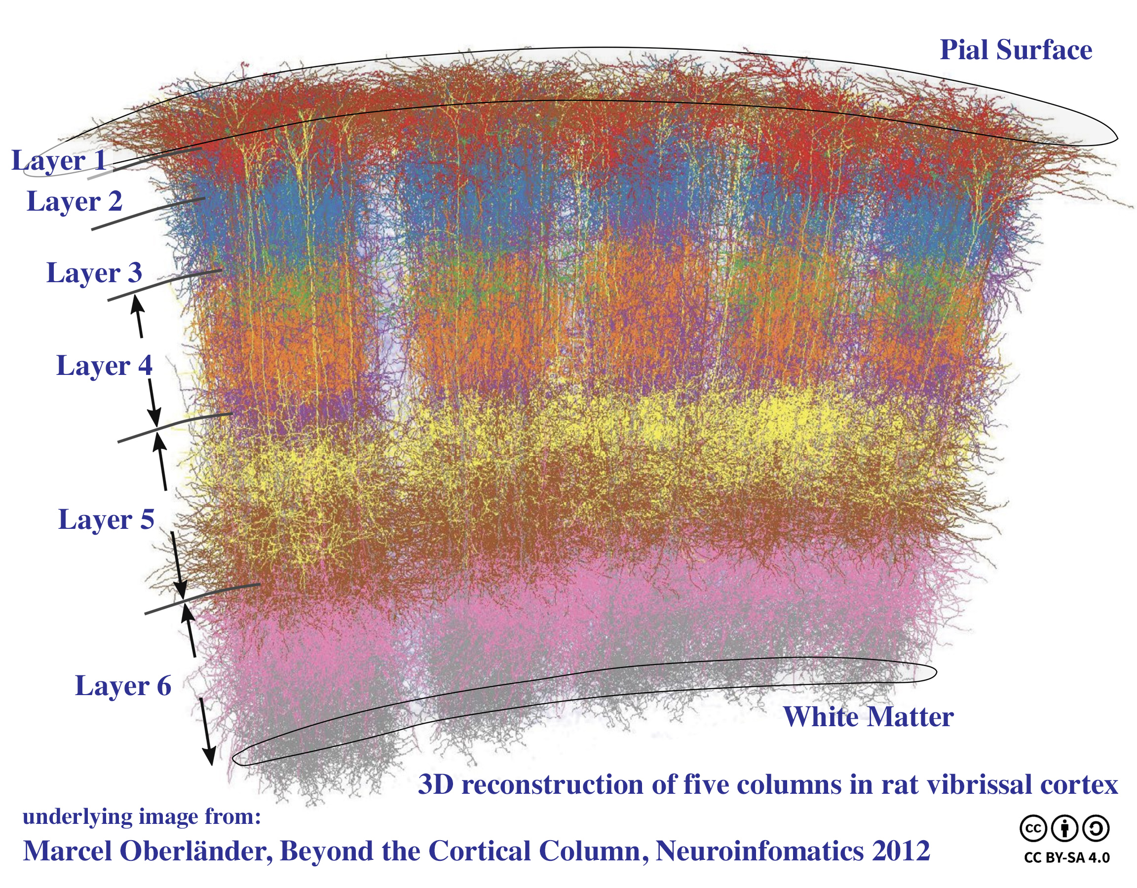
The neocortex, also called the neopallium, isocortex, or the six-layered cortex, is a set of layers of the mammalian cerebral cortex involved in higher-order brain functions such as sensory perception, cognition, generation of motor commands, spatial reasoning and language. The neocortex is further subdivided into the true isocortex and the proisocortex. In the human brain, the neocortex is the largest part of the cerebral cortex (the outer layer of the cerebrum). The neocortex makes up the largest part of the cerebral cortex, with the allocortex making up the rest. The neocortex is made up of six layers, labelled from the outermost inwards, I to VI. Etymology The term is from ''cortex'', Latin, "bark" or "rind", combined with ''neo-'', Greek, "new". ''Neopallium'' is a similar hybrid, from Latin ''pallium'', "cloak". ''Isocortex'' and ''allocortex'' are hybrids with Greek ''isos'', "same", and ''allos'', "other". Anatomy The neocortex is the most developed in its organisati ... [...More Info...] [...Related Items...] OR: [Wikipedia] [Google] [Baidu] |
Cortical Column
A cortical column is a group of neurons forming a cylindrical structure through the cerebral cortex of the brain perpendicular to the cortical surface. The structure was first identified by Mountcastle in 1957. He later identified minicolumns as the basic units of the neocortex which were arranged into columns. Each contains the same types of neurons, connectivity, and firing properties. Columns are also called hypercolumn, macrocolumn, functional column or sometimes cortical module,. Neurons within a minicolumn (microcolumn) encode similar features, whereas a hypercolumn "denotes a unit containing a full set of values for any given set of receptive field parameters". A cortical module is defined as either synonymous with a hypercolumn (Mountcastle) or as a tissue block of multiple overlapping hypercolumns. Cortical columns are proposed to be the canonical microcircuits for predictive coding, in which the process of cognition is implemented through a hierarchy of identical microc ... [...More Info...] [...Related Items...] OR: [Wikipedia] [Google] [Baidu] |
Proisocortex
Proisocortex or pro-isocortex is one of two subtypes of cortical areas in the areas belonging to the neocortex. The other subtype is termed the true isocortex. Proisocortical areas are transitional areas placed between areas of true isocortex and areas of periallocortex (which themselves are transitional between "true" allocortex and proisocortex). The histological structure of proisocortex is also transitional between true isocortex and either peripaleocortex or periarchicortex, depending on with which subtype of periallocortex the given proisocortical area borders. Proisocortex is found in the cingulate cortex (part of the limbic system), in Brodmann's areas 24, 25, 30 and 32, the insula and parahippocampal gyrus. The transitional cortical areas from isocortical side (i.e. proisocortex) and from allocortical side (i.e. periarchicortex and peripaleocortex) together are called mesocortex. The mesocortex is essentially the same as paralimbic cortex The paralimbic cor ... [...More Info...] [...Related Items...] OR: [Wikipedia] [Google] [Baidu] |
Rodent
Rodents (from Latin , 'to gnaw') are mammals of the order Rodentia (), which are characterized by a single pair of continuously growing incisors in each of the upper and lower jaws. About 40% of all mammal species are rodents. They are native to all major land masses except for New Zealand, Antarctica, and several oceanic islands, though they have subsequently been introduced to most of these land masses by human activity. Rodents are extremely diverse in their ecology and lifestyles and can be found in almost every terrestrial habitat, including human-made environments. Species can be arboreal, fossorial (burrowing), saltatorial/richochetal (leaping on their hind legs), or semiaquatic. However, all rodents share several morphological features, including having only a single upper and lower pair of ever-growing incisors. Well-known rodents include mice, rats, squirrels, prairie dogs, porcupines, beavers, guinea pigs, and hamsters. Rabbits, hares, and pikas, wh ... [...More Info...] [...Related Items...] OR: [Wikipedia] [Google] [Baidu] |
Long-finned Pilot Whale
The long-finned pilot whale (''Globicephala melas'') is a large species of oceanic dolphin. It shares the genus '' Globicephala'' with the short-finned pilot whale (''Globicephala macrorhynchus''). Long-finned pilot whales are known as such because of their unusually long pectoral fins. Taxonomy and naming Etymology Pilot whales get their name from the original belief that there was a "pilot" or lead individual in their groups.Olson, P.A. (2008). "Pilot whale ''Globicephala melas'' and ''G. muerorhynchus''". pp. 847–52 in ''Encyclopedia of Marine Mammals''. Perrin, W. F., Wursig, B., and Thewissen, J. G. M. (eds.). Academic Press; 2nd edition. Ridgway, S. H. (1998). ''Handbook of Marine Mammals: The second book of dolphins and the porpoises''. Volume 6, Elsevier. pp. 245–69. The name for the genus, "''Globicephala''" is derived from a combination of Latin ''globus'' ("globe") and Greek ''kephale'' ("head"). The specific name "''melas''" is Greek for "black". This species h ... [...More Info...] [...Related Items...] OR: [Wikipedia] [Google] [Baidu] |
Oceanic Dolphin
Oceanic dolphins or Delphinidae are a widely distributed family of dolphins that live in the sea. Close to forty extant species are recognised. They include several big species whose common names contain "whale" rather than "dolphin", such as the Globicephalinae (round-headed whales including the orca and pilot whale). Delphinidae is a family within the superfamily Delphinoidea, which also includes the porpoises (Phocoenidae) and the Monodontidae (beluga whale and narwhal). River dolphins are relatives of the Delphinoidea. Oceanic dolphins range in size from the and Maui's dolphin to the and orca, the largest known dolphin. Several species exhibit sexual dimorphism; the males are larger than females. They have streamlined bodies and two limbs that are modified into flippers. Though not quite as flexible as seals, some dolphins can travel at speeds 29 km/h (18 mph) for short distances. Most delphinids primarily eat fish, along with a smaller number of squid a ... [...More Info...] [...Related Items...] OR: [Wikipedia] [Google] [Baidu] |
Parahippocampal Gyrus
The parahippocampal gyrus (or hippocampal gyrus') is a grey matter cortical region of the brain that surrounds the hippocampus and is part of the limbic system. The region plays an important role in memory encoding and retrieval. It has been involved in some cases of hippocampal sclerosis. Asymmetry has been observed in schizophrenia. Structure The anterior part of the gyrus includes the perirhinal and entorhinal cortices. The term parahippocampal cortex is used to refer to an area that encompasses both the posterior parahippocampal gyrus and the medial portion of the fusiform gyrus. Function Scene recognition The parahippocampal place area (PPA) is a sub-region of the parahippocampal cortex that lies medially in the inferior temporo-occipital cortex. PPA plays an important role in the encoding and recognition of environmental scenes (rather than faces). fMRI studies indicate that this region of the brain becomes highly active when human subjects view topographical scene s ... [...More Info...] [...Related Items...] OR: [Wikipedia] [Google] [Baidu] |
Insular Cortex
The insular cortex (also insula and insular lobe) is a portion of the cerebral cortex folded deep within the lateral sulcus (the fissure separating the temporal lobe from the parietal and frontal lobes) within each hemisphere of the mammalian brain. The insulae are believed to be involved in consciousness and play a role in diverse functions usually linked to emotion or the regulation of the body's homeostasis. These functions include compassion, empathy, taste, perception, motor control, self-awareness, cognitive functioning, interpersonal experience, and awareness of homeostatic emotions such as hunger, pain and fatigue. In relation to these, it is involved in psychopathology. The insular cortex is divided into two parts: the anterior insula and the posterior insula in which more than a dozen field areas have been identified. The cortical area overlying the insula toward the lateral surface of the brain is the operculum (meaning ''lid''). The opercula are formed from part ... [...More Info...] [...Related Items...] OR: [Wikipedia] [Google] [Baidu] |
Brodmann Area 32
The Brodmann area 32, also known in the human brain as the dorsal anterior cingulate area 32, refers to a subdivision of the cytoarchitecturally defined cingulate cortex. In the human it forms an outer arc around the anterior cingulate gyrus. The cingulate sulcus defines approximately its inner boundary and the superior rostral sulcus (H) its ventral boundary; rostrally it extends almost to the margin of the frontal lobe. Cytoarchitecturally it is bounded internally by the ventral anterior cingulate area 24, externally by medial margins of the agranular frontal area 6, intermediate frontal area 8, granular frontal area 9, frontopolar area 10, and prefrontal area 11-1909. (Brodmann19-09). The dorsal region of the anterior cingulate gyrus is associated with rational thought processes, most notably active during the Stroop task. Guenon In the guenon, Brodmann area 32 is a subdivision of the cytoarchitecturally defined cingulate region of cerebral cortex. This area was named ... [...More Info...] [...Related Items...] OR: [Wikipedia] [Google] [Baidu] |
Brodmann Area 30
Brodmann area 30, also known as agranular retrolimbic area 30, is a subdivision of the cytoarchitecturally defined retrosplenial region of the cerebral cortex. In the human it is located in the isthmus of cingulate gyrus. Cytoarchitecturally it is bounded internally by the granular retrolimbic area 29 Brodmann area 29, also known as granular retrolimbic area 29 or granular retrosplenial cortex, is a cytoarchitecturally defined portion of the retrosplenial region of the cerebral cortex. In the human it is a narrow band located in the isthmus ..., dorsally by the ventral posterior cingulate area 23 and ventrolaterally by the ectorhinal area 36 (Brodmann-1909). See also * Brodmann area 30 Medial surface of cerebral hemisphere {{Neuroanatomy-stub ... [...More Info...] [...Related Items...] OR: [Wikipedia] [Google] [Baidu] |
Brodmann Area 25
Brodmann area 25 (BA25) is the subgenual area, area subgenualis or subgenual cingulatea area in the cerebral cortex of the brain and delineated based on its cytoarchitectonic characteristics. It is the 25th " Brodmann area" defined by Korbinian Brodmann (thus its name). BA25 is located in the cingulate region as a narrow band in the caudal portion of the subcallosal area adjacent to the paraterminal gyrus. The posterior parolfactory sulcus separates the paraterminal gyrus from BA25. Rostrally it is bound by the prefrontal area 11 of Brodmann. History Brodmann described this area as it is labeled now in 1909. Originally in 1905, Brodmann labeled the area as part of area 24. In 1909, he divided the area into area 24 and 25. Function This region is extremely rich in serotonin transporters and is considered as a governor for a vast network involving areas like hypothalamus and brain stem, which influences changes in appetite and sleep; the amygdala and insula, which affect th ... [...More Info...] [...Related Items...] OR: [Wikipedia] [Google] [Baidu] |
Brodmann Area 24
Brodmann area 24 is part of the anterior cingulate in the human brain. Human In the human this area is known as ventral anterior cingulate area 24, and it refers to a subdivision of the cytoarchitecturally defined cingulate cortex region of cerebral cortex (area cingularis anterior ventralis). It occupies most of the anterior cingulate gyrus in an arc around the genu of the corpus callosum. Its outer border corresponds approximately to the cingulate sulcus. Cytoarchitecturally it is bounded internally by the pregenual area 33, externally by the dorsal anterior cingulate area 32, and caudally by the ventral posterior cingulate area 23 and the dorsal posterior cingulate area 31. Guenon In the guenon this area is referred to as area 24 of Brodmann-1905. It includes portions of the cingulate gyrus and the frontal lobe. The cortex is thin; it lacks the internal granular layer (IV) so that the densely distributed, plump pyramidal cells of sublayer 3b of the external pyramid ... [...More Info...] [...Related Items...] OR: [Wikipedia] [Google] [Baidu] |
Brodmann Area
A Brodmann area is a region of the cerebral cortex, in the human or other primate brain, defined by its cytoarchitecture, or histological structure and organization of cells. History Brodmann areas were originally defined and numbered by the German anatomist Korbinian Brodmann based on the cytoarchitectural organization of neurons he observed in the cerebral cortex using the Nissl method of cell staining. Brodmann published his maps of cortical areas in humans, monkeys, and other species in 1909, along with many other findings and observations regarding the general cell types and laminar organization of the mammalian cortex. The same Brodmann area number in different species does not necessarily indicate homologous areas. A similar, but more detailed cortical map was published by Constantin von Economo and Georg N. Koskinas in 1925. Present importance Brodmann areas have been discussed, debated, refined, and renamed exhaustively for nearly a century and remain the most w ... [...More Info...] [...Related Items...] OR: [Wikipedia] [Google] [Baidu] |


