|
Lacrimals
The lacrimal bone is a small and fragile bone of the facial skeleton; it is roughly the size of the little fingernail. It is situated at the front part of the medial wall of the Orbit (anatomy), orbit. It has two surfaces and four borders. Several bony landmarks of the lacrimal bone function in the process of lacrimation or crying. Specifically, the lacrimal bone helps form the nasolacrimal canal necessary for tear translocation. A depression on the anterior inferior portion of the bone, the lacrimal fossa, houses the membranous lacrimal sac. Tears or lacrimal fluid, from the lacrimal glands, collect in this sac during excessive lacrimation. The fluid then flows through the nasolacrimal duct and into the nasopharynx. This drainage results in what is commonly referred to a runny nose during excessive crying or tear production. Injury or fracture of the lacrimal bone can result in posttraumatic obstruction of the lacrimal pathways. Structure Lateral or orbital surface The lateral or ... [...More Info...] [...Related Items...] OR: [Wikipedia] [Google] [Baidu] |
Ethmoid Bone
The ethmoid bone (; from grc, ἡθμός, hēthmós, sieve) is an unpaired bone in the skull that separates the nasal cavity from the brain. It is located at the roof of the nose, between the two orbits. The cubical bone is lightweight due to a spongy construction. The ethmoid bone is one of the bones that make up the orbit of the eye. Structure The ethmoid bone is an anterior cranial bone located between the eyes. It contributes to the medial wall of the orbit, the nasal cavity, and the nasal septum. The ethmoid has three parts: cribriform plate, ethmoidal labyrinth, and perpendicular plate. The cribriform plate forms the roof of the nasal cavity and also contributes to formation of the anterior cranial fossa, the ethmoidal labyrinth consists of a large mass on either side of the perpendicular plate, and the perpendicular plate forms the superior two-thirds of the nasal septum. Between the orbital plate and the nasal conchae are the ethmoidal sinuses or ethmoidal air cells, w ... [...More Info...] [...Related Items...] OR: [Wikipedia] [Google] [Baidu] |
Frontal Bone
The frontal bone is a bone in the human skull. The bone consists of two portions.'' Gray's Anatomy'' (1918) These are the vertically oriented squamous part, and the horizontally oriented orbital part, making up the bony part of the forehead, part of the bony orbital cavity holding the eye, and part of the bony part of the nose respectively. The name comes from the Latin word ''frons'' (meaning " forehead"). Structure of the frontal bone The frontal bone is made up of two main parts. These are the squamous part, and the orbital part. The squamous part marks the vertical, flat, and also the biggest part, and the main region of the forehead. The orbital part is the horizontal and second biggest region of the frontal bone. It enters into the formation of the roofs of the orbital and nasal cavities. Sometimes a third part is included as the nasal part of the frontal bone, and sometimes this is included with the squamous part. The nasal part is between the brow ridges, and ends ... [...More Info...] [...Related Items...] OR: [Wikipedia] [Google] [Baidu] |
Ethmoid
The ethmoid bone (; from grc, ἡθμός, hēthmós, sieve) is an unpaired bone in the skull that separates the nasal cavity from the brain. It is located at the roof of the nose, between the two orbits. The cubical bone is lightweight due to a spongy construction. The ethmoid bone is one of the bones that make up the orbit of the eye. Structure The ethmoid bone is an anterior cranial bone located between the eyes. It contributes to the medial wall of the orbit, the nasal cavity, and the nasal septum. The ethmoid has three parts: cribriform plate, ethmoidal labyrinth, and perpendicular plate. The cribriform plate forms the roof of the nasal cavity and also contributes to formation of the anterior cranial fossa, the ethmoidal labyrinth consists of a large mass on either side of the perpendicular plate, and the perpendicular plate forms the superior two-thirds of the nasal septum. Between the orbital plate and the nasal conchae are the ethmoidal sinuses or ethmoidal air cells ... [...More Info...] [...Related Items...] OR: [Wikipedia] [Google] [Baidu] |
Orbit (anatomy)
In anatomy, the orbit is the cavity or socket of the skull in which the eye and its appendages are situated. "Orbit" can refer to the bony socket, or it can also be used to imply the contents. In the adult human, the volume of the orbit is , of which the eye occupies . The orbital contents comprise the eye, the orbital and retrobulbar fascia, extraocular muscles, cranial nerves II, III, IV, V, and VI, blood vessels, fat, the lacrimal gland with its sac and duct, the eyelids, medial and lateral palpebral ligaments, cheek ligaments, the suspensory ligament, septum, ciliary ganglion and short ciliary nerves. Structure The orbits are conical or four-sided pyramidal cavities, which open into the midline of the face and point back into the head. Each consists of a base, an apex and four walls."eye, human."Encyclopædia Britannica from Encyclopædia Britannica 2006 Ultimate Reference Suite DVD 2009 Openings There are two important foramina, or windows, two important ... [...More Info...] [...Related Items...] OR: [Wikipedia] [Google] [Baidu] |
Lamina Papyracea
The orbital lamina of ethmoid bone, (or lamina papyracea or orbital lamina) is a smooth, oblong bone plate which forms the lateral surface of the labyrinth of the ethmoid bone in the skull. The plate covers in the middle and posterior ethmoidal cells and forms a large part of the medial wall of the orbit. It articulates above with the orbital plate of the frontal bone, below with the maxilla and the orbital process of palatine bone, in front with the lacrimal, and behind with the sphenoid. Its name lamina papyracea is an appropriate description, as this part of the ethmoid bone is paper-thin and fractures easily. A fracture here could cause entrapment of the medial rectus muscle The medial rectus muscle is a muscle in the orbit near the eye. It is one of the extraocular muscles. It originates from the common tendinous ring, and inserts into the anteromedial surface of the eye. It is supplied by the inferior division of th .... Additional Images File:Slide4fen.JPG, Orbital ... [...More Info...] [...Related Items...] OR: [Wikipedia] [Google] [Baidu] |
Albertosaurus
''Albertosaurus'' (; meaning "Alberta lizard") is a genus of tyrannosaurid theropod dinosaurs that lived in western North America during the Late Cretaceous Period, about 71 million years ago. The type species, ''A. sarcophagus'', was apparently restricted in range to the modern-day Canadian province of Alberta, after which the genus is named, although an indeterminate species ("cf. ''Albertosaurus'' sp.") has been discovered in the Corral de Enmedio and Packard Formations in Mexico. Scientists disagree on the content of the genus, with some recognizing ''Gorgosaurus libratus'' as a second species. As a tyrannosaurid, ''Albertosaurus'' was a bipedal predator with tiny, two-fingered hands and a massive head that had dozens of large, sharp teeth. It may have been an apex predator in its local ecosystem. While ''Albertosaurus'' was large for a theropod, it was much smaller than its larger and more famous relative ''Tyrannosaurus rex'', growing up to in length and w ... [...More Info...] [...Related Items...] OR: [Wikipedia] [Google] [Baidu] |
Ceratosaurus
''Ceratosaurus'' (from Greek κέρας/κέρατος, ' meaning "horn" and σαῦρος ' meaning "lizard") was a carnivorous theropod dinosaur in the Late Jurassic period ( Kimmeridgian to Tithonian). The genus was first described in 1884 by American paleontologist Othniel Charles Marsh based on a nearly complete skeleton discovered in Garden Park, Colorado, in rocks belonging to the Morrison Formation. The type species is ''Ceratosaurus nasicornis''. The Garden Park specimen remains the most complete skeleton known from the genus, and only a handful of additional specimens have been described since. Two additional species, ''Ceratosaurus dentisulcatus'' and ''Ceratosaurus magnicornis'', were described in 2000 from two fragmentary skeletons from the Cleveland-Lloyd Quarry of Utah and from the vicinity of Fruita, Colorado. The validity of these additional species has been questioned, however, and all three skeletons possibly represent different growth stages of the sa ... [...More Info...] [...Related Items...] OR: [Wikipedia] [Google] [Baidu] |
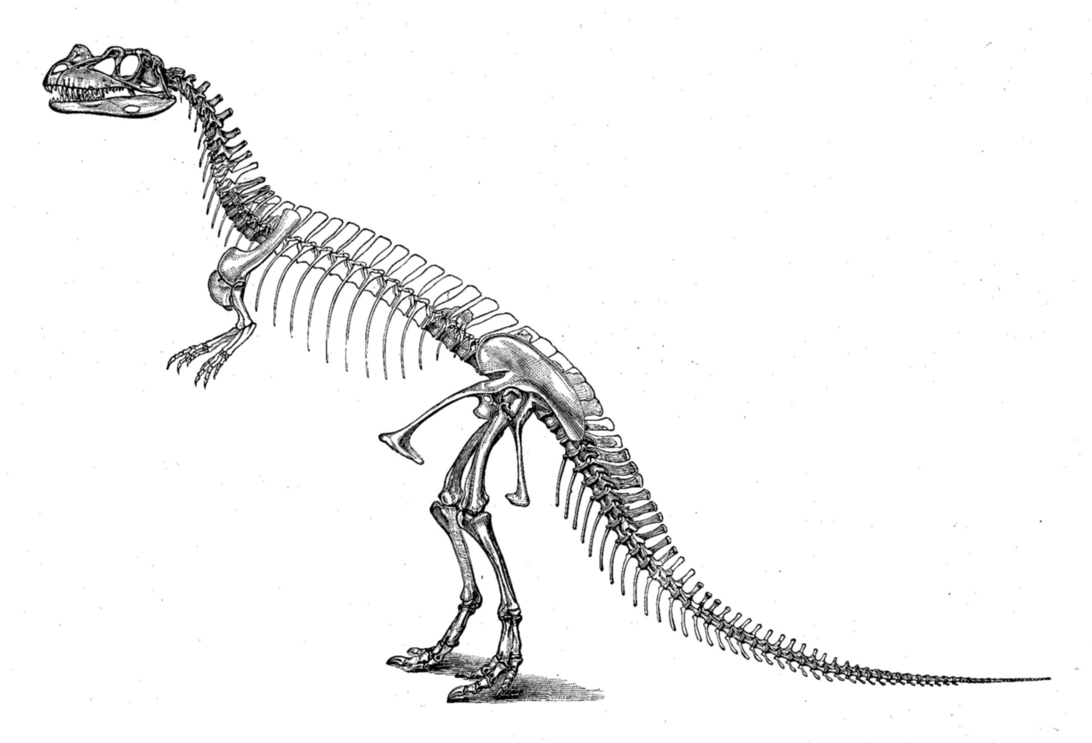
Allosaurus
''Allosaurus'' () is a genus of large carnosaurian theropod dinosaur that lived 155 to 145 million years ago during the Late Jurassic epoch ( Kimmeridgian to late Tithonian). The name "''Allosaurus''" means "different lizard" alluding to its unique (at the time of its discovery) concave vertebrae. It is derived from the Greek (') ("different, other") and (') ("lizard / generic reptile"). The first fossil remains that could definitively be ascribed to this genus were described in 1877 by paleontologist Othniel Charles Marsh. As one of the first well-known theropod dinosaurs, it has long attracted attention outside of paleontological circles. ''Allosaurus'' was a large bipedal predator. Its skull was light, robust and equipped with dozens of sharp, serrated teeth. It averaged in length for ''A. fragilis'', with the largest specimens estimated as being long. Relative to the large and powerful hindlimbs, its three-fingered forelimbs were small, and the body was ... [...More Info...] [...Related Items...] OR: [Wikipedia] [Google] [Baidu] |
Antorbital Fenestra
An antorbital fenestra (plural: fenestrae) is an opening in the skull that is in front of the eye sockets. This skull character is largely associated with archosauriforms, first appearing during the Triassic Period. Among extant archosaurs, birds still possess antorbital fenestrae, whereas crocodylians have lost them. The loss in crocodylians is believed to be related to the structural needs of their skulls for the bite force and feeding behaviours that they employ.Preushscoft, H., Witzel, U. 2002. Biomechanical Investigations on the Skulls of Reptiles and Mammals. Senckenbergiana Lethaea 82:207–222.Rayfield, E.J., Milner, A.C., Xuan, V.B., Young, P.G. 2007. Functional Morphology of Spinosaur "Crocodile Mimic" Dinosaurs. JVP. 27(4):892–901. In some archosaur species, the opening has closed but its location is still marked by a depression, or fossa, on the surface of the skull called the antorbital fossa. The antorbital fenestra houses a paranasal sinus that is confluent with ... [...More Info...] [...Related Items...] OR: [Wikipedia] [Google] [Baidu] |
Amphibian
Amphibians are four-limbed and ectothermic vertebrates of the class Amphibia. All living amphibians belong to the group Lissamphibia. They inhabit a wide variety of habitats, with most species living within terrestrial, fossorial, arboreal or freshwater aquatic ecosystems. Thus amphibians typically start out as larvae living in water, but some species have developed behavioural adaptations to bypass this. The young generally undergo metamorphosis from larva with gills to an adult air-breathing form with lungs. Amphibians use their skin as a secondary respiratory surface and some small terrestrial salamanders and frogs lack lungs and rely entirely on their skin. They are superficially similar to reptiles like lizards but, along with mammals and birds, reptiles are amniotes and do not require water bodies in which to breed. With their complex reproductive needs and permeable skins, amphibians are often ecological indicators; in recent decades there has been a dramatic ... [...More Info...] [...Related Items...] OR: [Wikipedia] [Google] [Baidu] |
Nasal Bone
The nasal bones are two small oblong bones, varying in size and form in different individuals; they are placed side by side at the middle and upper part of the face and by their junction, form the bridge of the upper one third of the nose. Each has two surfaces and four borders. Structure The two nasal bones are joined at the midline internasal suture and make up the bridge of the nose. Surfaces The ''outer surface'' is concavo-convex from above downward, convex from side to side; it is covered by the procerus and nasalis muscles, and perforated about its center by a foramen, for the transmission of a small vein. The ''inner surface'' is concave from side to side, and is traversed from above downward, by a groove for the passage of a branch of the nasociliary nerve. Articulations The nasal articulates with four bones: two of the cranium, the frontal and ethmoid, and two of the face, the opposite nasal and the maxilla. Other animals In primitive bony fish and tetr ... [...More Info...] [...Related Items...] OR: [Wikipedia] [Google] [Baidu] |
.jpg)


.png)