|
Intermammary Sulcus
The intermammary cleft, intermammary sulcus, or sulcus intermammarius is a Surface anatomy, surface feature of males and females that marks the division of the two breasts with the sternum (breastbone) in the middle.Dr. Ted Eisenberg and Joyce K. Eisenberg, ''The Scoop on Breasts: A Plastic Surgeon Busts the Myths'', Incompra Press, 2012, The International Federation of Associations of Anatomists (IFAA) uses the terms "sulcus intermammarius" or "intermammary cleft" when referring to the area between the breasts.Muhammad Adil Abbas Khan, Ammar Asrar Javed and Nigel MercerCleavage classification: categorizing a vital feminine aesthetic landmark Plastic and Aesthetic Research (PAR), 016-01-15 Etymology "Intermammary" ("wikt:inter-, inter", between + "wikt:mamma, mamma", breasts + "wikt:-ery, ry", place) means something that is located or performed between the breasts (example: intermammary intercourse). "Sulcus" is a Latin word that means a furrow or groove, commonly used to mean a ... [...More Info...] [...Related Items...] OR: [Wikipedia] [Google] [Baidu] |
Breast
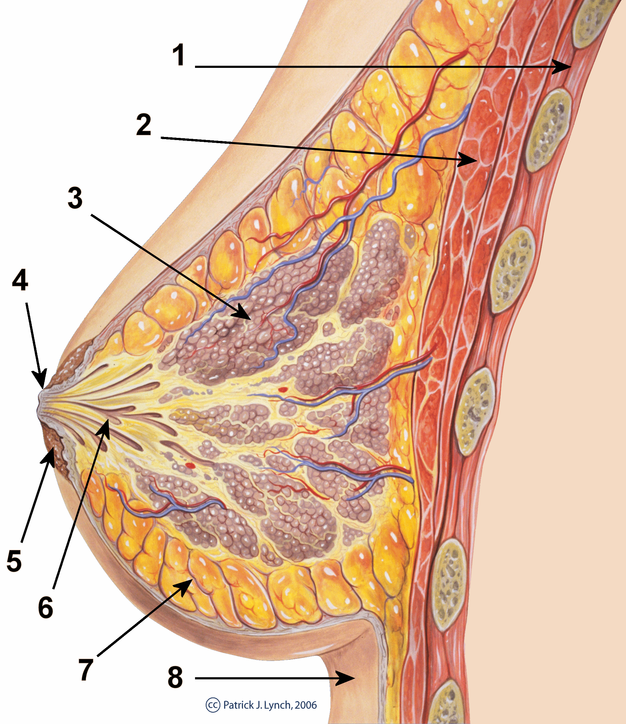
The breasts are two prominences located on the upper ventral region of the torso among humans and other primates. Both sexes develop breasts from the same embryology, embryological tissues. The relative size and development of the breasts is a major secondary sex distinction between females and males. There is also considerable Bra size, variation in size between individuals. Permanent Breast development, breast growth during puberty is caused by estrogens in conjunction with the growth hormone. Female humans are the only mammals that permanently develop breasts at puberty; all other mammals develop their mammary tissue during the latter period of pregnancy. In females, the breast serves as the mammary gland, which produces and secretes milk to feed infants. Subcutaneous fat covers and envelops a network of lactiferous duct, ducts that converge on the nipple, and these tissue (biology), tissues give the breast its distinct size and globular shape. At the ends of the ducts are ... [...More Info...] [...Related Items...] OR: [Wikipedia] [Google] [Baidu] |
Nipple
The nipple is a raised region of tissue on the surface of the breast from which, in lactating females, breast milk, milk from the mammary gland leaves the body through the lactiferous ducts to Breastfeeding, nurse an infant. The milk can flow through the nipple passively, or it can be ejected by smooth muscles, smooth muscular contraction, muscle contractions that occur along with the ductal system. The nipple is surrounded by the areola, which is often a darker colour than the surrounding skin. Male mammals also have nipples but without the same level of function or prominence. A nipple is often called a ''teat'' when referring to non-humans. "Nipple" or "teat" can also be used to describe the flexible mouthpiece of a baby bottle. In humans, the nipples of both males and females can be sexually stimulated as part of sexual arousal. In many cultures, female nipples are sexualized, or regarded as sex objects and evaluated in terms of their physical characteristics and sex appeal. ... [...More Info...] [...Related Items...] OR: [Wikipedia] [Google] [Baidu] |
Anatomical Variation
An anatomical variation, anatomical variant, or anatomical variability is a presentation of body structure with Morphology (biology), morphological features different from those that are typically described in the majority of individuals. Anatomical variations are categorized into three types including morphometric (size or shape), consistency (present or absent), and spatial (proximal/distal or right/left). Variations are seen as normal in the sense that they are found consistently among different individuals, are mostly without symptoms, and are termed anatomical variations rather than abnormalities. Anatomical variations are mainly caused by genetics and may vary considerably between different populations. The rate of variation considerably differs between single organ (anatomy), organs, particularly in muscles. Knowledge of anatomical variations is important in order to distinguish them from pathological conditions. A very early paper published in 1898, presented anatomic var ... [...More Info...] [...Related Items...] OR: [Wikipedia] [Google] [Baidu] |
Sternalis Muscle
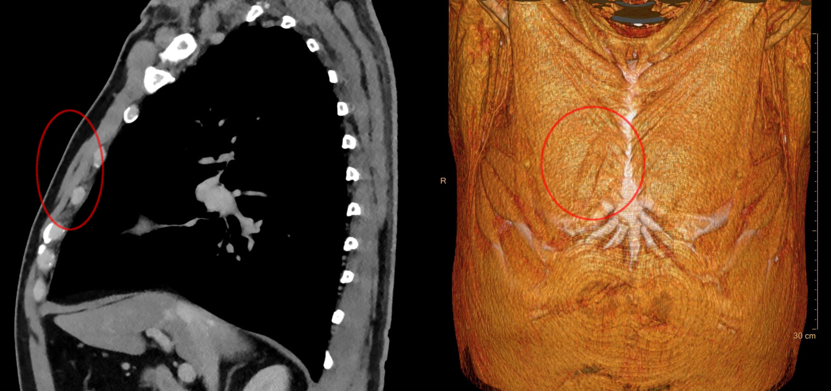
The rectus sternalis muscle is an anatomical variation that lies in front of the sternal end of the pectoralis major parallel to the margin of the sternum. The sternalis muscle may be a variation of the pectoralis major or of the rectus abdominis. Structure The sternalis is a muscle that runs along the anterior aspect of the body of the sternum. It lies superficially and parallel to the sternum. Its origin and insertion are variable. The sternalis muscle often originates from the upper part of the sternum and can display varying insertions such as the pectoral fascia, lower ribs, costal cartilages, rectus sheath, aponeurosis of the abdominal external oblique muscle. It may be present unilaterally or bilaterally. There is still a great deal of disagreement about its innervation and its embryonic origin. In a review, it was reported that the muscle was innervated by the external or internal thoracic nerves in 55% of the cases, by the intercostal nerves in 43% of the cases, whi ... [...More Info...] [...Related Items...] OR: [Wikipedia] [Google] [Baidu] |
Pectoral Fascia
The pectoral fascia is a thin lamina, covering the surface of the pectoralis major, and sending numerous prolongations between its fasciculi: it is attached, in the middle line, to the front of the sternum; above, to the clavicle; laterally and below it is continuous with the fascia of the shoulder, axilla, and thorax. It is very thin over the upper part of the pectoralis major, but thicker in the interval between it and the latissimus dorsi, where it closes in the axillary space and forms the ''axillary fascia''; it divides at the lateral margin of the latissimus dorsi into two layers, one of which passes in front of, and the other behind it; these proceed as far as the spinous processes of the thoracic vertebrae, to which they are attached. As the fascia leaves the lower edge of the pectoralis major to cross the floor of the axilla it sends a layer upward under cover of the muscle; this lamina splits to envelop the pectoralis minor, at the upper edge of which it is continuous ... [...More Info...] [...Related Items...] OR: [Wikipedia] [Google] [Baidu] |
Superficial Fascia
A fascia (; : fasciae or fascias; adjective fascial; ) is a generic term for macroscopic membranous bodily structures. Fasciae are classified as superficial, visceral or deep, and further designated according to their anatomical location. The knowledge of fascial structures is essential in surgery, as they create borders for infectious processes (for example Psoas abscess) and haematoma. An increase in pressure may result in a compartment syndrome, where a prompt fasciotomy may be necessary. For this reason, profound descriptions of fascial structures are available in anatomical literature from the 19th century. Function Fasciae were traditionally thought of as passive structures that transmit mechanical tension generated by muscular activities or external forces throughout the body. An important function of muscle fasciae is to reduce friction of muscular force. In doing so, fasciae provide a supportive and movable wrapping for nerves and blood vessels as they pass through ... [...More Info...] [...Related Items...] OR: [Wikipedia] [Google] [Baidu] |
Polythelia
A supernumerary nipple is an additional instance of nipple occurring in mammals, including humans and monkeys. They are often mistaken for moles. Studies variously report the prevalence of supernumerary nipples as approximately 1 in 18 and 1 in 40.Supernumerary nipples: prevalence, size, sex and side predilection – a prospective clinical study H. Schmidt, European Journal of Pediatrics volume 157, pages 821–823 (1998).Occurrence of Supernumerary Nipples in Newborns Francis Mimouni, MD; Paul Merlob, MD; Salomon H. Reisner, MB, ChB, Am J ... [...More Info...] [...Related Items...] OR: [Wikipedia] [Google] [Baidu] |
Costal Margin
The costal margin, also known as the costal arch, is the lower edge of the chest (thorax) formed by the bottom edge of the rib cage. Structure The costal margin is the medial margin formed by the cartilages of the seventh to tenth ribs. It attaches to the body and xiphoid process of the sternum. The thoracic diaphragm attaches to the costal margin. The costal angle is the angle between the left and right costal margins where they join the sternum. Function The costal margins somewhat protect the higher abdominal organs, such as the liver. Clinical significance The costal margin may be used for tissue harvesting of cartilage for use elsewhere in the body, such as to treat microtia. Different abdominal organs may be palpated just below the costal margin, such as the liver on the right side of the body. Pain across the costal margin is most commonly caused by costochondritis. The costal paradox, also known as Hoover's sign and the costal margin paradox, is a sign ... [...More Info...] [...Related Items...] OR: [Wikipedia] [Google] [Baidu] |
Xyphoid Process
The xiphoid process (), also referred to as the ensiform process, xiphisternum, or metasternum, constitutes a small cartilaginous process (extension) located in the inferior segment of the sternum, typically ossified in adult humans. Both the Greek-derived term ''xiphoid'' and its Latin equivalent, ''ensiform'', connote a "swordlike" or "sword-shaped" morphology. Structure The xiphoid process is anatomically situated at the level of the 9th thoracic vertebra (T9) and corresponds to the T7 dermatome. Development In neonates and young infants, particularly smaller infants, the tip of the xiphoid process may be seen as a palpable lump situated just below the sternal notch. Between the ages of 15 and 29, the xiphoid process typically undergoes fusion with the body of the sternum through a fibrous joint. Unlike the synovial articulation of major joints, this joint does not permit movement. Ossification of the xiphoid process typically occurs around the age of 40. Variation The xipho ... [...More Info...] [...Related Items...] OR: [Wikipedia] [Google] [Baidu] |
Sternum
The sternum (: sternums or sterna) or breastbone is a long flat bone located in the central part of the chest. It connects to the ribs via cartilage and forms the front of the rib cage, thus helping to protect the heart, lungs, and major blood vessels from injury. Shaped roughly like a necktie, it is one of the largest and longest flat bones of the body. Its three regions are the manubrium, the body, and the xiphoid process. The word ''sternum'' originates from Ancient Greek στέρνον (''stérnon'') 'chest'. Structure The sternum is a narrow, flat bone, forming the middle portion of the front of the chest. The top of the sternum supports the clavicles (collarbones) and its edges join with the costal cartilages of the first two pairs of ribs. The inner surface of the sternum is also the attachment of the sternopericardial ligaments. Its top is also connected to the sternocleidomastoid muscle. The sternum consists of three main parts, listed from the top: * Man ... [...More Info...] [...Related Items...] OR: [Wikipedia] [Google] [Baidu] |
Manubrium
The sternum (: sternums or sterna) or breastbone is a long flat bone located in the central part of the chest. It connects to the ribs via cartilage and forms the front of the rib cage, thus helping to protect the heart, human lung, lungs, and major blood vessels from injury. Shaped roughly like a necktie, it is one of the largest and longest flat bones of the body. Its three regions are the manubrium, the body, and the xiphoid process. The word ''sternum'' originates from Ancient Greek στέρνον (''stérnon'') 'chest'. Structure The sternum is a narrow, flat bone, forming the middle portion of the front of the chest. The top of the sternum supports the clavicles (collarbones) and its edges join with the costal cartilages of the first two pairs of ribs. The inner surface of the sternum is also the attachment of the sternopericardial ligaments. Its top is also connected to the sternocleidomastoid muscle. The sternum consists of three main parts, listed from the top: * Manubr ... [...More Info...] [...Related Items...] OR: [Wikipedia] [Google] [Baidu] |


