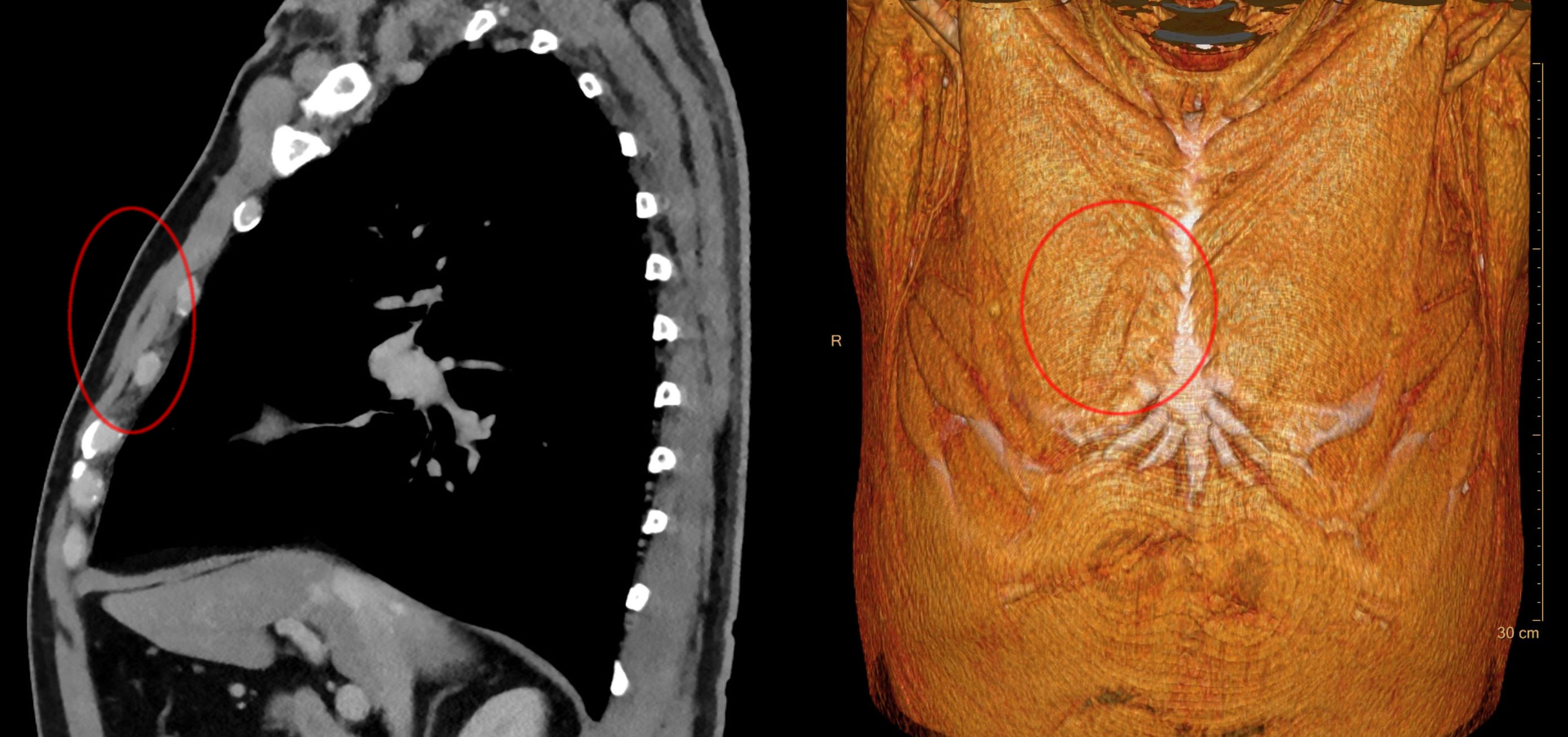
Sternalis Muscle on:
[Wikipedia]
[Google]
[Amazon]
The rectus sternalis muscle is an
 The presence of the sternalis is asymptomatic but aesthetic complaints have been reported as it was reported to cause chest
Image:PSM_V25_D748_Rectus_sternalis_and_the_great_pectoral_chest_muscle.jpg, The sternalis muscle as it lies on top the pectoralis major.
Image:Braus 1921 137.png, Sternalis muscle visible in an individual who also has an accessory abdominal pectoralis major, and an axillary arch of the
anatomical variation
An anatomical variation, anatomical variant, or anatomical variability is a presentation of body structure with Morphology (biology), morphological features different from those that are typically described in the majority of individuals. Anatomica ...
that lies in front of the sternal end of the pectoralis major parallel to the margin of the sternum
The sternum (: sternums or sterna) or breastbone is a long flat bone located in the central part of the chest. It connects to the ribs via cartilage and forms the front of the rib cage, thus helping to protect the heart, lungs, and major bl ...
. The sternalis muscle may be a variation of the pectoralis major or of the rectus abdominis
The rectus abdominis muscle, () also known as the "abdominal muscle" or simply better known as the "abs", is a pair of segmented skeletal muscle on the ventral aspect of a person, person's abdomen. The paired muscle is separated at the midline b ...
.
Structure
The sternalis is amuscle
Muscle is a soft tissue, one of the four basic types of animal tissue. There are three types of muscle tissue in vertebrates: skeletal muscle, cardiac muscle, and smooth muscle. Muscle tissue gives skeletal muscles the ability to muscle contra ...
that runs along the anterior aspect of the body of the sternum. It lies superficially and parallel to the sternum. Its origin and insertion are variable. The sternalis muscle often originates from the upper part of the sternum and can display varying insertions such as the pectoral fascia, lower ribs, costal cartilage
Costal cartilage, also known as rib cartilage, are bars of hyaline cartilage that serve to prolong the ribs forward and contribute to the elasticity of the walls of the thorax. Costal cartilage is only found at the anterior ends of the ribs, pr ...
s, rectus sheath
The rectus sheath (also called the rectus fascia.) is a tough fibrous compartment formed by the aponeuroses of the transverse abdominal, transverse abdominal muscle, and the internal oblique, internal and external oblique muscles. It contains the ...
, aponeurosis of the abdominal external oblique muscle
The abdominal external oblique muscle (also external oblique muscle or exterior oblique) is the largest and outermost of the three flat Abdomen#Muscles, abdominal muscles of the lateral anterior abdomen.
Structure
The external oblique is situat ...
. It may be present unilaterally or bilaterally. There is still a great deal of disagreement about its innervation and its embryonic origin.
In a review, it was reported that the muscle was innervated by the external or internal thoracic nerves in 55% of the cases, by the intercostal nerves
The intercostal nerves are part of the somatic nervous system, and arise from the anterior rami of the thoracic spinal nerves from T1 to T11. The intercostal nerves are distributed chiefly to the thoracic pleura and abdominal peritoneum, and dif ...
in 43% of the cases, while the remaining cases were supplied by both nerves. However, innervation by the pectoral nerves has also been reported. This appears to indicate that the sternalis is not always derived from the same embryonic origin.
Prevalence
Cadaveric studies have shown that the sternalis muscle has a mean prevalence of around 7.8% in the population, with a significantly higher incidence of unilateral rather than bilateral presentation and a slightly higher incidence in females. The prevalence among subpopulations ranges from 0.5% to 23.5%a possible reason for this sizable range could be that small, ill-defined, or tendinous fibers may or may not be accepted as constituting a sternalis muscle.Variations
A 2014 study classified the sternalis into three types depending on morphology. * Type I (single head and single belly) * Type II (double-headed/multi-headed) * Type III (double-bellied/multi-bellied) Type I, the single head and single belly was seen in the majority of reported cases (58.5%), type II in 18.1%, and type III in 23.4%. In addition to the above classification, triple-bellied/double-headed sternalis has also been reported.Function
There is no apparent physiological function of the sternalis muscle. However, there are many theories for a function. It may function as aproprioceptive
Proprioception ( ) is the sense of self-movement, force, and body position.
Proprioception is mediated by proprioceptors, a type of sensory receptor, located within muscles, tendons, and joints. Most animals possess multiple subtypes of propri ...
sensor for thoracic wall movements. It may also take part in the movement of the shoulder joint
The shoulder joint (or glenohumeral joint from Greek ''glene'', eyeball, + -''oid'', 'form of', + Latin ''humerus'', shoulder) is structurally classified as a synovial joint, synovial ball-and-socket joint and functionally as a diarthrosis and m ...
or have an additional role in elevation of the chest wall.
Clinical significance
 The presence of the sternalis is asymptomatic but aesthetic complaints have been reported as it was reported to cause chest
 asymmetry
Asymmetry is the absence of, or a violation of, symmetry (the property of an object being invariant to a transformation, such as reflection). Symmetry is an important property of both physical and abstract systems and it may be displayed in pre ...
or deviation of the nipple-areola complex. The presence of the sternalis may cause alterations in the electrocardiogram or confusion in mammography
Mammography (also called mastography; DICOM modality: MG) is the process of using low-energy X-rays (usually around 30 kVp) to examine the human breast for diagnosis and screening. The goal of mammography is the early detection of breast cance ...
. However, there is a potential benefit of the muscle as it can be used as a flap in a reconstructive surgery
Reconstructive surgery is surgery performed to restore normal appearance and function to body parts malformed by a disease or medical condition.
Description
Reconstructive surgery is a term with training, clinical, and reimbursement implicat ...
of the head and neck and the anterior chest wall.
History
The sternalis was first reported by Carbolius in 1604 and the name was first given by Turner in 1867.Turner W (1867) On the musculus sternalis. J Anat Physiol 1(2):246–378 Different terminologies have been given to the sternalis due to its highly varied morphology and the disagreement on its embryonic origin. The sternalis was referred to as the ''rectus sternalis,'' ''sternalis brutorum, musculus sternalis, episternalis, parasternalis, presternalis, rectus sterni, rectus thoracis, rectus thoracicus superficialis, superficial rectus abdominis, japonicas, and thoracicus'' depending on studies''.''Additional images
latissimus dorsi
The latissimus dorsi () is a large, flat muscle on the back that stretches to the sides, behind the arm, and is partly covered by the trapezius on the back near the midline.
The word latissimus dorsi (plural: ''latissimi dorsi'') comes from L ...
.
Image:Musculus sternalis rechts - CT axial und Volume rendering - 001.jpg, Musculus sternalis seen at axial CT and volume rendering.
References