|
Hair Follicle Receptors
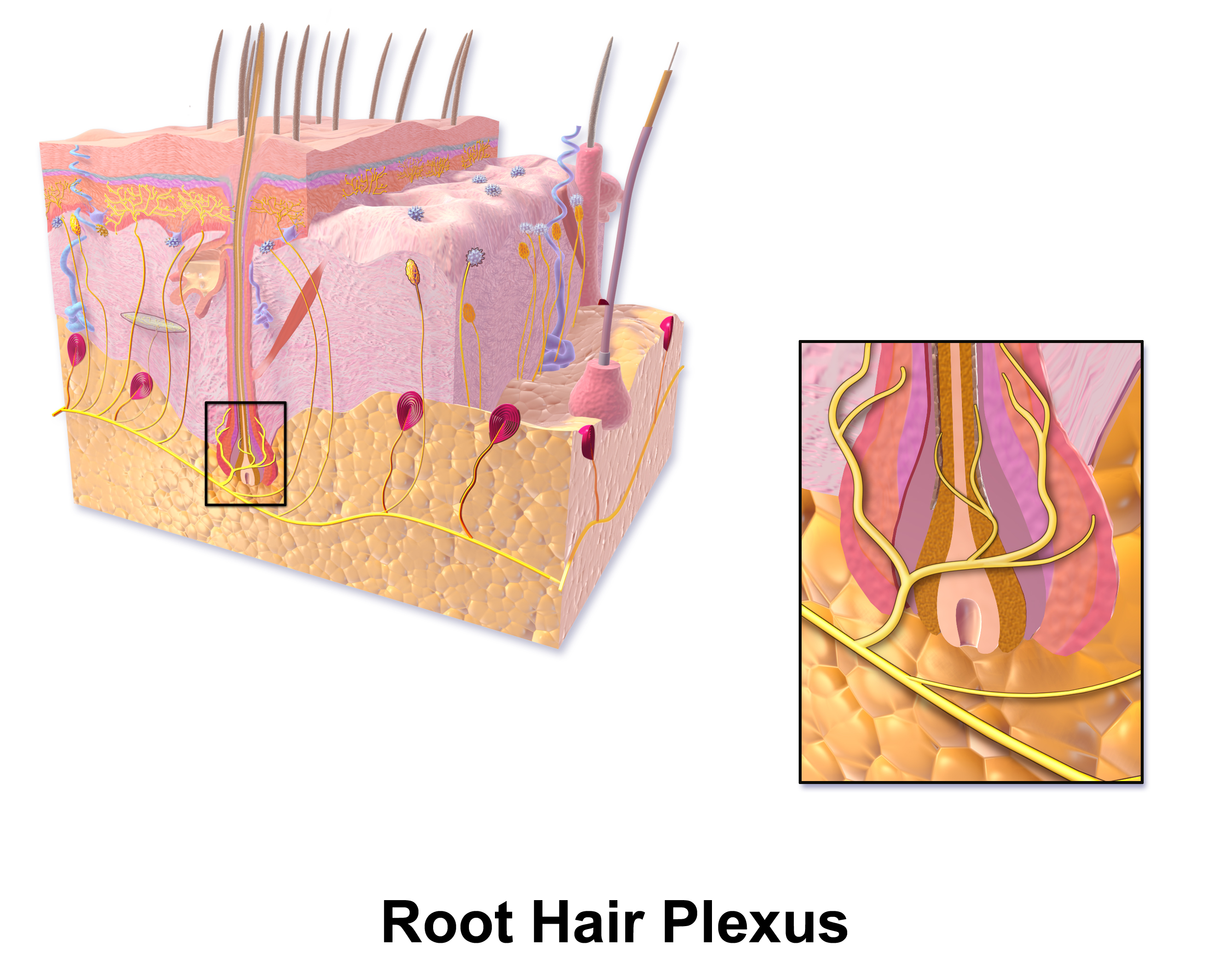
A hair plexus or root hair plexus is a special group of nerve fiber endings and serves as a very sensitive mechanoreceptor for touch sensation. Hair contains a number of different types of nerve endings. They are specialized for the detection of different kinds of stimuli and thus different types of neuron innervate these structures within the skin. In particular there are neurons innervating the hair that detect, deflection of the hair (i.e. to detect stroking), and pulling of the hair (i.e. noxious stimuli). The hair follicles are innervated by at least 5 classes of low threshold mechanical receptors. They are mechanoreceptors conveying touch sensation with cell bodies located inside of either dorsal root ganglia or trigeminal root ganglia. For most of the body (excluding the head and neck), crude touch and noxious stimuli from these receptors are further conveyed by the spinothalamic tract whereas discriminative and light touch are conveyed to via the dorsal column medial lem ... [...More Info...] [...Related Items...] OR: [Wikipedia] [Google] [Baidu] |
Blausen 0806 Skin RootHairPlexus
Blausen Medical Communications, Inc. (BMC) is the creator and owner of a library of two- and three-dimensional medical and scientific images and animations, a developer of information technology allowing access to that content, and a business focused on licensing and distributing the content. It was founded by Bruce Blausen in Houston, Texas, in 1991, and is privately held. Background Blausen Medical Communications is a privately held company founded by Bruce Blausen in Houston, Texas in 1991. BMC created and owns a library of medical and scientific images and animations, and has developed information technology tools allowing access to the library; as well, it licenses and otherwise works to distribute the content. As of this date, BMC's animation library comprised approximately 1,500 animations and over 27,000 two- and three-dimensional images designed for point-of-care patient education, which could be accessed by consumers or professional caregivers (primarily via hospital o ... [...More Info...] [...Related Items...] OR: [Wikipedia] [Google] [Baidu] |
Nerve Fiber
An axon (from Greek ἄξων ''áxōn'', axis) or nerve fiber (or nerve fibre: see spelling differences) is a long, slender projection of a nerve cell, or neuron, in vertebrates, that typically conducts electrical impulses known as action potentials away from the nerve cell body. The function of the axon is to transmit information to different neurons, muscles, and glands. In certain sensory neurons ( pseudounipolar neurons), such as those for touch and warmth, the axons are called afferent nerve fibers and the electrical impulse travels along these from the periphery to the cell body and from the cell body to the spinal cord along another branch of the same axon. Axon dysfunction can be the cause of many inherited and acquired neurological disorders that affect both the peripheral and central neurons. Nerve fibers are classed into three typesgroup A nerve fibers, group B nerve fibers, and group C nerve fibers. Groups A and B are myelinated, and group C are unmyelinated. T ... [...More Info...] [...Related Items...] OR: [Wikipedia] [Google] [Baidu] |
Mechanoreceptor
A mechanoreceptor, also called mechanoceptor, is a sensory receptor that responds to mechanical pressure or distortion. Mechanoreceptors are located on sensory neurons that convert mechanical pressure into action potential, electrical signals that, in animals, are sent to the central nervous system. Vertebrate mechanoreceptors Cutaneous mechanoreceptors Cutaneous mechanoreceptors respond to mechanical stimuli that result from physical interaction, including pressure and vibration. They are located in the skin, like other cutaneous receptors. They are all innervated by Aβ fibers, except the mechanorecepting free nerve endings, which are innervated by A delta fiber, Aδ fibers. Cutaneous mechanoreceptors can be categorized by what kind of sensation they perceive, by the rate of adaptation, and by morphology. Furthermore, each has a different receptive field. By sensation * The Slowly Adapting type 1 (SA1) mechanoreceptor, with the Merkel corpuscle end-organ (also known as M ... [...More Info...] [...Related Items...] OR: [Wikipedia] [Google] [Baidu] |
Dorsal Root Ganglion
A dorsal root ganglion (or spinal ganglion; also known as a posterior root ganglion) is a cluster of neurons (a ganglion) in a dorsal root of a spinal nerve. The cell bodies of sensory neurons known as first-order neurons are located in the dorsal root ganglia. The axons of dorsal root ganglion neurons are known as afferents. In the peripheral nervous system, afferents refer to the axons that relay sensory information into the central nervous system (i.e. the brain and the spinal cord). Structure The neurons comprising the dorsal root ganglion are of the pseudo-unipolar type, meaning they have a cell body (soma) with two branches that act as a single axon, often referred to as a ''distal process'' and a ''proximal process''. Unlike the majority of neurons found in the central nervous system, an action potential in posterior root ganglion neuron may initiate in the ''distal process'' in the periphery, bypass the cell body, and continue to propagate along the ''proximal proc ... [...More Info...] [...Related Items...] OR: [Wikipedia] [Google] [Baidu] |
Trigeminal Ganglion
The trigeminal ganglion (also known as: Gasserian ganglion, semilunar ganglion, or Gasser's ganglion) is the sensory ganglion of each trigeminal nerve (CN V). The trigeminal ganglion is located within the trigeminal cave (Meckel's cave), a cavity formed by dura mater. Anatomy The trigeminal ganglion contains cell bodies of the pseudo-unipolar sensory neurons of the trigeminal nerve which extend their axons both distally/peripherally into the three divisions of the trigeminal nerve on the one end, and proximally/centrally to the brainstem on the other end; the trigeminal root extends from the trigeminal ganglion to the ventrolateral aspect of the pons. The trigeminal ganglion is situated within the trigeminal cave (or Meckel's cave), a cerebrospinal fluid-filled cavity formed by a double layer of dura mater overlying the trigeminal impression near the apex of the petrous part of the temporal bone. Structure The trigeminal ganglion is somewhat crescent-shaped, with its co ... [...More Info...] [...Related Items...] OR: [Wikipedia] [Google] [Baidu] |
Spinothalamic Tract
The spinothalamic tract is a nerve tract in the anterolateral system in the spinal cord. This tract is an ascending sensory pathway to the thalamus. From the ventral posterolateral nucleus in the thalamus, sensory information is relayed upward to the somatosensory cortex of the postcentral gyrus. The spinothalamic tract consists of two adjacent pathways: anterior and lateral. The anterior spinothalamic tract carries information about crude touch. The lateral spinothalamic tract conveys pain and temperature. In the spinal cord, the spinothalamic tract has somatotopic organization. This is the segmental organization of its cervical, thoracic, lumbar, and sacral components, which is arranged from most medial to most lateral respectively. The pathway crosses over ( decussates) at the level of the spinal cord, rather than in the brainstem like the dorsal column-medial lemniscus pathway and lateral corticospinal tract. It is one of the three tracts which make up the anterola ... [...More Info...] [...Related Items...] OR: [Wikipedia] [Google] [Baidu] |
Dorsal Column–medial Lemniscus Pathway
The dorsal column–medial lemniscus pathway (DCML) (also known as the posterior column-medial lemniscus pathway (PCML) is the major sensory pathway of the central nervous system that conveys sensations of fine touch, vibration, two-point discrimination, and proprioception (body position) from the skin and joints. It transmits this information to the somatosensory cortex of the postcentral gyrus in the parietal lobe of the brain.O'Sullivan, S. B., & Schmitz, T. J. (2007). Physical Rehabilitation (5th Edition ed.). Philadelphia: F.A. Davis Company. The pathway receives information from sensory receptors throughout the body, and carries this in the gracile fasciculus and the cuneate fasciculus, tracts that make up the white matter dorsal columns (also known as the posterior funiculi) of the spinal cord. At the level of the medulla oblongata, the fibers of the tracts decussate and are continued in the medial lemniscus, on to the thalamus and relayed from there through the inte ... [...More Info...] [...Related Items...] OR: [Wikipedia] [Google] [Baidu] |
Spinal Trigeminal Nucleus
The spinal trigeminal nucleus is a nucleus in the medulla that receives information about deep/crude touch, pain, and temperature from the ipsilateral face. In addition to the trigeminal nerve (CN V), the facial (CN VII), glossopharyngeal (CN IX), and vagus nerves (CN X) also convey pain information from their areas to the spinal trigeminal nucleus. Thus the spinal trigeminal nucleus receives afferents from V, VII, [...More Info...] [...Related Items...] OR: [Wikipedia] [Google] [Baidu] |


