|
Dorsum Sellae
The dorsum sellae is part of the sphenoid bone in the skull. Together with the basilar part of the occipital bone it forms the clivus. In the sphenoid bone, the anterior boundary of the sella turcica is completed by two small eminences, one on either side, called the middle clinoid processes, while the posterior boundary is formed by a square-shaped plate of bone, the dorsum sellae, ending at its superior angles in two tubercles, the posterior clinoid processes The posterior clinoid processes are the tubercles of the sphenoid bone situated at the superior angles of the dorsum sellae (one on each angle) which represents the posterior boundary of the sella turcica. They vary considerably in size and form. ..., the size and form of which vary considerably in different individuals. Additional images File:Gray569.png, Tentorium cerebelli from above. File:Slide2iiii.JPG, Dorsum sellae References External links * * Bones of the head and neck {{musculoskeletal-s ... [...More Info...] [...Related Items...] OR: [Wikipedia] [Google] [Baidu] |
Sphenoid Bone
The sphenoid bone is an unpaired bone of the neurocranium. It is situated in the middle of the skull towards the front, in front of the basilar part of occipital bone, basilar part of the occipital bone. The sphenoid bone is one of the seven bones that articulate to form the orbit (anatomy), orbit. Its shape somewhat resembles that of a butterfly, bat or wasp with its wings extended. The name presumably originates from this shape, since () means in Ancient Greek. Structure It is divided into the following parts: * a median portion, known as the body of sphenoid bone, containing the sella turcica, which houses the pituitary gland as well as the paired paranasal sinuses, the sphenoidal sinuses * two Greater wing of sphenoid bone, greater wings on the lateral side of the body and two Lesser wing of sphenoid bone, lesser wings from the anterior side. * Pterygoid processes of the sphenoides, directed downwards from the junction of the body and the greater wings. Two sphenoidal co ... [...More Info...] [...Related Items...] OR: [Wikipedia] [Google] [Baidu] |
Base Of The Skull
The base of skull, also known as the cranial base or the cranial floor, is the most inferior area of the skull. It is composed of the endocranium and the lower parts of the calvaria. Structure Structures found at the base of the skull are for example: Bones There are five bones that make up the base of the skull: *Ethmoid bone *Sphenoid bone *Occipital bone *Frontal bone *Temporal bone Sinuses * Occipital sinus * Superior sagittal sinus * Superior petrosal sinus Foramina of the skull * Foramen cecum *Optic foramen * Foramen lacerum * Foramen rotundum * Foramen magnum * Foramen ovale *Jugular foramen * Internal auditory meatus * Mastoid foramen * Sphenoidal emissary foramen * Foramen spinosum Sutures * Frontoethmoidal suture * Sphenofrontal suture * Sphenopetrosal suture * Sphenoethmoidal suture * Petrosquamous suture * Sphenosquamosal suture Other * Sphenoidal lingula * Subarcuate fossa * Dorsum sellae * Jugular process * Petro-occipital fissure * Condylar canal * J ... [...More Info...] [...Related Items...] OR: [Wikipedia] [Google] [Baidu] |
Sphenoid Bone
The sphenoid bone is an unpaired bone of the neurocranium. It is situated in the middle of the skull towards the front, in front of the basilar part of occipital bone, basilar part of the occipital bone. The sphenoid bone is one of the seven bones that articulate to form the orbit (anatomy), orbit. Its shape somewhat resembles that of a butterfly, bat or wasp with its wings extended. The name presumably originates from this shape, since () means in Ancient Greek. Structure It is divided into the following parts: * a median portion, known as the body of sphenoid bone, containing the sella turcica, which houses the pituitary gland as well as the paired paranasal sinuses, the sphenoidal sinuses * two Greater wing of sphenoid bone, greater wings on the lateral side of the body and two Lesser wing of sphenoid bone, lesser wings from the anterior side. * Pterygoid processes of the sphenoides, directed downwards from the junction of the body and the greater wings. Two sphenoidal co ... [...More Info...] [...Related Items...] OR: [Wikipedia] [Google] [Baidu] |
Human Skull
The skull, or cranium, is typically a bony enclosure around the brain of a vertebrate. In some fish, and amphibians, the skull is of cartilage. The skull is at the head end of the vertebrate. In the human, the skull comprises two prominent parts: the neurocranium and the facial skeleton, which evolved from the first pharyngeal arch. The skull forms the frontmost portion of the axial skeleton and is a product of cephalization and vesicular enlargement of the brain, with several special senses structures such as the eyes, ears, nose, tongue and, in fish, specialized tactile organs such as barbels near the mouth. The skull is composed of three types of bone: cranial bones, facial bones and ossicles, which is made up of a number of fused flat and irregular bones. The cranial bones are joined at firm fibrous junctions called sutures and contains many foramina, fossae, processes, and sinuses. In zoology, the openings in the skull are called fenestrae, the ... [...More Info...] [...Related Items...] OR: [Wikipedia] [Google] [Baidu] |
Basilar Part Of Occipital Bone
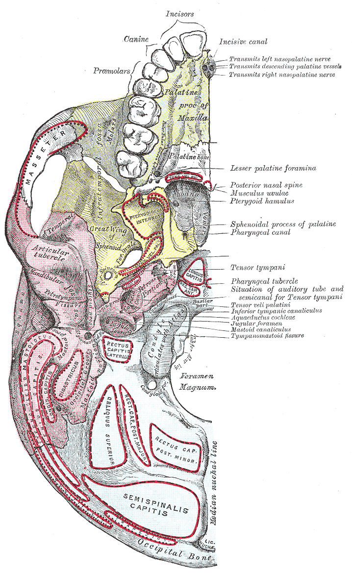
The basilar part of the occipital bone (also basioccipital) extends forward and upward from the foramen magnum, and presents in front an area more or less quadrilateral in outline. In the young skull, this area is rough and uneven, and is joined to the body of the sphenoid by a plate of cartilage. By the twenty-fifth year, this cartilaginous plate is ossified, and the occipital and sphenoid form a continuous bone. Surfaces On its ''lower surface'', about 1 cm. in front of the foramen magnum, is the pharyngeal tubercle which gives attachment to the fibrous raphe of the pharynx. On either side of the middle line the longus capitis and rectus capitis anterior are inserted, and immediately in front of the foramen magnum the anterior atlantooccipital membrane is attached. The ''upper surface'', which constitutes the lower half of the clivus, presents a broad, shallow groove which inclines upward and forward from the foramen magnum; it supports the medulla oblongata, and ... [...More Info...] [...Related Items...] OR: [Wikipedia] [Google] [Baidu] |
Clivus (anatomy)
The clivus (, Latin for "slope") or Blumenbach clivus is a part of the occipital bone at the base of the skull, extending anteriorly from the foramen magnum. It is related to the pons and the abducens nerve (CN VI). The term is also used for the clivus ocularis, an unrelated feature of the retina. Structure The clivus is a shallow depression behind the dorsum sellae of the sphenoid bone, extending inferiorly to the foramen magnum. It slopes gradually to the anterior part of the basilar occipital bone at its junction with the sphenoid bone. Synchondrosis of these two bones forms the clivus. On axial planes, it sits just posterior to the sphenoid sinuses. It is medial to the foramen lacerum and proximal to the anastomosis of the internal carotid artery with the Circle of Willis. (The artery reaches the middle cranial fossa above the foramen lacerum). It is anterior to the basilar artery. On sagittal plane, it can be divided into two surfaces, the pharyngeal (inferior) surf ... [...More Info...] [...Related Items...] OR: [Wikipedia] [Google] [Baidu] |
Sella Turcica
The sella turcica (Latin for 'Turkish saddle') is a saddle-shaped depression in the body of the sphenoid bone of the human skull and of the skulls of other hominids including chimpanzees, gorillas and orangutans. It serves as a cephalometric landmark. The pituitary gland or hypophysis is located within the most inferior aspect of the sella turcica, the hypophyseal fossa. Structure The sella turcica is located in the sphenoid bone behind the chiasmatic groove and the tuberculum sellae. It belongs to the middle cranial fossa. The sella turcica's most inferior portion is known as the hypophyseal fossa (the "seat of the saddle"), and contains the pituitary gland (hypophysis). In front of the hypophyseal fossa is the tuberculum sellae. Completing the formation of the saddle posteriorly is the dorsum sellae, which is continuous with the clivus, inferoposteriorly. The dorsum sellae is terminated laterally by the posterior clinoid processes. Development It is widely believed th ... [...More Info...] [...Related Items...] OR: [Wikipedia] [Google] [Baidu] |
Middle Clinoid Processes
The middle clinoid process is a small, bilaterally paired elevation on either side of the tuberculum sellae, at the anterior boundary of the sella turcica. A (larger) anterior clinoid process is situated lateral to each middle clinoid process. The diaphragma sellae (i.e. the dura forming the roof of the cavernous sinus) and the dura of the floor of the hypophyseal fossa (sella turcica) attach onto the middle clinoid processes. On each side of the body, the internal carotid artery passes between the anterior and middle clinoid processes. Etymology Clinoid likely comes from the Greek root ''klinein'' or the Latin Latin ( or ) is a classical language belonging to the Italic languages, Italic branch of the Indo-European languages. Latin was originally spoken by the Latins (Italic tribe), Latins in Latium (now known as Lazio), the lower Tiber area aroun ... ''clinare'', both meaning "sloped" as in "inclined." References Bones of the head and neck {{musculoskelet ... [...More Info...] [...Related Items...] OR: [Wikipedia] [Google] [Baidu] |
Tubercle (anatomy)
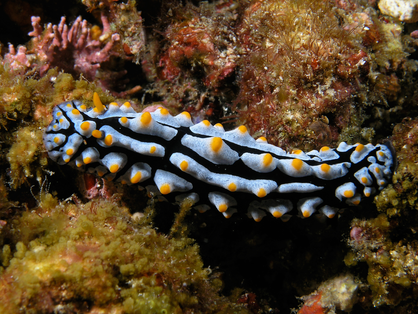
In anatomy, a tubercle (literally 'small tuber', Latin for 'lump') is any round nodule, small eminence, or warty outgrowth found on external or internal organs of a plant or an animal. In plants A tubercle is generally a wart-like projection, but it has slightly different meaning depending on which family of plants or animals it is used to refer to. In the case of certain orchids and cacti, it denotes a round nodule, small eminence, or warty outgrowth found on the lip. They are also known as podaria (singular ''podarium''). When referring to some members of the pea family, it is used to refer to the wart-like excrescences that are found on the roots. In fungi In mycology, a tubercle is used to refer to a mass of hyphae from which a mushroom is made. In animals When it is used in relation to certain dorid nudibranchs such as '' Peltodoris nobilis'', it means the nodules on the dorsum of the animal. The tubercles in nudibranchs can present themselves in different w ... [...More Info...] [...Related Items...] OR: [Wikipedia] [Google] [Baidu] |
Posterior Clinoid Processes
The posterior clinoid processes are the tubercles of the sphenoid bone situated at the superior angles of the dorsum sellae (one on each angle) which represents the posterior boundary of the sella turcica. They vary considerably in size and form. The posterior clinoid processes deepen the sella turcica, and give attachment to (the attached border of) the tentorium cerebelli, and the dura forming the floor of the hypophyseal fossa (sella turcica). The petroclinoid ligament The petroclinoid ligament is a fold of dura matter. It extends between the posterior clinoid process and anterior clinoid process and the petrosal part of the temporal bone of the skull. There are two separate bands of the ligament; named the anterior and posterior petroclinoid ligaments respectively. The anterior petroclinoid ligament is considered to be an extension of the tentorium cerebelli and the posterior petroclinoid ligament arises from the posteromedial extensions of the tentorial notch. The anterio ... [...More Info...] [...Related Items...] OR: [Wikipedia] [Google] [Baidu] |


