|
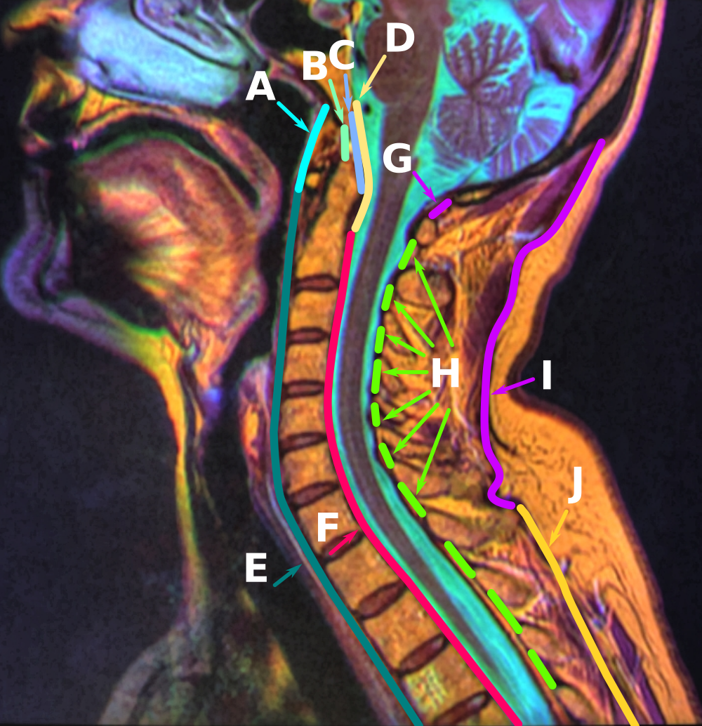
Clivus (anatomy)
The clivus (, Latin for "slope") or Blumenbach clivus is a part of the occipital bone at the base of the skull, extending anteriorly from the foramen magnum. It is related to the pons and the abducens nerve (CN VI). The term is also used for the clivus ocularis, an unrelated feature of the retina. Structure The clivus is a shallow depression behind the dorsum sellae of the sphenoid bone, extending inferiorly to the foramen magnum. It slopes gradually to the anterior part of the basilar occipital bone at its junction with the sphenoid bone. Synchondrosis of these two bones forms the clivus. On axial planes, it sits just posterior to the sphenoid sinuses. It is medial to the foramen lacerum and proximal to the anastomosis of the internal carotid artery with the Circle of Willis. (The artery reaches the middle cranial fossa above the foramen lacerum). It is anterior to the basilar artery. On sagittal plane, it can be divided into two surfaces, the pharyngeal (inferior) surf ... [...More Info...] [...Related Items...] OR: [Wikipedia] [Google] [Baidu] |
Latin Language
Latin ( or ) is a classical language belonging to the Italic languages, Italic branch of the Indo-European languages. Latin was originally spoken by the Latins (Italic tribe), Latins in Latium (now known as Lazio), the lower Tiber area around Rome, Italy. Through the expansion of the Roman Republic, it became the dominant language in the Italian Peninsula and subsequently throughout the Roman Empire. It has greatly influenced many languages, Latin influence in English, including English, having contributed List of Latin words with English derivatives, many words to the English lexicon, particularly after the Christianity in Anglo-Saxon England, Christianization of the Anglo-Saxons and the Norman Conquest. Latin Root (linguistics), roots appear frequently in the technical vocabulary used by fields such as theology, List of Latin and Greek words commonly used in systematic names, the sciences, List of medical roots, suffixes and prefixes, medicine, and List of Latin legal terms ... [...More Info...] [...Related Items...] OR: [Wikipedia] [Google] [Baidu] |
Sagittal Plane
The sagittal plane (; also known as the longitudinal plane) is an anatomical plane that divides the body into right and left sections. It is perpendicular to the transverse and coronal planes. The plane may be in the center of the body and divide it into two equal parts ( mid-sagittal), or away from the midline and divide it into unequal parts (para-sagittal). The term ''sagittal'' was coined by Gerard of Cremona. Variations in terminology Examples of sagittal planes include: * The terms '' median plane'' or ''mid-sagittal plane'' are sometimes used to describe the sagittal plane running through the midline. This plane cuts the body into halves (assuming bilateral symmetry), passing through midline structures such as the navel and spine. It is one of the planes which, combined with the umbilical plane, defines the four quadrants of the human abdomen. * The term ''parasagittal'' is used to describe any plane parallel or adjacent to a given sagittal plane. Specific named pa ... [...More Info...] [...Related Items...] OR: [Wikipedia] [Google] [Baidu] |
Chordoma
Chordoma is a rare slow-growing neoplasm (cancer) that arises from cellular remnants of the notochord in the bones of the skull base and spine. The evidence for the notochordal origin of chordoma is the location of the tumors (along the neuraxis), the similar immunohistochemical staining patterns, expression of T-box transcription factor T, brachyury, and the demonstration that notochordal cells are preferentially left behind in the clivus (anatomy), clivus and Sacrococcygeal symphysis, sacrococcygeal regions when the remainder of the notochord regresses during fetal life. In layman's terms, chordoma is a type of bone cancer, and is classified as a sarcoma. Chordomas are sometimes mistakenly referred to as a brain, brainstem or spinal cord tumors due to their location near those critical structures, but they are not derived from nervous tissue. Presentation Chordomas can arise from bone in the skull base and anywhere along the spine. The two most common locations are cranially ... [...More Info...] [...Related Items...] OR: [Wikipedia] [Google] [Baidu] |
Palsy
Palsy is a medical term which refers to various types of paralysisDan Agin, ''More Than Genes: What Science Can Tell Us About Toxic Chemicals, Development, and the Risk to Our Children'' (2009), p. 172. or paresis, often accompanied by weakness and the loss of feeling and uncontrolled body movements such as shaking. The word originates from the Anglo-Norman ''paralisie'', ''parleisie'' ''et al.'', from the accusative form of Latin ''paralysis'', from Ancient Greek παράλυσις (''parálusis''), from παραλύειν (''paralúein'', "to disable on one side"), from παρά (''pará'', "beside") + λύειν (''lúein'', "loosen"). The word is longstanding in the English language, having appeared in the play '' Grim the Collier of Croydon'', reported to have been written as early as 1599: In some editions, the Bible passage of Luke 5:18 is translated to refer to "a man which was taken with a palsy". More modern editions simply refer to a man who is paralysed. Although th ... [...More Info...] [...Related Items...] OR: [Wikipedia] [Google] [Baidu] |
Intracranial Pressure
Intracranial pressure (ICP) is the pressure exerted by fluids such as cerebrospinal fluid (CSF) inside the skull and on the brain tissue. ICP is measured in millimeters of mercury ( mmHg) and at rest, is normally 7–15 mmHg for a supine adult. This equals to 9–20 cmH2O, which is a common scale used in lumbar punctures. The body has various mechanisms by which it keeps the ICP stable, with CSF pressures varying by about 1 mmHg in normal adults through shifts in production and absorption of CSF. Changes in ICP are attributed to volume changes in one or more of the constituents contained in the cranium. CSF pressure has been shown to be influenced by abrupt changes in intrathoracic pressure during coughing (which is induced by contraction of the diaphragm and abdominal wall muscles, the latter of which also increases intra-abdominal pressure), the valsalva maneuver, and communication with the vasculature ( venous and arterial systems). Intracranial hypertension (IH), ... [...More Info...] [...Related Items...] OR: [Wikipedia] [Google] [Baidu] |
Saunders (imprint)
Saunders is an American academic publisher based in the United States. It is currently an imprint of Elsevier. Formerly independent, the W. B. Saunders company was acquired by CBS in 1968, who added it to their publishing division Holt, Rinehart & Winston. When CBS left the publishing field in 1986, it sold the academic publishing units to Harcourt Brace Jovanovich. Harcourt was acquired by Reed Elsevier in 2001. . Northern Illinois University Libraries. Retrieved May 2, 2015. W. B. Saunders published the Kinsey Reports
[...More Info...] [...Related Items...] OR: [Wikipedia] [Google] [Baidu] |
Apical Ligament Of Dens
The ligament of apex dentis (or apical odontoid ligament) is a ligament that spans between the second cervical vertebra in the neck and the skull. It lies as a fibrous cord in the triangular interval between the alar ligaments, which extends from the tip of the odontoid process on the axis to the anterior margin of the foramen magnum, being intimately blended with the deep portion of the anterior atlantooccipital membrane and superior crus of the transverse ligament of the atlas. It is regarded as a rudimentary intervertebral fibrocartilage, and in it traces of the notochord The notochord is an elastic, rod-like structure found in chordates. In vertebrates the notochord is an embryonic structure that disintegrates, as the vertebrae develop, to become the nucleus pulposus in the intervertebral discs of the verteb ... may persist. References External links * Ligaments of the head and neck Bones of the vertebral column {{ligament-stub ... [...More Info...] [...Related Items...] OR: [Wikipedia] [Google] [Baidu] |
Ossification
Ossification (also called osteogenesis or bone mineralization) in bone remodeling is the process of laying down new bone material by cells named osteoblasts. It is synonymous with bone tissue formation. There are two processes resulting in the formation of normal, healthy bone tissue: Intramembranous ossification is the direct laying down of bone into the primitive connective tissue ( mesenchyme), while endochondral ossification involves cartilage as a precursor. In fracture healing, endochondral osteogenesis is the most commonly occurring process, for example in fractures of long bones treated by plaster of Paris, whereas fractures treated by open reduction and internal fixation with metal plates, screws, pins, rods and nails may heal by intramembranous osteogenesis. Heterotopic ossification is a process resulting in the formation of bone tissue that is often atypical, at an extraskeletal location. Calcification is often confused with ossification. Calcificatio ... [...More Info...] [...Related Items...] OR: [Wikipedia] [Google] [Baidu] |
Craniopharyngeal Canal
The craniopharyngeal canal is a human anatomical feature sometimes found in the sphenoid bone opening to the sella turcica. It is a canal (a passage or channel) sometimes found extending from the anterior part of the fossa hypophyseos of the sphenoid bone to the under surface of the skull, and marks the original position of Rathke's pouch; while at the junction of the septum of the nose with the palate The palate () is the roof of the mouth in humans and other mammals. It separates the oral cavity from the nasal cavity. A similar structure is found in crocodilians, but in most other tetrapods, the oral and nasal cavities are not truly sep ... traces of the stomodeal end are occasionally present. This canal is found in 0.4% of individuals. References *''Dorland's Illustrated Medical Dictionary'', 27th ed. 1988 W.B. Saunders Company. Philadelphia, PA. Bones of the head and neck Anatomical variations {{musculoskeletal-stub ... [...More Info...] [...Related Items...] OR: [Wikipedia] [Google] [Baidu] |
Fossa Navicularis Magna
Fossa navicularis magna (also known as ''pharyngeal fossa'' or ''phyaryngeal fovela'') is a variant bony depression found at the midline of the occipital part of clivus. This fossa was first described by Tourtual. Its prevalence ranges from 0.9 to 5.3%. Structure Fossa navicularis magna is located on the anterior surface or pharyngeal surface of the clivus. Its position when present is between the spheno-occipital synchondrosis and the foramen magnum. Size of this fossa varies considerably and its depth ranges from 3.49 to 4.94 mm. A histological study reported the presence of loose connective tissue containing collagen and elastic fibers within the fossa navicularis magna. Development Two theories have been proposed to explain the formation of fossa navicularis magna. It is believed that the fossa is formed as a remnant of the notochord or residue of the channels for emissary veins. Clinical significance Different pathologies were found associated with fossa navicularis ma ... [...More Info...] [...Related Items...] OR: [Wikipedia] [Google] [Baidu] |
Anatomical Variation
An anatomical variation, anatomical variant, or anatomical variability is a presentation of body structure with Morphology (biology), morphological features different from those that are typically described in the majority of individuals. Anatomical variations are categorized into three types including morphometric (size or shape), consistency (present or absent), and spatial (proximal/distal or right/left). Variations are seen as normal in the sense that they are found consistently among different individuals, are mostly without symptoms, and are termed anatomical variations rather than abnormalities. Anatomical variations are mainly caused by genetics and may vary considerably between different populations. The rate of variation considerably differs between single organ (anatomy), organs, particularly in muscles. Knowledge of anatomical variations is important in order to distinguish them from pathological conditions. A very early paper published in 1898, presented anatomic var ... [...More Info...] [...Related Items...] OR: [Wikipedia] [Google] [Baidu] |


