|
Cavernous Sinus
The cavernous sinus within the human head is one of the dural venous sinuses creating a cavity called the lateral sellar compartment bordered by the temporal bone of the skull and the sphenoid bone, lateral to the sella turcica. Structure The cavernous sinus is one of the dural venous sinuses of the head. It is a network of veins that sit in a cavity. It sits on both sides of the sphenoidal bone and pituitary gland, approximately 1 × 2 cm in size in an adult. The carotid siphon of the internal carotid artery, and cranial nerves III, IV, V (branches V1 and V2) and VI all pass through this blood filled space. Both sides of cavernous sinus are connected to each other via intercavernous sinuses. The cavernous sinus lies in between the inner and outer layers of dura mater. Nearby structures * Above: optic tract, optic chiasma, internal carotid artery. * Inferiorly: foramen lacerum, and the junction of the body and greater wing of sphenoid bone. * Medially: p ... [...More Info...] [...Related Items...] OR: [Wikipedia] [Google] [Baidu] [Amazon] |
Middle Cerebral Vein
The middle cerebral veins - are the superficial and deep veins - that run along the lateral sulcus. The superficial middle cerebral vein is also known as the superficial Sylvian vein, and the deep middle cerebral vein is also known as the deep Sylvian vein. The lateral sulcus or lateral fissure, is also known as the Sylvian fissure. Superficial middle cerebral vein The superficial middle cerebral vein (superficial Sylvian vein) begins on the lateral surface of the cerebral hemisphere, hemisphere. It runs along the lateral sulcus to empty into either the cavernous sinus, or the sphenoparietal sinus. It is adherent to the deep surface of the arachnoid mater bridging the lateral sulcus. It drains the adjacent cortex. Anastomoses At its posterior extremity, the superficial middle cerebral vein is connected with the superior sagittal sinus via the superior anastomotic vein, and with the transverse sinus via the inferior anastomotic vein. Deep middle cerebral vein The deep middl ... [...More Info...] [...Related Items...] OR: [Wikipedia] [Google] [Baidu] [Amazon] |
Optic Tract
In neuroanatomy, the optic tract () is a part of the visual system in the brain. It is a continuation of the optic nerve that relays information from the optic chiasm to the ipsilateral lateral geniculate nucleus (LGN), pretectal nuclei, and superior colliculus. It is composed of two individual tracts, the left optic tract and the right optic tract, each of which conveys visual information exclusive to its respective contralateral half of the visual field. Each of these tracts is derived from a combination of temporal and nasal retinal fibers from each eye that corresponds to one half of the visual field. In more specific terms, the optic tract contains fibers from the ipsilateral temporal hemiretina and contralateral nasal hemiretina. Anatomy Arterial supply The optic tract receives arterial supply from the anterior choroidal artery, and posterior communicating artery. Function Visual function The optic tract carries retinal information relating to the whole visua ... [...More Info...] [...Related Items...] OR: [Wikipedia] [Google] [Baidu] [Amazon] |
Inferior Cerebral Veins
The inferior cerebral veins are veins that drain the undersurface of the cerebral hemispheres and empty into the cavernous and transverse sinuses. Those on the orbital surface of the frontal lobe join the superior cerebral veins, and through these open into the superior sagittal sinus. Those of the temporal lobe anastomose with the middle cerebral and basal veins, and join the cavernous, sphenoparietal, and superior petrosal sinus The superior petrosal sinus is one of the dural venous sinuses located beneath the brain. It receives blood from the cavernous sinus and passes backward and laterally to drain into the transverse sinus. The sinus receives superior petrosal veins, ...es. Image File:Slide6Neo.JPG, Meninges and superficial cerebral veins. Deep dissection. Superior view. References Veins of the head and neck {{circulatory-stub ... [...More Info...] [...Related Items...] OR: [Wikipedia] [Google] [Baidu] [Amazon] |
Superficial Middle Cerebral Vein
The middle cerebral veins - are the superficial and deep veins - that run along the lateral sulcus. The superficial middle cerebral vein is also known as the superficial Sylvian vein, and the deep middle cerebral vein is also known as the deep Sylvian vein. The lateral sulcus or lateral fissure, is also known as the Sylvian fissure. Superficial middle cerebral vein The superficial middle cerebral vein (superficial Sylvian vein) begins on the lateral surface of the hemisphere. It runs along the lateral sulcus to empty into either the cavernous sinus, or the sphenoparietal sinus. It is adherent to the deep surface of the arachnoid mater bridging the lateral sulcus. It drains the adjacent cortex. Anastomoses At its posterior extremity, the superficial middle cerebral vein is connected with the superior sagittal sinus via the superior anastomotic vein, and with the transverse sinus via the inferior anastomotic vein. Deep middle cerebral vein The deep middle cerebral vein (d ... [...More Info...] [...Related Items...] OR: [Wikipedia] [Google] [Baidu] [Amazon] |
Sphenoparietal Sinus
The sphenoparietal sinus is a paired dural venous sinus situated along the posterior edge of the lesser wing of either sphenoid bone. It drains into the cavernous sinus. Anatomy A sphenoparietal sinus is situated under each lesser wing of the sphenoid bone near the posterior edge of this bone, between the anterior cranial fossa and middle cranial fossa. It terminates by draining into the anterior part of the cavernous sinus. Tributaries A sphenoparietal sinus receives small veins from the adjacent dura and sometimes the frontal ramus of the middle meningeal vein, communicating rami from the superficial middle cerebral vein, temporal lobe veins, and the anterior temporal diploic veins The diploic veins are large, thin-walled valveless veins that channel in the diploë between the inner and outer layers of the cortical bone in the skull, first identified in dogs by the anatomist Guillaume Dupuytren. A single layer of endotheli .... References Additional images File: ... [...More Info...] [...Related Items...] OR: [Wikipedia] [Google] [Baidu] [Amazon] |
Superior Ophthalmic Vein
The superior ophthalmic vein is a vein of the orbit that drains venous blood from structures of the upper orbit. It is formed by the union of the angular vein, and supraorbital vein. It passes backwards within the orbit alongside the ophthalmic artery, then exits the orbit through the superior orbital fissure to drain into the cavernous sinus. The superior ophthalmic vein can be a path for the spread of infection from the danger triangle of the face to the cavernous sinus and the pterygoid plexus. It may also be affected by an arteriovenous fistula of the cavernous sinus. Structure The superior ophthalmic vein - together with the inferior ophthalmic vein - represents the principal drainage system of the orbit (with the superior ophthalmic vein being the larger of the two). The superior ophthalmic vein drains venous blood from structures of the upper orbit. The superior ophthalmic vein forms/represents a connection between facial veins, and intracranial veins. It is valveles ... [...More Info...] [...Related Items...] OR: [Wikipedia] [Google] [Baidu] [Amazon] |
Petrous Temporal Bone
The petrous part of the temporal bone is pyramid-shaped and is wedged in at the base of the skull between the sphenoid bone, sphenoid and occipital bones. Directed medially, forward, and a little upward, it presents a base, an apex, three surfaces, and three angles, and houses in its interior the components of the inner ear. The petrous portion is among the most basal elements of the skull and forms part of the endocranium. Petrous comes from the Latin word ''petrosus'', meaning "stone-like, hard". It is one of the densest bones in the body. In other mammals, it is a separate bone, the petrosal bone. The petrous bone is important for studies of ancient DNA from skeletal remains, as it tends to contain extremely well-preserved DNA. Base The base is fused with the internal surfaces of the squamous part of temporal bone, squamous, Tympanic part of the temporal bone, tympanic, and mastoid part of the temporal bone, mastoid parts. Apex The apex, which is rough and uneven, is rece ... [...More Info...] [...Related Items...] OR: [Wikipedia] [Google] [Baidu] [Amazon] |
Superior Orbital Fissure
The superior orbital fissure is a foramen or cleft of the skull between the lesser and greater wings of the sphenoid bone. It gives passage to multiple structures, including the oculomotor nerve, trochlear nerve, ophthalmic nerve, abducens nerve, ophthalmic veins, and sympathetic fibres from the cavernous plexus. Structure The superior orbital fissure is usually 22 mm wide in adults, and is much larger medially. Its boundaries are formed by the (caudal surface of the) lesser wing of the sphenoid bone, and (medial border of the) greater wing of the sphenoid bone. Contents The superior orbital fissure is traversed by the following structures: * (superior and inferior divisions of the) oculomotor nerve (CN III) * trochlear nerve (CN IV) * lacrimal, frontal, and nasociliary branches of ophthalmic nerve (CN V1) * abducens nerve (CN VI) * superior ophthalmic vein and superior division of the inferior ophthalmic vein * sympathetic fibres from the cavernous nerve plex ... [...More Info...] [...Related Items...] OR: [Wikipedia] [Google] [Baidu] [Amazon] |
Uncus
The uncus is an anterior extremity of the parahippocampal gyrus. It is separated from the apex of the temporal lobe by a sulcus called the rhinal sulcus. Although superficially continuous with the hippocampal gyrus, the uncus forms morphologically a part of the rhinencephalon. An important landmark that crosses the inferior surface of the uncus is the band of Giacomini or ''tail'' of the dentate gyrus. The term comes from the Latin word uncus, meaning ''hook'', and it was coined by Félix Vicq-d'Azyr (1748–1794). Clinical significance The part of the olfactory cortex that is on the temporal lobe covers the area of the uncus, which leads into the two significant clinical aspects: herniations and seizures * Herniations of the brain can occur if increased intracranial pressure due to a tumor, hemorrhage, or edema pushes the uncus over the tentorial notch against the brainstem and related cranial nerves. This can compress the oculomotor nerve (CN III). This causes pr ... [...More Info...] [...Related Items...] OR: [Wikipedia] [Google] [Baidu] [Amazon] |
Temporal Lobe
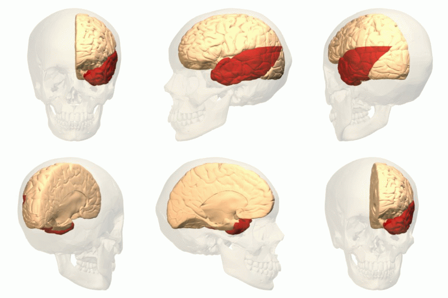
The temporal lobe is one of the four major lobes of the cerebral cortex in the brain of mammals. The temporal lobe is located beneath the lateral fissure on both cerebral hemispheres of the mammalian brain. The temporal lobe is involved in processing sensory input into derived meanings for the appropriate retention of visual memory, language comprehension, and emotion association. ''Temporal'' refers to the head's temples. Structure The temporal lobe consists of structures that are vital for declarative or long-term memory. Declarative (denotative) or explicit memory is conscious memory divided into semantic memory (facts) and episodic memory (events). The medial temporal lobe structures are critical for long-term memory, and include the hippocampal formation, perirhinal cortex, parahippocampal, and entorhinal neocortical regions. The hippocampus is critical for memory formation, and the surrounding medial temporal cortex is currently theorized to be critical f ... [...More Info...] [...Related Items...] OR: [Wikipedia] [Google] [Baidu] [Amazon] |
Sphenoidal Air Sinus
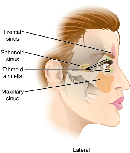
The sphenoid sinus is a paired paranasal sinus in the body of the sphenoid bone. It is one pair of the four paired paranasal sinuses.Illustrated Anatomy of the Head and Neck, Fehrenbach and Herring, Elsevier, 2012, page 64 The two sphenoid sinuses are separated from each other by a septum. Each sphenoid sinus communicates with the nasal cavity via the opening of sphenoidal sinus. The two sphenoid sinuses vary in size and shape, and are usually asymmetrical. Structure On average, a sphenoid sinus measures 2.2 cm vertical height, 2 cm in transverse breadth; and 2.2 cm antero-posterior depth. Each spehoid sinus is in the body of sphenoid bone, just under the sella turcica. The sphenoid sinuses are separated from each other medially by the septum of sphenoidal sinuses, which is usually asymmetrical. An opening of sphenoidal sinus forms a passage between each sphenoidal sinus and the nasal cavity. Posteriorly, an opening of sphenoidal sinus opens into the sphenoidal si ... [...More Info...] [...Related Items...] OR: [Wikipedia] [Google] [Baidu] [Amazon] |
Hypophysis Cerebri
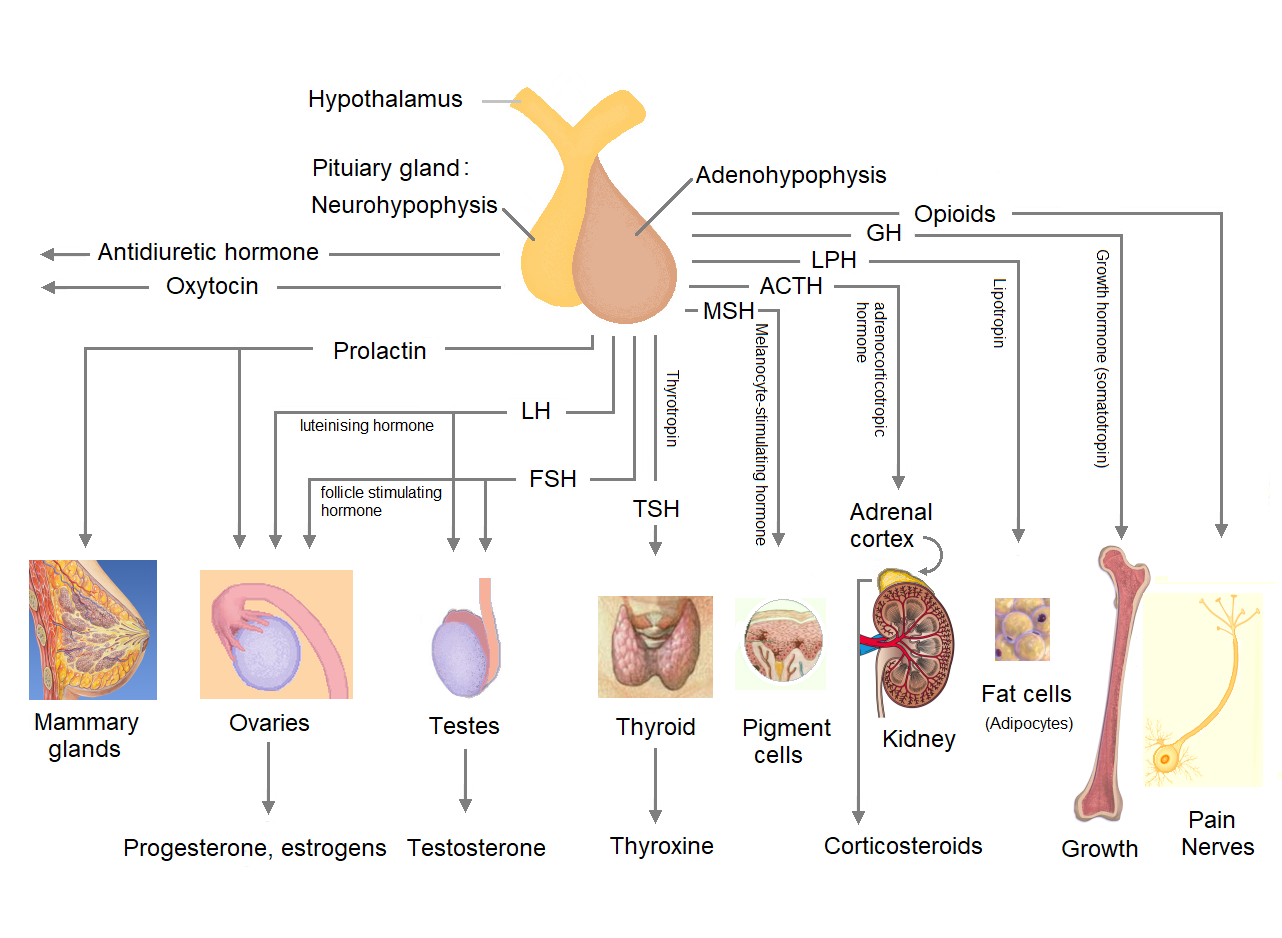
The pituitary gland or hypophysis is an endocrine gland in vertebrates. In humans, the pituitary gland is located at the base of the brain, protruding off the bottom of the hypothalamus. The pituitary gland and the hypothalamus control much of the body's endocrine system. It is seated in part of the sella turcica a depression in the sphenoid bone, known as the hypophyseal fossa. The human pituitary gland is oval shaped, about 1 cm in diameter, in weight on average, and about the size of a kidney bean. Digital version. There are two main lobes of the pituitary, an anterior lobe, and a posterior lobe joined and separated by a small intermediate lobe. The anterior lobe (adenohypophysis) is the glandular part that produces and secretes several hormones. The posterior lobe (neurohypophysis) secretes neurohypophysial hormones produced in the hypothalamus. Both lobes have different origins and they are both controlled by the hypothalamus. Hormones secreted from the pituitary gland h ... [...More Info...] [...Related Items...] OR: [Wikipedia] [Google] [Baidu] [Amazon] |


