|
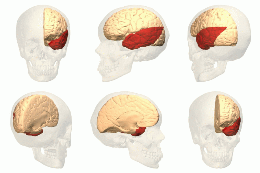
Inferior Cerebral Veins
The inferior cerebral veins are veins that drain the undersurface of the cerebral hemispheres and empty into the cavernous and transverse sinuses. Those on the orbital surface of the frontal lobe join the superior cerebral veins, and through these open into the superior sagittal sinus. Those of the temporal lobe anastomose with the middle cerebral and basal veins, and join the cavernous, sphenoparietal, and superior petrosal sinus The superior petrosal sinus is one of the dural venous sinuses located beneath the brain. It receives blood from the cavernous sinus and passes backward and laterally to drain into the transverse sinus. The sinus receives superior petrosal veins, ...es. Image File:Slide6Neo.JPG, Meninges and superficial cerebral veins. Deep dissection. Superior view. References Veins of the head and neck {{circulatory-stub ... [...More Info...] [...Related Items...] OR: [Wikipedia] [Google] [Baidu] |
Cerebral Hemisphere
The vertebrate cerebrum (brain) is formed by two cerebral hemispheres that are separated by a groove, the longitudinal fissure. The brain can thus be described as being divided into left and right cerebral hemispheres. Each of these hemispheres has an outer layer of grey matter, the cerebral cortex, that is supported by an inner layer of white matter. In eutherian (placental) mammals, the hemispheres are linked by the corpus callosum, a very large bundle of axon, nerve fibers. Smaller commissures, including the anterior commissure, the posterior commissure and the fornix (neuroanatomy), fornix, also join the hemispheres and these are also present in other vertebrates. These commissures transfer information between the two hemispheres to coordinate localized functions. There are three known poles of the cerebral hemispheres: the ''occipital lobe, occipital pole'', the ''frontal lobe, frontal pole'', and the ''temporal lobe, temporal pole''. The central sulcus is a prominent fissu ... [...More Info...] [...Related Items...] OR: [Wikipedia] [Google] [Baidu] |
Cerebrum
The cerebrum (: cerebra), telencephalon or endbrain is the largest part of the brain, containing the cerebral cortex (of the two cerebral hemispheres) as well as several subcortical structures, including the hippocampus, basal ganglia, and olfactory bulb. In the human brain, the cerebrum is the uppermost region of the central nervous system. The cerebrum prenatal development, develops prenatally from the forebrain (prosencephalon). In mammals, the Dorsum (biology), dorsal telencephalon, or Pallium (neuroanatomy), pallium, develops into the cerebral cortex, and the ventral telencephalon, or Pallium (neuroanatomy), subpallium, becomes the basal ganglia. The cerebrum is also divided into approximately symmetric Lateralization of brain function, left and right cerebral hemispheres. With the assistance of the cerebellum, the cerebrum controls all voluntary actions in the human body. Structure The cerebrum is the largest part of the brain. Depending upon the position of the animal, ... [...More Info...] [...Related Items...] OR: [Wikipedia] [Google] [Baidu] |
Dural Venous Sinuses
The dural venous sinuses (also called dural sinuses, cerebral sinuses, or cranial sinuses) are venous sinuses (channels) found between the periosteal and meningeal layers of dura mater in the brain. They receive blood from the cerebral veins, and cerebrospinal fluid (CSF) from the subarachnoid space via arachnoid granulations. They mainly empty into the internal jugular vein. Cranial venous sinuses communicate with veins outside the skull through emissary veins. These communications help to keep the pressure of blood in the sinuses constant. The major dural venous sinuses included the superior sagittal sinus, inferior sagittal sinus, transverse sinus, straight sinus, sigmoid sinus and cavernous sinus. These sinuses play a crucial role in cerebral venous drainage. A dural venous sinus, in human anatomy, is any of the channels of a branching complex sinus network that lies between layers of the dura mater, the outermost covering of the brain, and functions to collect oxygen-de ... [...More Info...] [...Related Items...] OR: [Wikipedia] [Google] [Baidu] |
Cerebral Arteries
The cerebral arteries describe three main pairs of artery, arteries and their branches, which perfusion, perfuse the cerebrum of the brain. The three main arteries are the: * ''Anterior cerebral artery'' (ACA), which supplies blood to the medial portion of the brain, including the superior parts of the Frontal lobe, frontal and anterior Parietal lobe, parietal lobes * ''Middle cerebral artery'' (MCA), which supplies blood to the majority of the lateral portion of the brain, including the Temporal lobe, temporal and lateral-parietal lobes. It is the largest of the cerebral arteries and is often affected in strokes * ''Posterior cerebral artery'' (PCA), which supplies blood to the posterior portion of the brain, including the occipital lobe, thalamus, and midbrain Both the ACA and MCA originate from the cerebral portion of internal carotid artery, while PCA branches from the intersection of the posterior communicating artery and the anterior portion of the basilar artery. The three p ... [...More Info...] [...Related Items...] OR: [Wikipedia] [Google] [Baidu] |
Cavernous
The cavernous sinus within the human head is one of the dural venous sinuses creating a cavity called the lateral sellar compartment bordered by the temporal bone of the skull and the sphenoid bone, lateral to the sella turcica. Structure The cavernous sinus is one of the dural venous sinuses of the head. It is a network of veins that sit in a cavity. It sits on both sides of the sphenoidal bone and pituitary gland, approximately 1 × 2 cm in size in an adult. The carotid siphon of the internal carotid artery, and cranial nerves III, IV, V (branches V1 and V2) and VI all pass through this blood filled space. Both sides of cavernous sinus are connected to each other via intercavernous sinuses. The cavernous sinus lies in between the inner and outer layers of dura mater. Nearby structures * Above: optic tract, optic chiasma, internal carotid artery. * Inferiorly: foramen lacerum, and the junction of the body and greater wing of sphenoid bone. * Medially: pituitar ... [...More Info...] [...Related Items...] OR: [Wikipedia] [Google] [Baidu] |
Transverse Sinuses
The transverse sinuses (left and right lateral sinuses), within the human head, are two areas beneath the brain which allow blood to drain from the back of the head. They run laterally in a groove along the interior surface of the occipital bone. They drain from the confluence of sinuses (by the internal occipital protuberance) to the sigmoid sinuses, which ultimately connect to the internal jugular vein. ''See diagram (at right)'': labeled under the brain as "" (for Latin: ''sinus transversus'' https://www.ncbi.nlm.nih.gov/books/NBK482257] Structure The transverse sinuses are of large size and begin at the internal occipital protuberance; one, generally the right, being the direct continuation of the |
Frontal Lobe
The frontal lobe is the largest of the four major lobes of the brain in mammals, and is located at the front of each cerebral hemisphere (in front of the parietal lobe and the temporal lobe). It is parted from the parietal lobe by a Sulcus (neuroanatomy), groove between tissues called the central sulcus and from the temporal lobe by a deeper groove called the lateral sulcus (Sylvian fissure). The most anterior rounded part of the frontal lobe (though not well-defined) is known as the frontal pole, one of the three Cerebral hemisphere#Poles, poles of the cerebrum. The frontal lobe is covered by the frontal cortex. The frontal cortex includes the premotor cortex and the primary motor cortex – parts of the motor cortex. The front part of the frontal cortex is covered by the prefrontal cortex. The nonprimary motor cortex is a functionally defined portion of the frontal lobe. There are four principal Gyrus, gyri in the frontal lobe. The precentral gyrus is directly anterior to the ... [...More Info...] [...Related Items...] OR: [Wikipedia] [Google] [Baidu] |
Superior Cerebral Veins
The superior cerebral veins are several cerebral veins that drain the superolateral and superomedial surfaces of the cerebral hemispheres into the superior sagittal sinus. There are 8-12 cerebral veins. They are predominantly found in the sulci between the gyri, but can also be found running across the gyri. Anatomy Fate The superior cerebral veins drain into the superior sagittal sinus The superior sagittal sinus (also known as the superior longitudinal sinus), within the human head, is an unpaired dural venous sinus lying along the attached margin of the falx cerebri. It allows blood to drain from the lateral aspects of the a ... individually. The anterior veins run at near right angles to the sinus while the posterior and larger veins are directed at oblique angles, opening into the sinus in a direction opposed to the current (anterior to posterior) of the blood contained within it. Additional images File:Slide6Neo.JPG, Meninges and superficial cerebral veins. Deep di ... [...More Info...] [...Related Items...] OR: [Wikipedia] [Google] [Baidu] |
Superior Sagittal Sinus
The superior sagittal sinus (also known as the superior longitudinal sinus), within the human head, is an unpaired dural venous sinus lying along the attached margin of the falx cerebri. It allows blood to drain from the lateral aspects of the anterior cerebral hemispheres to the confluence of sinuses. Cerebrospinal fluid drains through arachnoid granulations into the superior sagittal sinus and is returned to the venous circulation. Structure It is triangular in section. It is narrower anteriorly, and gradually increases in size as it passes posterior-ward. It commences at the foramen cecum, through which it receives emissary veins from the nasal cavity. It passes posterior-ward along its entire course. It is accommodated within a groove which runs across the inner surface of the frontal bone, the adjacent margins of the two parietal lobes, and the superior division of the cruciate eminence of the occipital lobe. Near the internal occipital protuberance, it deviates to e ... [...More Info...] [...Related Items...] OR: [Wikipedia] [Google] [Baidu] |
Temporal Lobe
The temporal lobe is one of the four major lobes of the cerebral cortex in the brain of mammals. The temporal lobe is located beneath the lateral fissure on both cerebral hemispheres of the mammalian brain. The temporal lobe is involved in processing sensory input into derived meanings for the appropriate retention of visual memory, language comprehension, and emotion association. ''Temporal'' refers to the head's temples. Structure The temporal lobe consists of structures that are vital for declarative or long-term memory. Declarative (denotative) or explicit memory is conscious memory divided into semantic memory (facts) and episodic memory (events). The medial temporal lobe structures are critical for long-term memory, and include the hippocampal formation, perirhinal cortex, parahippocampal, and entorhinal neocortical regions. The hippocampus is critical for memory formation, and the surrounding medial temporal cortex is currently theorized to be critical f ... [...More Info...] [...Related Items...] OR: [Wikipedia] [Google] [Baidu] |
Middle Cerebral Veins
The middle cerebral veins - are the superficial and deep veins - that run along the lateral sulcus. The superficial middle cerebral vein is also known as the superficial Sylvian vein, and the deep middle cerebral vein is also known as the deep Sylvian vein. The lateral sulcus or lateral fissure, is also known as the Sylvian fissure. Superficial middle cerebral vein The superficial middle cerebral vein (superficial Sylvian vein) begins on the lateral surface of the hemisphere. It runs along the lateral sulcus to empty into either the cavernous sinus, or the sphenoparietal sinus. It is adherent to the deep surface of the arachnoid mater bridging the lateral sulcus. It drains the adjacent cortex. Anastomoses At its posterior extremity, the superficial middle cerebral vein is connected with the superior sagittal sinus via the superior anastomotic vein, and with the transverse sinus via the inferior anastomotic vein. Deep middle cerebral vein The deep middle cerebral vein (dee ... [...More Info...] [...Related Items...] OR: [Wikipedia] [Google] [Baidu] |
Basal Veins
Basal or basilar is a term meaning ''base'', ''bottom'', or ''minimum''. Science * Basal (anatomy), an anatomical term of location for features associated with the base of an organism or structure * Basal (medicine), a minimal level that is necessary for health or life, such as a minimum insulin dose * Basal (phylogenetics), a relative position in a phylogenetic tree closer to the root Places * Basal, Hungary, a village in Hungary * Basal, Pakistan, a village in the Attock District Other * Basal plate (other) * Basal sliding, the act of a glacier sliding over the bed before it due to meltwater increasing the water pressure underneath the glacier causing it to be lifted from its bed * Basal conglomerate, see conglomerate (geology) Conglomerate () is a sedimentary rock made up of rounded gravel-sized pieces of rock surrounded by finer-grained sediments (such as sand, silt, or clay). The larger fragments within conglomerate are called clasts, while the finer sediment ... [...More Info...] [...Related Items...] OR: [Wikipedia] [Google] [Baidu] |

