|
Bradyopsia
Bradyopsia, also known as "prolonged electro-retinal response suppression" is a visual condition in which the photoreceptor cells in the retina have a slower-than-normal recovery of light sensitivity after exposure to light. It is inherited as an autosomal recessive disease. It is uncommon with only a few dozen patients described in the medical literature as of 2025. Because of the subtle nature of the symptoms, because many ophthalmologists and optometrists are unaware of it, and because non-standard electroretinogram (ERG) testing is needed to confirm the diagnosis, many cases are likely to be undiagnosed. Symptoms and signs Patients with bradyopsia can have nearly normal visual acuity (20/25 to 20/40) when tested with stationary, high-contrast standard visual acuity charts such as the Snellen chart with a dimly lit background. However, the acuity may vary from visit to visit and can be as poor 20/200 when tested with a bright background. The visual acuity improves with ... [...More Info...] [...Related Items...] OR: [Wikipedia] [Google] [Baidu] |
RGS9BP
Regulator of G protein signaling 9 binding protein is a protein that in humans is encoded by the RGS9BP gene. Function The protein encoded by this gene functions as a regulator of G protein-coupled receptor signaling in phototransduction. Studies in bovine and mouse show that this gene is expressed only in the retina, and is localized in the rod outer segment membranes. This protein is associated with a heterotetrameric complex, specifically interacting with the regulator of G-protein signaling 9, and appears to function as the membrane anchor for the other largely soluble interacting partners. Mutations in this gene are associated with prolonged electroretinal response suppression (PERRS), also known as bradyopsia Bradyopsia, also known as "prolonged electro-retinal response suppression" is a visual condition in which the photoreceptor cells in the retina have a slower-than-normal recovery of light sensitivity after exposure to light. It is inherited as an .... References ... [...More Info...] [...Related Items...] OR: [Wikipedia] [Google] [Baidu] |
RGS9
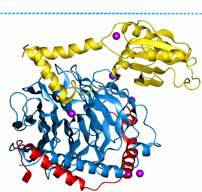
Regulator of G-protein signalling 9, also known as RGS9, is a human gene, which codes for a protein involved in regulation of signal transduction inside cells. Members of the RGS family, such as RGS9, are signaling proteins that suppress the activity of G proteins by promoting their deactivation. upplied by OMIMref name="entrez" /> There are two splice isoforms of RGS9 with quite different properties and patterns of expression. RGS9-1 is mainly found in the eye and is involved in regulation of phototransduction in rod and cone cells of the retina; genetic mutations in RGS9-1 cause the eye disease bradyopsia. RGS9-2 is found in the brain, and regulates dopamine and opioid signaling in the basal ganglia. RGS9-2 is of particular interest as the most important RGS protein involved in terminating signalling by the mu opioid receptor (although RGS4 and RGS17 are also involved), and is thought to be important in the development of tolerance to opioid drugs. RGS9-deficient mice exhi ... [...More Info...] [...Related Items...] OR: [Wikipedia] [Google] [Baidu] |
Autosomal Recessive Disease
A genetic disorder is a health problem caused by one or more abnormalities in the genome. It can be caused by a mutation in a single gene (monogenic) or multiple genes (polygenic) or by a chromosome abnormality. Although polygenic disorders are the most common, the term is mostly used when discussing disorders with a single genetic cause, either in a gene or chromosome. The mutation responsible can occur spontaneously before embryonic development (a ''de novo'' mutation), or it can be inherited from two parents who are carriers of a faulty gene (autosomal recessive inheritance) or from a parent with the disorder (autosomal dominant inheritance). When the genetic disorder is inherited from one or both parents, it is also classified as a hereditary disease. Some disorders are caused by a mutation on the X chromosome and have X-linked inheritance. Very few disorders are inherited on the Y chromosome or mitochondrial DNA (due to their size). There are well over 6,000 known genetic ... [...More Info...] [...Related Items...] OR: [Wikipedia] [Google] [Baidu] |
Optical Coherence Tomography
Optical coherence tomography (OCT) is a high-resolution imaging technique with most of its applications in medicine and biology. OCT uses coherent near-infrared light to obtain micrometer-level depth resolved images of biological tissue or other scattering media. It uses interferometry techniques to detect the amplitude and time-of-flight of reflected light. OCT uses transverse sample scanning of the light beam to obtain two- and three-dimensional images. Short-coherence-length light can be obtained using a superluminescent diode (SLD) with a broad spectral bandwidth or a broadly tunable laser with narrow linewidth. The first demonstration of OCT imaging (in vitro) was published by a team from MIT and Harvard Medical School in a 1991 article in the journal ''Science (journal), Science''. The article introduced the term "OCT" to credit its derivation from optical coherence-domain reflectometry, in which the axial resolution is based on temporal coherence. The first demonstrat ... [...More Info...] [...Related Items...] OR: [Wikipedia] [Google] [Baidu] |
Vertebrate Visual Opsin
Vertebrate visual opsins are a subclass of ciliary opsins and mediate vision in vertebrates. They include the opsins in human rod and cone cells. They are often abbreviated to ''opsin'', as they were the first opsins discovered and are still the most widely studied opsins. Opsins Opsin refers strictly to the apoprotein (without bound retinal). When an opsin binds retinal to form a holoprotein, it is referred to as Retinylidene protein. However, the distinction is often ignored, and opsin may refer loosely to both (regardless of whether retinal is bound). Opsins are G-protein-coupled receptors (GPCRs) and must bind retinal — typically 11-''cis''-retinal — in order to be photosensitive, since the retinal acts as the chromophore. When the Retinylidene protein absorbs a photon, the retinal isomerizes and is released by the opsin. The process that follows the isomerization and renewal of retinal is known as the visual cycle. Free 11-''cis''-retinal is photosensitive and ... [...More Info...] [...Related Items...] OR: [Wikipedia] [Google] [Baidu] |
Rhodopsin
Rhodopsin, also known as visual purple, is a protein encoded by the ''RHO'' gene and a G-protein-coupled receptor (GPCR). It is a light-sensitive receptor protein that triggers visual phototransduction in rod cells. Rhodopsin mediates dim light vision and thus is extremely sensitive to light. When rhodopsin is exposed to light, it immediately photobleaches. In humans, it is fully regenerated in about 30 minutes, after which the rods are more sensitive. Defects in the rhodopsin gene cause eye diseases such as retinitis pigmentosa and congenital stationary night blindness. History Rhodopsin was discovered by Franz Christian Boll in 1876. The name rhodopsin derives from Ancient Greek () for "rose", due to its pinkish color, and () for "sight". It was coined in 1878 by the German physiologist Wilhelm Friedrich Kühne (1837–1900). When George Wald discovered that rhodopsin is a holoprotein, consisting of retinal and an apoprotein, he called it opsin, which tod ... [...More Info...] [...Related Items...] OR: [Wikipedia] [Google] [Baidu] |
G Protein
G proteins, also known as guanine nucleotide-binding proteins, are a Protein family, family of proteins that act as molecular switches inside cells, and are involved in transmitting signals from a variety of stimuli outside a cell (biology), cell to its interior. Their activity is regulated by factors that control their ability to bind to and hydrolyze guanosine triphosphate (GTP) to guanosine diphosphate (GDP). When they are bound to GTP, they are 'on', and, when they are bound to GDP, they are 'off'. G proteins belong to the larger group of enzymes called GTPases. There are two classes of G proteins. The first function as monomeric small GTPases (small G-proteins), while the second function as heterotrimeric G protein protein complex, complexes. The latter class of complexes is made up of ''G alpha subunit, alpha'' (Gα), ''beta'' (Gβ) and ''gamma'' (Gγ) protein subunit, subunits. In addition, the beta and gamma subunits can form a stable Protein dimer, dimeric complex re ... [...More Info...] [...Related Items...] OR: [Wikipedia] [Google] [Baidu] |
Visual Phototransduction
Visual phototransduction is the sensory transduction process of the visual system by which light is detected by photoreceptor cells ( rods and cones) in the vertebrate retina. A photon is absorbed by a retinal chromophore (each bound to an opsin), which initiates a signal cascade through several intermediate cells, then through the retinal ganglion cells (RGCs) comprising the optic nerve. Overview Light enters the eye, passes through the optical media, then the inner neural layers of the retina before finally reaching the photoreceptor cells in the outer layer of the retina. The light may be absorbed by a chromophore bound to an opsin, which photoisomerizes the chromophore, initiating both the visual cycle, which "resets" the chromophore, and the phototransduction cascade, which transmits the visual signal to the brain. The cascade begins with graded polarisation (an analog signal) of the excited photoreceptor cell, as its membrane potential increases from a resting po ... [...More Info...] [...Related Items...] OR: [Wikipedia] [Google] [Baidu] |
Dominance (genetics)
In genetics, dominance is the phenomenon of one variant (allele) of a gene on a chromosome masking or overriding the effect of a different variant of the same gene on the other copy of the chromosome. The first variant is termed dominant and the second is called recessive. This state of having two different variants of the same gene on each chromosome is originally caused by a mutation in one of the genes, either new (''de novo'') or inherited. The terms autosomal dominant or autosomal recessive are used to describe gene variants on non-sex chromosomes ( autosomes) and their associated traits, while those on sex chromosomes (allosomes) are termed X-linked dominant, X-linked recessive or Y-linked; these have an inheritance and presentation pattern that depends on the sex of both the parent and the child (see Sex linkage). Since there is only one Y chromosome, Y-linked traits cannot be dominant or recessive. Additionally, there are other forms of dominance, such as incomp ... [...More Info...] [...Related Items...] OR: [Wikipedia] [Google] [Baidu] |
Electroretinography
Electroretinography measures the electrical responses of various cell types in the retina, including the Photoreceptor cell, photoreceptors (rod cell, rods and cone cell, cones), inner retinal cells (Retinal bipolar cell, bipolar and amacrine cell, amacrine cells), and the Retinal ganglion cell, ganglion cells. Electrodes are placed on the surface of the cornea (DTL silver/nylon fiber string or ERG jet) or on the skin beneath the eye (sensor strips) to measure retinal responses. Retinal pigment epithelium (RPE) responses are measured with an Electrooculography, EOG test with skin-contact electrodes placed near the Canthus, canthi. During a recording, the patient's eyes are exposed to standardized stimulus (physiology), stimuli and the resulting signal is displayed showing the time course of the signal's amplitude (voltage). Signals are very small, and typically are measured in microvolts or nanovolts. The ERG is composed of electrical potentials contributed by different cell type ... [...More Info...] [...Related Items...] OR: [Wikipedia] [Google] [Baidu] |
Fundus (eye)
The fundus of the eye is the interior surface of the eye opposite the lens and includes the retina, optic disc, macula, fovea, and posterior pole.Cassin, B. and Solomon, S. ''Dictionary of Eye Terminology''. Gainesville, Florida: Triad Publishing Company, 1990. The fundus can be examined by ophthalmoscopy and/or fundus photography. Variation The color of the fundus varies both between and within species. In one study of primates the retina is blue, green, yellow, orange, and red; only the human fundus (from a lightly pigmented blond person) is red. The major differences noted among the "higher" primate species were size and regularity of the border of macular area, size and shape of the optic disc, apparent 'texturing' of retina, and pigmentation of retina. Clinical significance Medical signs that can be detected from observation of eye fundus (generally by funduscopy) include hemorrhages, exudates, cotton wool spots, blood vessel abnormalities ( tortuosity, puls ... [...More Info...] [...Related Items...] OR: [Wikipedia] [Google] [Baidu] |
Electroretinogram
Electroretinography measures the electrical responses of various cell types in the retina, including the photoreceptors ( rods and cones), inner retinal cells ( bipolar and amacrine cells), and the ganglion cells. Electrodes are placed on the surface of the cornea (DTL silver/nylon fiber string or ERG jet) or on the skin beneath the eye (sensor strips) to measure retinal responses. Retinal pigment epithelium (RPE) responses are measured with an EOG test with skin-contact electrodes placed near the canthi. During a recording, the patient's eyes are exposed to standardized stimuli and the resulting signal is displayed showing the time course of the signal's amplitude (voltage). Signals are very small, and typically are measured in microvolts or nanovolts. The ERG is composed of electrical potentials contributed by different cell types within the retina, and the stimulus conditions (flash or pattern stimulus, whether a background light is present, and the colors of the stimulus ... [...More Info...] [...Related Items...] OR: [Wikipedia] [Google] [Baidu] |


