|
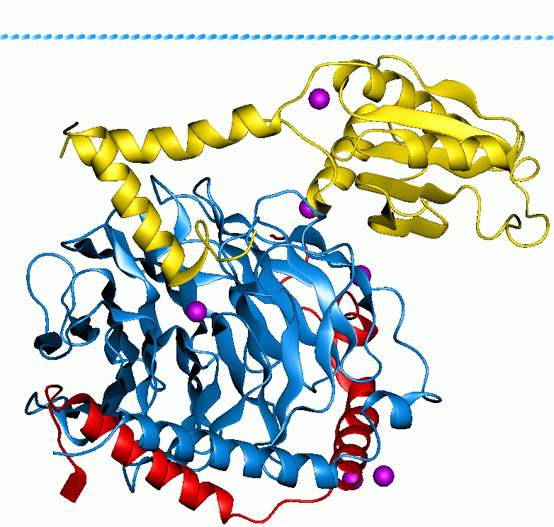
RGS9
Regulator of G-protein signalling 9, also known as RGS9, is a human gene, which codes for a protein involved in regulation of signal transduction inside cells. Members of the RGS family, such as RGS9, are signaling proteins that suppress the activity of G proteins by promoting their deactivation. upplied by OMIMref name="entrez" /> There are two splice isoforms of RGS9 with quite different properties and patterns of expression. RGS9-1 is mainly found in the eye and is involved in regulation of phototransduction in rod and cone cells of the retina; genetic mutations in RGS9-1 cause the eye disease bradyopsia. RGS9-2 is found in the brain, and regulates dopamine and opioid signaling in the basal ganglia. RGS9-2 is of particular interest as the most important RGS protein involved in terminating signalling by the mu opioid receptor (although RGS4 and RGS17 are also involved), and is thought to be important in the development of tolerance to opioid drugs. RGS9-deficient mice exhi ... [...More Info...] [...Related Items...] OR: [Wikipedia] [Google] [Baidu] |
Bradyopsia
Bradyopsia, also known as "prolonged electro-retinal response suppression" is a visual condition in which the photoreceptor cells in the retina have a slower-than-normal recovery of light sensitivity after exposure to light. It is inherited as an autosomal recessive disease. It is uncommon with only a few dozen patients described in the medical literature as of 2025. Because of the subtle nature of the symptoms, because many ophthalmologists and optometrists are unaware of it, and because non-standard electroretinogram (ERG) testing is needed to confirm the diagnosis, many cases are likely to be undiagnosed. Symptoms and signs Patients with bradyopsia can have nearly normal visual acuity (20/25 to 20/40) when tested with stationary, high-contrast standard visual acuity charts such as the Snellen chart with a dimly lit background. However, the acuity may vary from visit to visit and can be as poor 20/200 when tested with a bright background. The visual acuity improves with ... [...More Info...] [...Related Items...] OR: [Wikipedia] [Google] [Baidu] |
GNB5
Guanine nucleotide-binding protein subunit beta-5 is a protein that in humans is encoded by the ''GNB5'' gene. Alternatively spliced transcript variants encoding different isoforms exist. Function Heterotrimeric guanine nucleotide-binding proteins (G proteins), which integrate signals between receptors and effector proteins, are composed of an alpha, a beta, and a gamma subunit. These subunits are encoded by families of related genes. This gene encodes a beta subunit. Beta subunits are important regulators of alpha subunits, as well as of certain signal transduction receptors and effectors. GNB5 has been shown to differentially control RGS protein stability and membrane anchor binding, and therefore is involved in the control of complex neuronal G protein signaling pathways. Interactions GNB5 has been shown to interact with: * GNG7, * GNG13, * RGS7 and * RGS9 Regulator of G-protein signalling 9, also known as RGS9, is a human gene, which codes for a protein involve ... [...More Info...] [...Related Items...] OR: [Wikipedia] [Google] [Baidu] |
Regulator Of G Protein Signalling
Regulators of G protein signaling (RGS) are protein structural domains or the proteins that contain these domains, that function to activate the GTPase activity of heterotrimeric G-protein α-subunits. RGS proteins are multi-functional, GTPase-accelerating proteins that promote GTP hydrolysis by the α-subunit of heterotrimeric G proteins, thereby inactivating the G protein and rapidly switching off G protein-coupled receptor signaling pathways. Upon activation by receptors, G proteins exchange GDP for GTP, are released from the receptor, and dissociate into a free, active GTP-bound α-subunit and βγ-dimer, both of which activate downstream effectors. The response is terminated upon GTP hydrolysis by the α-subunit (), which can then re-bind the βγ-dimer ( ) and the receptor. RGS proteins markedly reduce the lifespan of GTP-bound α-subunits by stabilising the G protein transition state. Whereas receptors stimulate GTP binding, RGS proteins stimulate GTP hydrolysis. RGS prot ... [...More Info...] [...Related Items...] OR: [Wikipedia] [Google] [Baidu] |
RGS4
Regulator of G protein signaling 4 also known as RGP4 is a protein that in humans is encoded by the ''RGS4'' gene. RGP4 regulates G protein signaling. Function Regulator of G protein signalling (RGS) family members are regulatory molecules that act as GTPase activating proteins (GAPs) for G alpha subunits of heterotrimeric G proteins. RGS proteins are able to deactivate G protein subunits of the Gi alpha, Go alpha and Gq alpha subtypes. They drive G proteins into their inactive GDP-bound forms. Regulator of G protein signaling 4 belongs to this family. All RGS proteins share a conserved 120-amino acid sequence termed the RGS domain which conveys GAP activity. Regulator of G protein signaling 4 protein is 37% identical to RGS1 and 97% identical to rat Rgs4. This protein negatively regulates signaling upstream or at the level of the heterotrimeric G protein and is localized in the cytoplasm. Clinical significance A number of studies associate the RGS4 gene with schizop ... [...More Info...] [...Related Items...] OR: [Wikipedia] [Google] [Baidu] |
RGS17
Regulator of G-protein signaling 17 is a protein that in humans is encoded by the ''RGS17'' gene. Function This gene encodes a member of the regulator of G-protein signaling family. This protein contains a conserved, 120 amino acid motif called the Regulator of G protein signaling, RGS domain and a cysteine-rich region. The protein attenuates the signaling activity of G-proteins by binding to activated, GTP-bound G alpha subunits and acting as a GTPase activating protein (GAP), increasing the rate of conversion of the GTP to GDP. This hydrolysis allows the G alpha subunits to bind G beta/gamma subunit heterodimers, forming inactive G-protein heterotrimers, thereby terminating the signal. Along with RGS4, RGS9 and RGS14, RGS17 plays an important role in termination of signalling by mu opioid receptors and development of tolerance to opioid analgesic drugs. Clinical significance RGS17 is a putative lung cancer susceptibility gene in the lung cancer associated locus on chromoso ... [...More Info...] [...Related Items...] OR: [Wikipedia] [Google] [Baidu] |
Gene
In biology, the word gene has two meanings. The Mendelian gene is a basic unit of heredity. The molecular gene is a sequence of nucleotides in DNA that is transcribed to produce a functional RNA. There are two types of molecular genes: protein-coding genes and non-coding genes. During gene expression (the synthesis of Gene product, RNA or protein from a gene), DNA is first transcription (biology), copied into RNA. RNA can be non-coding RNA, directly functional or be the intermediate protein biosynthesis, template for the synthesis of a protein. The transmission of genes to an organism's offspring, is the basis of the inheritance of phenotypic traits from one generation to the next. These genes make up different DNA sequences, together called a genotype, that is specific to every given individual, within the gene pool of the population (biology), population of a given species. The genotype, along with environmental and developmental factors, ultimately determines the phenotype ... [...More Info...] [...Related Items...] OR: [Wikipedia] [Google] [Baidu] |
Signal Transduction
Signal transduction is the process by which a chemical or physical signal is transmitted through a cell as a biochemical cascade, series of molecular events. Proteins responsible for detecting stimuli are generally termed receptor (biology), receptors, although in some cases the term sensor is used. The changes elicited by ligand (biochemistry), ligand binding (or signal sensing) in a receptor give rise to a biochemical cascade, which is a chain of biochemical events known as a Cell signaling#Signaling pathways, signaling pathway. When signaling pathways interact with one another they form networks, which allow cellular responses to be coordinated, often by combinatorial signaling events. At the molecular level, such responses include changes in the transcription (biology), transcription or translation (biology), translation of genes, and post-translational modification, post-translational and conformational changes in proteins, as well as changes in their location. These molecula ... [...More Info...] [...Related Items...] OR: [Wikipedia] [Google] [Baidu] |
G Protein
G proteins, also known as guanine nucleotide-binding proteins, are a Protein family, family of proteins that act as molecular switches inside cells, and are involved in transmitting signals from a variety of stimuli outside a cell (biology), cell to its interior. Their activity is regulated by factors that control their ability to bind to and hydrolyze guanosine triphosphate (GTP) to guanosine diphosphate (GDP). When they are bound to GTP, they are 'on', and, when they are bound to GDP, they are 'off'. G proteins belong to the larger group of enzymes called GTPases. There are two classes of G proteins. The first function as monomeric small GTPases (small G-proteins), while the second function as heterotrimeric G protein protein complex, complexes. The latter class of complexes is made up of ''G alpha subunit, alpha'' (Gα), ''beta'' (Gβ) and ''gamma'' (Gγ) protein subunit, subunits. In addition, the beta and gamma subunits can form a stable Protein dimer, dimeric complex re ... [...More Info...] [...Related Items...] OR: [Wikipedia] [Google] [Baidu] |
Photoreceptor Cell
A photoreceptor cell is a specialized type of neuroepithelial cell found in the retina that is capable of visual phototransduction. The great biological importance of photoreceptors is that they convert light (visible electromagnetic radiation) into signals that can stimulate biological processes. To be more specific, photoreceptor proteins in the cell absorb photons, triggering a change in the cell's membrane potential. There are currently three known types of photoreceptor cells in mammalian eyes: rod cell, rods, cone cell, cones, and intrinsically photosensitive retinal ganglion cells. The two classic photoreceptor cells are rods and cones, each contributing information used by the visual system to form an image of the environment, Visual perception, sight. Rods primarily mediate scotopic vision (dim conditions) whereas cones primarily mediate photopic vision (bright conditions), but the processes in each that supports phototransduction is similar. The intrinsically photosen ... [...More Info...] [...Related Items...] OR: [Wikipedia] [Google] [Baidu] |
Retina
The retina (; or retinas) is the innermost, photosensitivity, light-sensitive layer of tissue (biology), tissue of the eye of most vertebrates and some Mollusca, molluscs. The optics of the eye create a focus (optics), focused two-dimensional image of the visual world on the retina, which then processes that image within the retina and sends nerve impulses along the optic nerve to the visual cortex to create visual perception. The retina serves a function which is in many ways analogous to that of the photographic film, film or image sensor in a camera. The neural retina consists of several layers of neurons interconnected by Chemical synapse, synapses and is supported by an outer layer of pigmented epithelial cells. The primary light-sensing cells in the retina are the photoreceptor cells, which are of two types: rod cell, rods and cone cell, cones. Rods function mainly in dim light and provide monochromatic vision. Cones function in well-lit conditions and are responsible fo ... [...More Info...] [...Related Items...] OR: [Wikipedia] [Google] [Baidu] |
Basal Ganglia
The basal ganglia (BG) or basal nuclei are a group of subcortical Nucleus (neuroanatomy), nuclei found in the brains of vertebrates. In humans and other primates, differences exist, primarily in the division of the globus pallidus into external and internal regions, and in the division of the striatum. Positioned at the base of the forebrain and the top of the midbrain, they have strong connections with the cerebral cortex, thalamus, brainstem and other brain areas. The basal ganglia are associated with a variety of functions, including regulating voluntary motor control, motor movements, procedural memory, procedural learning, habituation, habit formation, conditional learning, eye movements, cognition, and emotion. The main functional components of the basal ganglia include the striatum, consisting of both the dorsal striatum (caudate nucleus and putamen) and the ventral striatum (nucleus accumbens and olfactory tubercle), the globus pallidus, the ventral pallidum, the substa ... [...More Info...] [...Related Items...] OR: [Wikipedia] [Google] [Baidu] |


