|
Agenesis Of The Corpus Callosum
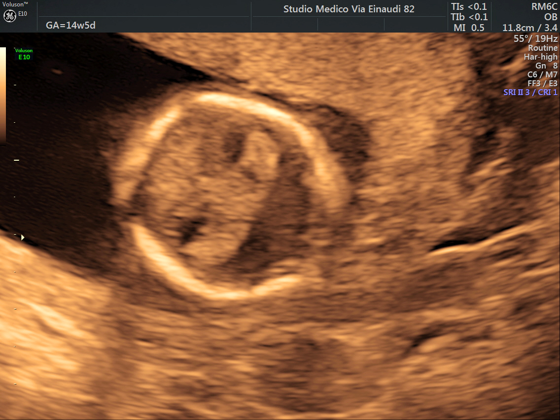
Agenesis of the corpus callosum (ACC) is a rare birth defect in which there is a complete or partial absence of the corpus callosum. It occurs when the development of the corpus callosum, the band of white matter connecting the two hemispheres in the brain, in the embryo is disrupted. The result of this is that the fibers that would otherwise form the corpus callosum are instead longitudinally oriented along the ipsilateral ventricular wall and form structures called Probst bundles. In addition to agenesis, other degrees of callosal defects exist, including hypoplasia (underdevelopment or thinness), hypogenesis (partial agenesis) or dysgenesis (malformation). ACC is found in many syndromes and can often present alongside hypoplasia of the cerebellar vermis. When this is the case, there can also be an enlarged fourth ventricle or hydrocephalus; this is called Dandy–Walker malformation. Signs and symptoms Signs and symptoms of ACC and other callosal disorders vary greatly ... [...More Info...] [...Related Items...] OR: [Wikipedia] [Google] [Baidu] |
Corpus Callosum
The corpus callosum (Latin for "tough body"), also callosal commissure, is a wide, thick nerve tract, consisting of a flat bundle of commissural fibers, beneath the cerebral cortex in the brain. The corpus callosum is only found in placental mammals. It spans part of the longitudinal fissure, connecting the left and right cerebral hemispheres, enabling communication between them. It is the largest white matter structure in the human brain, about in length and consisting of 200–300 million axonal projections. A number of separate nerve tracts, classed as subregions of the corpus callosum, connect different parts of the hemispheres. The main ones are known as the genu, the rostrum, the trunk or body, and the splenium. Structure The corpus callosum forms the floor of the longitudinal fissure that separates the two cerebral hemispheres. Part of the corpus callosum forms the roof of the lateral ventricles. The corpus callosum has four main parts – individual nerve ... [...More Info...] [...Related Items...] OR: [Wikipedia] [Google] [Baidu] |
Rare Disease
A rare disease is any disease that affects a small percentage of the population. In some parts of the world, an orphan disease is a rare disease whose rarity means there is a lack of a market large enough to gain support and resources for discovering treatments for it, except by the government granting economically advantageous conditions to creating and selling such treatments. Orphan drugs are ones so created or sold. Most rare diseases are genetic and thus are present throughout the person's entire life, even if symptoms do not immediately appear. Many rare diseases appear early in life, and about 30% of children with rare diseases will die before reaching their fifth birthdays. With only four diagnosed patients in 27 years, ribose-5-phosphate isomerase deficiency is considered the rarest known genetic disease. No single cut-off number has been agreed upon for which a disease is considered rare. A disease may be considered rare in one part of the world, or in a particular g ... [...More Info...] [...Related Items...] OR: [Wikipedia] [Google] [Baidu] |
Dysautonomic
Dysautonomia or autonomic dysfunction is a condition in which the autonomic nervous system (ANS) does not work properly. This may affect the functioning of the heart, bladder, intestines, sweat glands, pupils, and blood vessels. Dysautonomia has many causes, not all of which may be classified as neuropathic. A number of conditions can feature dysautonomia, such as Parkinson's disease, multiple system atrophy, dementia with Lewy bodies, Ehlers-Danlos syndromes, autoimmune autonomic ganglionopathy and autonomic neuropathy, HIV/AIDS, autonomic failure, and postural orthostatic tachycardia syndrome. The diagnosis is achieved through functional testing of the ANS, focusing on the affected organ system. Investigations may be performed to identify underlying disease processes that may have led to the development of symptoms or autonomic neuropathy. Symptomatic treatment is available for many symptoms associated with dysautonomia, and some disease processes can be directly treated. Si ... [...More Info...] [...Related Items...] OR: [Wikipedia] [Google] [Baidu] |
Acrocallosal Syndrome
Acrocallosal syndrome (also known as ACLS) is an extremely rare autosomal recessive syndrome characterized by corpus callosum agenesis, polydactyly, multiple dysmorphic features, motor and intellectual disabilities, and other symptoms. The syndrome was first described by Albert Schinzel in 1979. Mutations in ''KIF7'' are causative for ACLS, and mutations in ''GLI3'' are associated with a similar syndrome. Signs and symptoms Acrocallosal syndrome (ACLS, ACS, Schinzel-type, Hallux-duplication) is a rare, heterogeneous autosomal recessive disorder first discovered by Albert Schinzel (1979) in a 3-year-old boy. Characteristics of this syndrome include agenesis of the corpus callosum, macrocephaly, hypertelorism, poor motor skills, intellectual disability, extra fingers and toes (particularly hallux duplication), and cleft palate. Seizures may also occur. Mechanism Mutations in the ''KIF7'' gene are causative for ACLS. KIF7 is a 1343 amino acid protein with a kinesin motor, c ... [...More Info...] [...Related Items...] OR: [Wikipedia] [Google] [Baidu] |
Schizencephaly
Schizencephaly () is a rare birth defect characterized by abnormal clefts lined with grey matter that form the ependyma of the cerebral ventricles to the pia mater. These clefts can occur bilaterally or unilaterally. Common clinical features of this malformation include epilepsy, motor deficits, and psychomotor retardation. Presentation Schizencephaly can be distinguished from porencephaly by the fact that in schizencephaly, the fluid-filled component is entirely lined by heterotopic grey matter, while a porencephalic cyst is lined mostly by white matter. Individuals with clefts in both hemispheres, or bilateral clefts, are often developmentally delayed and have delayed speech and language skills and corticospinal dysfunction. Individuals with smaller, unilateral clefts (clefts in one hemisphere) may be weak or paralyzed on one side of the body and may have average or near-average intelligence. Patients with schizencephaly may also have varying degrees of microcephaly, Cognit ... [...More Info...] [...Related Items...] OR: [Wikipedia] [Google] [Baidu] |
Grey Matter Heterotopia
MRI of a child experiencing seizures. There are small foci of grey matter Heterotopia (medicine)">heterotopia in the corpus callosum, deep to the Cortical dysplasia, dysplastic cortex. (double arrows) Gray matter heterotopias are neurological disorders caused by clumps of gray matter (nodules of neurons) Ectopia (medicine), located in the wrong part of the brain. A grey matter heterotopia is characterized as a type of focal cortical dysplasia. The neurons in heterotopia appear to be normal, except for their mislocation; nuclear studies have shown glucose metabolism equal to that of normally positioned gray matter. The condition causes a variety of symptoms, but usually includes some degree of epilepsy or recurring seizures, and often affects the brain's ability to function on higher levels. Symptoms range from nonexistent to profound; the condition is occasionally discovered as an incidentaloma when brain imaging performed for an unrelated problem and has no apparent ill eff ... [...More Info...] [...Related Items...] OR: [Wikipedia] [Google] [Baidu] |
Neuronal Migration Disorders
Neuronal migration disorder (NMD) refers to a heterogenous group of disorders that, it is supposed, share the same etiopathological mechanism: a variable degree of disruption in the migration of neuroblast In vertebrates, a neuroblast or primitive nerve cell is a postmitotic cell that does not divide further, and which will develop into a neuron after a migration phase. In invertebrates such as ''Drosophila,'' neuroblasts are neural progenitor cell ...s during neurogenesis. The neuronal migration disorders are termed dysgenesis (embryology), cerebral dysgenesis disorders, brain malformations caused by primary alterations during neurogenesis; on the other hand, brain malformations are highly diverse and refer to any insult to the brain during its formation and maturation due to intrinsic or extrinsic causes that ultimately will alter the normal brain anatomy. However, there is some controversy in the terminology because virtually any malformation will involve neuroblast migration, ... [...More Info...] [...Related Items...] OR: [Wikipedia] [Google] [Baidu] |
Hydrocephalus
Hydrocephalus is a condition in which an accumulation of cerebrospinal fluid (CSF) occurs within the brain. This typically causes increased pressure inside the skull. Older people may have headaches, double vision, poor balance, urinary incontinence, personality changes, or mental impairment. In babies, it may be seen as a rapid increase in head size. Other symptoms may include vomiting, sleepiness, seizures, and downward pointing of the eyes. Hydrocephalus can occur due to birth defects or be acquired later in life. Associated birth defects include neural tube defects and those that result in aqueductal stenosis. Other causes include meningitis, brain tumors, traumatic brain injury, intraventricular hemorrhage, and subarachnoid hemorrhage. The four types of hydrocephalus are communicating, noncommunicating, ''ex vacuo'', and normal pressure. Diagnosis is typically made by physical examination and medical imaging. Hydrocephalus is typically treated by the surgical ... [...More Info...] [...Related Items...] OR: [Wikipedia] [Google] [Baidu] |
Holoprosencephaly
Holoprosencephaly (HPE) is a cephalic disorder in which the prosencephalon (the forebrain of the embryo) fails to develop into two hemispheres, typically occurring between the 18th and 28th day of gestation. Normally, the forebrain is formed and the face begins to develop in the fifth and sixth weeks of human pregnancy. The condition also occurs in other species. Holoprosencephaly is estimated to occur in approximately 1 in every 250 conceptions and most cases are not compatible with life and result in fetal death in utero due to deformities to the skull and brain. However, holoprosencephaly is still estimated to occur in approximately 1 in every 8,000 live births. When the embryo's forebrain does not divide to form bilateral cerebral hemispheres (the left and right halves of the brain), it causes defects in the development of the face and in brain structure and function. The severity of holoprosencephaly is highly variable. In less severe cases, babies are born with normal ... [...More Info...] [...Related Items...] OR: [Wikipedia] [Google] [Baidu] |
Colpocephaly
Colpocephaly is a cephalic disorder involving the disproportionate enlargement of the occipital horns of the lateral ventricles and is usually diagnosed early after birth due to seizures. It is a nonspecific finding and is associated with multiple neurological syndromes, including agenesis of the corpus callosum, Chiari malformation, lissencephaly, and microcephaly. Although the exact cause of colpocephaly is not known yet, it is commonly believed to occur as a result of neuronal migration disorders during early brain development, intrauterine disturbances, perinatal injuries, and other central nervous system disorders. Individuals with colpocephaly have various degrees of motor disabilities, visual defects, spasticity, and moderate to severe intellectual disability. No specific treatment for colpocephaly exists, but patients may undergo certain treatments to improve their motor function or intellectual disability. Symptoms There are various symptoms of colpocephaly and pati ... [...More Info...] [...Related Items...] OR: [Wikipedia] [Google] [Baidu] |
Chiari Malformation
Chiari malformation (CM) is a structural defect in the cerebellum, characterized by a downward displacement of one or both cerebellar tonsils through the foramen magnum (the opening at the base of the skull). CMs can cause headaches, difficulty swallowing, vomiting, dizziness, neck pain, unsteady gait, poor hand coordination, numbness and tingling of the hands and feet, and speech problems. Less often, people may experience ringing or buzzing in the ears, weakness, slow heart rhythm, or fast heart rhythm, curvature of the spine ( scoliosis) related to spinal cord impairment, abnormal breathing, such as central sleep apnea, characterized by periods of breathing cessation during sleep, and, in severe cases, paralysis. This can sometimes lead to non-communicating hydrocephalus as a result of obstruction of cerebrospinal fluid (CSF) outflow. The cerebrospinal fluid outflow is caused by phase difference in outflow and influx of blood in the vasculature of the brain. The malforma ... [...More Info...] [...Related Items...] OR: [Wikipedia] [Google] [Baidu] |


