|
X-ray Spectrometer
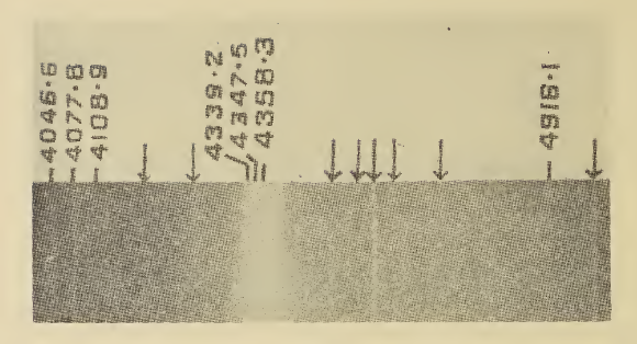
X-ray spectroscopy is a general term for several spectroscopic techniques for characterization of materials by using x-ray radiation. Characteristic X-ray spectroscopy When an electron from the inner shell of an atom is excited by the energy of a photon, it moves to a higher energy level. When it returns to the low energy level, the energy it previously gained by excitation is emitted as a photon of one of the wavelengths uniquely characteristic of the element. Analysis of the X-ray emission spectrum produces qualitative results about the elemental composition of the specimen. Comparison of the specimen's spectrum with the spectra of samples of known composition produces quantitative results (after some mathematical corrections for absorption, fluorescence and atomic number). Atoms can be excited by a high-energy beam of charged particles such as electrons (in an electron microscope for example), protons (see PIXE) or a beam of X-rays (see X-ray fluorescence, or XRF or also ... [...More Info...] [...Related Items...] OR: [Wikipedia] [Google] [Baidu] |
Spectroscopy
Spectroscopy is the field of study that measures and interprets electromagnetic spectra. In narrower contexts, spectroscopy is the precise study of color as generalized from visible light to all bands of the electromagnetic spectrum. Spectroscopy, primarily in the electromagnetic spectrum, is a fundamental exploratory tool in the fields of astronomy, chemistry, materials science, and physics, allowing the composition, physical structure and electronic structure of matter to be investigated at the atomic, molecular and macro scale, and over astronomical distances. Historically, spectroscopy originated as the study of the wavelength dependence of the absorption by gas phase matter of visible light dispersed by a prism. Current applications of spectroscopy include biomedical spectroscopy in the areas of tissue analysis and medical imaging. Matter waves and acoustic waves can also be considered forms of radiative energy, and recently gravitational waves have been associa ... [...More Info...] [...Related Items...] OR: [Wikipedia] [Google] [Baidu] |
Microprobe
A microprobe is an instrument that applies a stable and well-focused beam of charged particles (electrons or ions) to a sample. Types When the primary beam consists of accelerated electrons, the probe is termed an electron microprobe, when the primary beam consists of accelerated ions, the term ion microprobe is used. The term microprobe may also be applied to optical analytical techniques, when the instrument is set up to analyse micro samples or micro areas of larger specimens. Such techniques include micro Raman spectroscopy, micro infrared spectroscopy and micro LIBS. All of these techniques involve modified optical microscopes to locate the area to be analysed, direct the probe beam and collect the analytical signal. A laser microprobe is a mass spectrometer that uses ionization by a pulsed laser and subsequent mass analysis of the generated ions. Uses Scientists use this beam of charged particles to determine the elemental composition of solid materials (minerals, glas ... [...More Info...] [...Related Items...] OR: [Wikipedia] [Google] [Baidu] |
Binding Energy
In physics and chemistry, binding energy is the smallest amount of energy required to remove a particle from a system of particles or to disassemble a system of particles into individual parts. In the former meaning the term is predominantly used in condensed matter physics, atomic physics, and chemistry, whereas in nuclear physics the term '' separation energy'' is used. A bound system is typically at a lower energy level than its unbound constituents. According to relativity theory, a decrease in the total energy of a system is accompanied by a decrease in the total mass, where . Types There are several types of binding energy, each operating over a different distance and energy scale. The smaller the size of a bound system, the higher its associated binding energy. Mass–energy relation A bound system is typically at a lower energy level than its unbound constituents because its mass must be less than the total mass of its unbound constituents. For systems with low bi ... [...More Info...] [...Related Items...] OR: [Wikipedia] [Google] [Baidu] |
Crystal Field Excitations
Crystal field excitation is the electronic transition of an electron between two orbitals of an atom that is situated in a crystal field environment. They are often observed in coordination complexes of transition metals. Some examples of crystal field excitations are ''dd''-transitions on a copper atom that is surrounded by an octahedron of oxygen atoms, or ''ff''-transitions on the uranium Uranium is a chemical element; it has chemical symbol, symbol U and atomic number 92. It is a silvery-grey metal in the actinide series of the periodic table. A uranium atom has 92 protons and 92 electrons, of which 6 are valence electrons. Ura ... atom in uranium antimonide. References Crystallography {{Crystallography-stub ... [...More Info...] [...Related Items...] OR: [Wikipedia] [Google] [Baidu] |
Electronvolt
In physics, an electronvolt (symbol eV), also written electron-volt and electron volt, is the measure of an amount of kinetic energy gained by a single electron accelerating through an Voltage, electric potential difference of one volt in vacuum. When used as a Units of energy, unit of energy, the numerical value of 1 eV in joules (symbol J) is equal to the numerical value of the Electric charge, charge of an electron in coulombs (symbol C). Under the 2019 revision of the SI, this sets 1 eV equal to the exact value Historically, the electronvolt was devised as a standard unit of measure through its usefulness in Particle accelerator#Electrostatic particle accelerators, electrostatic particle accelerator sciences, because a particle with electric charge ''q'' gains an energy after passing through a voltage of ''V''. Definition and use An electronvolt is the amount of energy gained or lost by a single electron when it moves through an Voltage, electric potential differenc ... [...More Info...] [...Related Items...] OR: [Wikipedia] [Google] [Baidu] |
Soft X-ray
An X-ray (also known in many languages as Röntgen radiation) is a form of high-energy electromagnetic radiation with a wavelength shorter than those of ultraviolet rays and longer than those of gamma rays. Roughly, X-rays have a wavelength ranging from 10 nanometers to 10 picometers, corresponding to frequencies in the range of 30 petahertz to 30 exahertz ( to ) and photon energies in the range of 100 eV to 100 keV, respectively. X-rays were discovered in 1895 by the German scientist Wilhelm Conrad Röntgen, who named it ''X-radiation'' to signify an unknown type of radiation.Novelline, Robert (1997). ''Squire's Fundamentals of Radiology''. Harvard University Press. 5th edition. . X-rays can penetrate many solid substances such as construction materials and living tissue, so X-ray radiography is widely used in medical diagnostics (e.g., checking for broken bones) and materials science (e.g., identification of some chemical elements and ... [...More Info...] [...Related Items...] OR: [Wikipedia] [Google] [Baidu] |
Electron Configuration
In atomic physics and quantum chemistry, the electron configuration is the distribution of electrons of an atom or molecule (or other physical structure) in atomic or molecular orbitals. For example, the electron configuration of the neon atom is , meaning that the 1s, 2s, and 2p subshells are occupied by two, two, and six electrons, respectively. Electronic configurations describe each electron as moving independently in an orbital, in an average field created by the nuclei and all the other electrons. Mathematically, configurations are described by Slater determinants or configuration state functions. According to the laws of quantum mechanics, a level of energy is associated with each electron configuration. In certain conditions, electrons are able to move from one configuration to another by the emission or absorption of a quantum of energy, in the form of a photon. Knowledge of the electron configuration of different atoms is useful in understanding the structu ... [...More Info...] [...Related Items...] OR: [Wikipedia] [Google] [Baidu] |
Raman Spectroscopy
Raman spectroscopy () (named after physicist C. V. Raman) is a Spectroscopy, spectroscopic technique typically used to determine vibrational modes of molecules, although rotational and other low-frequency modes of systems may also be observed. Raman spectroscopy is commonly used in chemistry to provide a structural fingerprint by which molecules can be identified. Raman spectroscopy relies upon inelastic scattering of photons, known as Raman scattering. A source of monochromatic light, usually from a laser in the visible spectrum, visible, near infrared, or ultraviolet, near ultraviolet range is used, although X-ray Raman scattering, X-rays can also be used. The laser light interacts with molecular vibrations, phonons or other excitations in the system, resulting in the energy of the laser photons being shifted up or down. The shift in energy gives information about the vibrational modes in the system. Time-resolved spectroscopy and infrared spectroscopy typically yields similar y ... [...More Info...] [...Related Items...] OR: [Wikipedia] [Google] [Baidu] |
Synchrotron
A synchrotron is a particular type of cyclic particle accelerator, descended from the cyclotron, in which the accelerating particle beam travels around a fixed closed-loop path. The strength of the magnetic field which bends the particle beam into its closed path increases with time during the accelerating process, being ''synchronized'' to the increasing kinetic energy of the particles. The synchrotron is one of the first accelerator concepts to enable the construction of large-scale facilities, since bending, beam focusing and acceleration can be separated into different components. The most powerful modern particle accelerators use versions of the synchrotron design. The largest synchrotron-type accelerator, also the largest particle accelerator in the world, is the Large Hadron Collider (LHC) near Geneva, Switzerland, completed in 2008 by the European Organization for Nuclear Research (CERN). It can accelerate beams of protons to an energy of 7 electron volt, teraelectro ... [...More Info...] [...Related Items...] OR: [Wikipedia] [Google] [Baidu] |
Diffraction Grating
In optics, a diffraction grating is an optical grating with a periodic structure that diffraction, diffracts light, or another type of electromagnetic radiation, into several beams traveling in different directions (i.e., different diffraction angles). The emerging coloration is a form of structural coloration. The directions or diffraction angles of these beams depend on the wave (light) Angle of incidence (optics), incident angle to the diffraction grating, the spacing or periodic distance between adjacent diffracting elements (e.g., parallel slits for a transmission grating) on the grating, and the wavelength of the incident light. The grating acts as a dispersion (optics), dispersive element. Because of this, diffraction gratings are commonly used in monochromators and spectrometers, but other applications are also possible such as optical encoders for high-precision motion control and wavefront measurement. For typical applications, a reflection (optics), reflective grati ... [...More Info...] [...Related Items...] OR: [Wikipedia] [Google] [Baidu] |
X-ray Tube
An X-ray tube is a vacuum tube that converts electrical input power into X-rays. The availability of this controllable source of X-rays created the field of radiography, the imaging of partly opaque objects with penetrating radiation. In contrast to other sources of ionizing radiation, X-rays are only produced as long as the X-ray tube is energized. X-ray tubes are also used in CT scanners, airport luggage scanners, X-ray crystallography, material and structure analysis, and for industrial inspection. Increasing demand for high-performance computed tomography (CT) scanning and angiography systems has driven development of very high-performance medical X-ray tubes. History X-ray tubes evolved from experimental Crookes tubes with which X-rays were first discovered on November 8, 1895, by the German physicist Wilhelm Conrad Röntgen. The first-generation ''cold cathode'' or ''Crookes'' X-ray tubes were used until the 1920s. These tubes work by ionisation of residual gas within the ... [...More Info...] [...Related Items...] OR: [Wikipedia] [Google] [Baidu] |
Cathode-ray Tube
A cathode-ray tube (CRT) is a vacuum tube containing one or more electron guns, which emit electron beams that are manipulated to display images on a phosphorescent screen. The images may represent electrical waveforms on an oscilloscope, a Film frame, frame of video on an Analog television, analog television set (TV), Digital imaging, digital raster graphics on a computer monitor, or other phenomena like radar targets. A CRT in a TV is commonly called a picture tube. CRTs have also been Williams tube, used as memory devices, in which case the screen is not intended to be visible to an observer. The term ''cathode ray'' was used to describe electron beams when they were first discovered, before it was understood that what was emitted from the cathode was a beam of electrons. In CRT TVs and computer monitors, the entire front area of the tube is scanned repeatedly and systematically in a fixed pattern called a raster scan, raster. In color devices, an image is produced by con ... [...More Info...] [...Related Items...] OR: [Wikipedia] [Google] [Baidu] |


