|
Wiggers Diagram
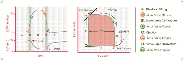
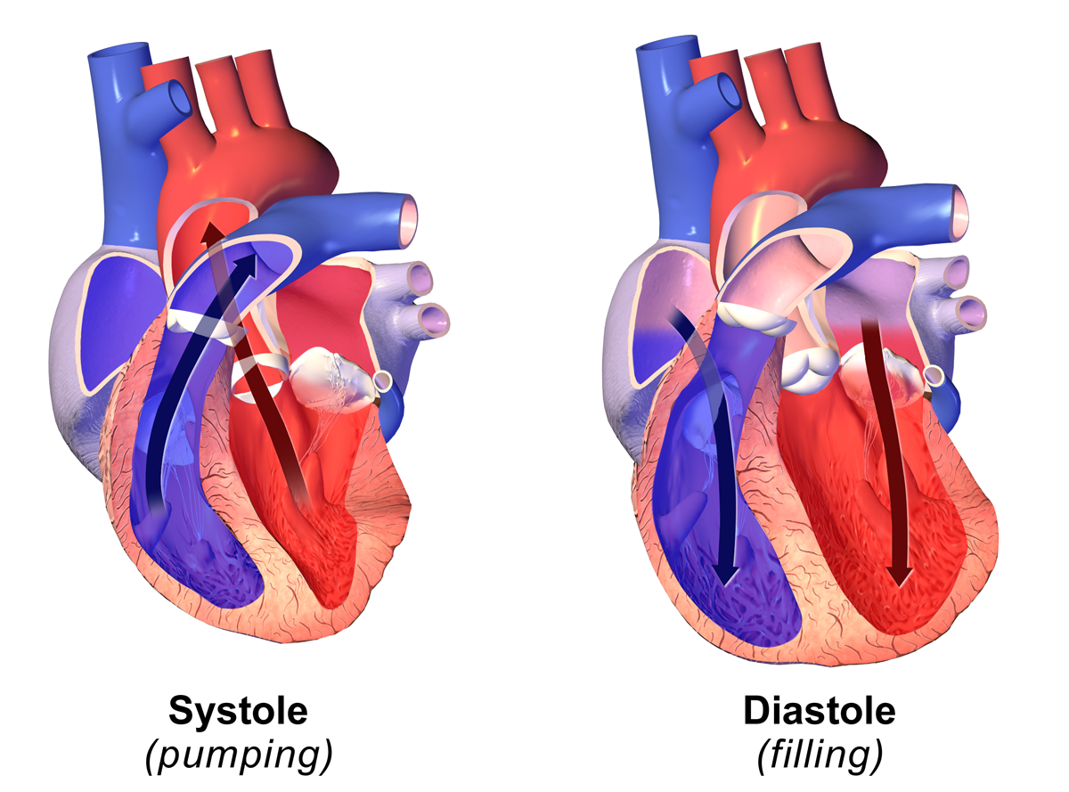
A Wiggers diagram, named after its developer, Carl Wiggers, is a unique diagram that has been used in teaching cardiac physiology for more than a century. In the Wiggers diagram, the X-axis is used to plot time subdivided into the cardiac phases, while the Y-axis typically contains the following on a single grid: * Blood pressure ** Aortic pressure ** Ventricular pressure ** Atrial pressure * Ventricular volume * Electrocardiogram * Arterial flow (optional) * Heart sounds (optional) The Wiggers diagram clearly illustrates the coordinated variation of these values as the heart beats, assisting one in understanding the entire cardiac cycle. Events Note that during isovolumetric/isovolumic contraction and relaxation, all the heart valves are closed; at no time are all the heart valves open. *S3 and S4 heart sounds are associated with pathologies and are not routinely heard. Additional images File:Heart systole.svg, Ventricular systole File:Heart diasystole.svg, Cardia ... [...More Info...] [...Related Items...] OR: [Wikipedia] [Google] [Baidu] |
Wiggers Diagram 2
Wiggers is a German and Dutch patronymic surname. The given name ''Wigger'' is a form of the Germanic Wichard, from Wîh- ("battle") and -hard ("strong"). at the Corpus of First Names in The Netherlands Variant spelling include ''Wichers'' and ''Wiggerts''. People with this surname include * Carl J. Wiggers (1883–1963), American physiologist **, a cardiac physiology diagram he devised * Charles Wiggers ( |
Systole (medicine)
Systole ( ) is the part of the cardiac cycle during which some chambers of the heart contract after refilling with blood. Its contrasting phase is diastole, the relaxed phase of the cardiac cycle when the chambers of the heart are refilling with blood. Etymology The term originates, via Neo-Latin, from Ancient Greek (''sustolē''), from (''sustéllein'' 'to contract'; from ''sun'' 'together' + ''stéllein'' 'to send'), and is similar to the use of the English term ''to squeeze''. Terminology, general explanation The mammalian heart has four chambers: the left atrium above the left ventricle (lighter pink, see graphic), which two are connected through the mitral (or bicuspid) valve; and the right atrium above the right ventricle (lighter blue), connected through the tricuspid valve. The atria are the receiving blood chambers for the circulation of blood and the ventricles are the discharging chambers. In late ventricular diastole, the atrial chambers contract and send ... [...More Info...] [...Related Items...] OR: [Wikipedia] [Google] [Baidu] |
Pressure–volume Loop Analysis In Cardiology
A plot of a system's Pressure-volume curves, pressure versus volume has long been used to measure the work done by the system and its efficiency. This analysis can be applied to heat engines and pumps, including the heart. A considerable amount of information on cardiac performance can be determined from the ''pressure vs. volume'' plot (pressure–volume diagram). A number of pressure–volume loop experiments, methods have been determined for measuring PV-loop values experimentally. Cardiac pressure–volume loops Real-time Ventricle (heart), left ventricular (LV) pressure–volume loops provide a framework for understanding cardiac mechanics in experimental animals and humans. Such loops can be generated by real-time measurement of pressure and volume within the left ventricle. Several physiologically relevant Hemodynamics, hemodynamic parameters such as stroke volume, cardiac output, ejection fraction, myocardial contractility, etc. can be determined from these loops. To ge ... [...More Info...] [...Related Items...] OR: [Wikipedia] [Google] [Baidu] |
Electrocardiography
Electrocardiography is the process of producing an electrocardiogram (ECG or EKG), a recording of the heart's electrical activity through repeated cardiac cycles. It is an electrogram of the heart which is a graph of voltage versus time of the electrical activity of the heart using electrodes placed on the skin. These electrodes detect the small electrical changes that are a consequence of cardiac muscle depolarization followed by repolarization during each cardiac cycle (heartbeat). Changes in the normal ECG pattern occur in numerous cardiac abnormalities, including: * Cardiac rhythmicity, Cardiac rhythm disturbances, such as atrial fibrillation and ventricular tachycardia; * Inadequate coronary artery blood flow, such as myocardial ischemia and myocardial infarction; * and electrolyte disturbances, such as hypokalemia. Traditionally, "ECG" usually means a 12-lead ECG taken while lying down as discussed below. However, other devices can record the electrical activity of ... [...More Info...] [...Related Items...] OR: [Wikipedia] [Google] [Baidu] |
Pathology
Pathology is the study of disease. The word ''pathology'' also refers to the study of disease in general, incorporating a wide range of biology research fields and medical practices. However, when used in the context of modern medical treatment, the term is often used in a narrower fashion to refer to processes and tests that fall within the contemporary medical field of "general pathology", an area that includes a number of distinct but inter-related medical specialties that diagnose disease, mostly through analysis of tissue (biology), tissue and human cell samples. Idiomatically, "a pathology" may also refer to the predicted or actual progression of particular diseases (as in the statement "the many different forms of cancer have diverse pathologies", in which case a more proper choice of word would be "Pathophysiology, pathophysiologies"). The suffix ''pathy'' is sometimes used to indicate a state of disease in cases of both physical ailment (as in cardiomyopathy) and psych ... [...More Info...] [...Related Items...] OR: [Wikipedia] [Google] [Baidu] |
Heart Valves
A heart valve is a biological one-way valve that allows blood to flow in one direction through the chambers of the heart. A mammalian heart usually has four valves. Together, the valves determine the direction of blood flow through the heart. Heart valves are opened or closed by a difference in blood pressure on each side. The mammalian heart has two atrioventricular valves separating the upper atria from the lower ventricles: the mitral valve in the left heart, and the tricuspid valve in the right heart. The two semilunar valves are at the entrance of the arteries leaving the heart. These are the aortic valve at the aorta, and the pulmonary valve at the pulmonary artery. The heart also has a coronary sinus valve and an inferior vena cava valve, not discussed here. Structure The heart valves and the chambers are lined with endocardium. Heart valves separate the atria from the ventricles, or the ventricles from a blood vessel. Heart valves are situated around the fi ... [...More Info...] [...Related Items...] OR: [Wikipedia] [Google] [Baidu] |
Isovolumetric/isovolumic Contraction
In cardiac physiology, isometric contraction is an event occurring in early systole during which the ventricles contract with no corresponding volume change ( isometrically). This short-lasting portion of the cardiac cycle takes place while all heart valves are closed. The inverse operation is isovolumetric relaxation diastole with all valves optimally closed. Description In a healthy young adult, blood enters the atria and flows to the ventricles via the opened atrioventricular valves (tricuspid and mitral valves). Atrial contraction rapidly follows, actively pumping about 30% of the returning blood. As diastole ends, the ventricles begin depolarizing and, while ventricular pressure starts to rise owing to contraction, the atrioventricular valves close in order to prevent backflow to the atria. At this stage, which corresponds to the R peak or the QRS complex seen on an ECG, the semilunar valves (aortic and pulmonary valves) are also closed. The net result is that, while cont ... [...More Info...] [...Related Items...] OR: [Wikipedia] [Google] [Baidu] |
Diastole
Diastole ( ) is the relaxed phase of the cardiac cycle when the chambers of the heart are refilling with blood. The contrasting phase is systole when the heart chambers are contracting. Atrial diastole is the relaxing of the atria, and ventricular diastole the relaxing of the ventricles. The term originates from the Greek word (''diastolē''), meaning "dilation", from (''diá'', "apart") + (''stéllein'', "to send"). Role in cardiac cycle A typical heart rate is 75 beats per minute (bpm), which means that the cardiac cycle that produces one heartbeat, lasts for less than one second. The cycle requires 0.3 sec in ventricular systole (contraction)—pumping blood to all body systems from the two ventricles; and 0.5 sec in diastole (dilation), re-filling the four chambers of the heart, for a total of 0.8 sec to complete the cycle. Early ventricular diastole During early ventricular diastole, pressure in the two ventricles begins to drop from the peak reached during systole ... [...More Info...] [...Related Items...] OR: [Wikipedia] [Google] [Baidu] |
ST Segment
In electrocardiography, the ST segment connects the QRS complex and the T wave and has a duration of 0.005 to 0.150 sec (5 to 150 ms). It starts at the J point (junction between the QRS complex and ST segment) and ends at the beginning of the T wave. However, since it is usually difficult to determine exactly where the ST segment ends and the T wave begins, the relationship between the ST segment and T wave should be examined together. The typical ST segment duration is usually around 0.08 sec (80 ms). It should be essentially level with the PR and TP segments. The ST segment represents the isoelectric period when the ventricles are in between depolarization and repolarization. Interpretation * The normal ST segment has a slight upward concavity. * Flat, downsloping, or depressed ST segments may indicate coronary ischemia. * ST elevation may indicate transmural myocardial infarction. An elevation of >1mm and longer than 80 milliseconds following the J-point. This measure has a ... [...More Info...] [...Related Items...] OR: [Wikipedia] [Google] [Baidu] |
QRS Complex
The QRS complex is the combination of three of the graphical deflections seen on a typical electrocardiogram (ECG or EKG). It is usually the central and most visually obvious part of the tracing. It corresponds to the depolarization of the right and left ventricles of the heart and contraction of the large ventricular muscles. In adults, the QRS complex normally lasts ; in children it may be shorter. The Q, R, and S waves occur in rapid succession, do not all appear in all leads, and reflect a single event and thus are usually considered together. A Q wave is any downward deflection immediately following the P wave. An R wave follows as an upward deflection, and the S wave is any downward deflection after the R wave. The T wave follows the S wave, and in some cases, an additional U wave follows the T wave. To measure the QRS interval start at the end of the PR interval (or beginning of the Q wave) to the end of the S wave. Normally this interval is 0.08 to 0.10 seconds. W ... [...More Info...] [...Related Items...] OR: [Wikipedia] [Google] [Baidu] |
Heart Valves
A heart valve is a biological one-way valve that allows blood to flow in one direction through the chambers of the heart. A mammalian heart usually has four valves. Together, the valves determine the direction of blood flow through the heart. Heart valves are opened or closed by a difference in blood pressure on each side. The mammalian heart has two atrioventricular valves separating the upper atria from the lower ventricles: the mitral valve in the left heart, and the tricuspid valve in the right heart. The two semilunar valves are at the entrance of the arteries leaving the heart. These are the aortic valve at the aorta, and the pulmonary valve at the pulmonary artery. The heart also has a coronary sinus valve and an inferior vena cava valve, not discussed here. Structure The heart valves and the chambers are lined with endocardium. Heart valves separate the atria from the ventricles, or the ventricles from a blood vessel. Heart valves are situated around the fi ... [...More Info...] [...Related Items...] OR: [Wikipedia] [Google] [Baidu] |
Carl J
Carl may refer to: *Carl, Georgia, city in USA * Carl, West Virginia, an unincorporated community *Carl (name), includes info about the name, variations of the name, and a list of people with the name * Carl², a TV series * "Carl", an episode of television series ''Aqua Teen Hunger Force'' * An informal nickname for a student or alum of Carleton College CARL may refer to: * Canadian Association of Research Libraries * Colorado Alliance of Research Libraries See also * Carle (other) *Charles *Carle, a surname * Karl (other) *Karle (other) Karle may refer to: Places * Karle (Svitavy District), a municipality and village in the Czech Republic * Karli, India, a town in Maharashtra, India ** Karla Caves, a complex of Buddhist cave shrines * Karle, Belgaum, a settlement in Belgaum ... {{disambig ja:カール zh:卡尔 ... [...More Info...] [...Related Items...] OR: [Wikipedia] [Google] [Baidu] |


