|
Translocations
In genetics, chromosome translocation is a phenomenon that results in unusual rearrangement of chromosomes. This includes "balanced" and "unbalanced" translocation, with three main types: "reciprocal", "nonreciprocal" and "Robertsonian" translocation. Reciprocal translocation is a chromosome abnormality caused by exchange of parts between non-homologous chromosomes. Two detached fragments of two different chromosomes are switched. Robertsonian translocation occurs when two non-homologous chromosomes get attached, meaning that given two healthy pairs of chromosomes, one of each pair "sticks" and blends together homogeneously. Each type of chromosomal translocation can result in disorders for growth, function and the development of an individuals body, often resulting from a change in their genome. A gene fusion may be created when the translocation joins two otherwise-separated genes. It is detected on cytogenetics or a karyotype of affected cells. Translocations can be balanced ... [...More Info...] [...Related Items...] OR: [Wikipedia] [Google] [Baidu] |
Robertsonian Translocation
Robertsonian translocation (ROB) is a Chromosome abnormality, chromosomal abnormality where the entire long arms of two different chromosome, chromosomes become fused to each other. It is the most common form of chromosomal translocation in humans, affecting 1 out of every 1,000 babies born. It does not usually cause medical problems, though some people may produce Gamete, gametes with an incorrect number of chromosomes, resulting in a risk of miscarriage. In rare cases this Chromosomal translocation, translocation results in Down syndrome and Patau syndrome. Robertsonian translocations result in a reduction in the number of chromosomes. A Robertsonian evolutionary fusion, which may have occurred in the common ancestor of humans and other great apes, is the reason humans have 46 chromosomes while all other primates have 48. Detailed DNA studies of chimpanzee, orangutan, gorilla and bonobo apes has determined that where human chromosome 2 is present in our DNA in all four great ape ... [...More Info...] [...Related Items...] OR: [Wikipedia] [Google] [Baidu] |
Fluorescence In Situ Hybridization
Fluorescence ''in situ'' hybridization (FISH) is a molecular cytogenetic technique that uses fluorescent probes that bind to only particular parts of a nucleic acid sequence with a high degree of sequence complementarity. It was developed by biomedical researchers in the early 1980s to detect and localize the presence or absence of specific DNA sequences on chromosomes. Fluorescence microscopy can be used to find out where the fluorescent probe is bound to the chromosomes. FISH is often used for finding specific features in DNA for use in genetic counseling, medicine, and species identification. FISH can also be used to detect and localize specific RNA targets (mRNA, lncRNA and miRNA) in cells, circulating tumor cells, and tissue samples. In this context, it can help define the spatial-temporal patterns of gene expression within cells and tissues. Probes – RNA and DNA In biology, a probe is a single strand of DNA or RNA that is complementary to a nucleotide sequence of ... [...More Info...] [...Related Items...] OR: [Wikipedia] [Google] [Baidu] |
Gene Fusion
In genetics, a fusion gene is a hybrid gene formed from two previously independent genes. It can occur as a result of translocation, interstitial deletion, or chromosomal inversion. Fusion genes have been found to be prevalent in all main types of human neoplasia. The identification of these fusion genes play a prominent role in being a diagnostic and prognostic marker. History The first fusion gene was described in cancer cells in the early 1980s. The finding was based on the discovery in 1960 by Peter Nowell and David Hungerford in Philadelphia of a small abnormal marker chromosome in patients with chronic myeloid leukemia—the first consistent chromosome abnormality detected in a human malignancy, later designated the Philadelphia chromosome. In 1973, Janet Rowley in Chicago showed that the Philadelphia chromosome had originated through a translocation between chromosomes 9 and 22, and not through a simple deletion of chromosome 22 as was previously thought. Several ... [...More Info...] [...Related Items...] OR: [Wikipedia] [Google] [Baidu] |
Karyotype
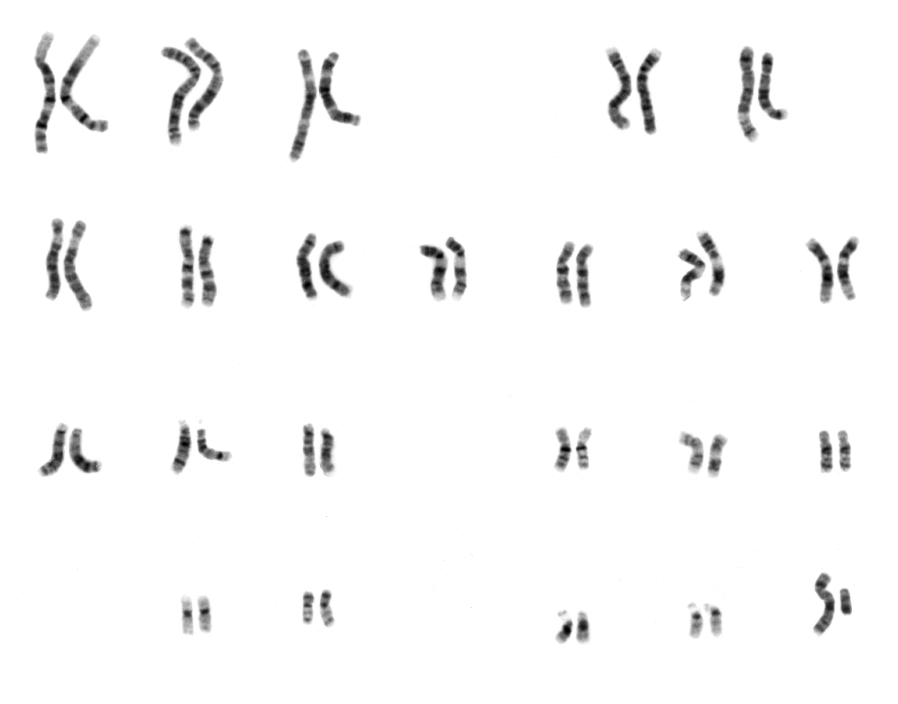
A karyotype is the general appearance of the complete set of chromosomes in the cells of a species or in an individual organism, mainly including their sizes, numbers, and shapes. Karyotyping is the process by which a karyotype is discerned by determining the chromosome complement of an individual, including the number of chromosomes and any abnormalities. A karyogram or idiogram is a graphical depiction of a karyotype, wherein chromosomes are generally organized in pairs, ordered by size and position of centromere for chromosomes of the same size. Karyotyping generally combines light microscopy and photography in the metaphase of the cell cycle, and results in a photomicrographic (or simply micrographic) karyogram. In contrast, a schematic karyogram is a designed graphic representation of a karyotype. In schematic karyograms, just one of the sister chromatids of each chromosome is generally shown for brevity, and in reality they are generally so close together that they look as ... [...More Info...] [...Related Items...] OR: [Wikipedia] [Google] [Baidu] |
Chromosome Abnormality
A chromosomal abnormality, chromosomal anomaly, chromosomal aberration, chromosomal mutation, or chromosomal disorder is a missing, extra, or irregular portion of chromosomal DNA. These can occur in the form of numerical abnormalities, where there is an atypical number of chromosomes, or as structural abnormalities, where one or more individual chromosomes are altered. Chromosome mutation was formerly used in a strict sense to mean a change in a chromosomal segment, involving more than one gene. Chromosome anomalies usually occur when there is an error in cell division following meiosis or mitosis. Chromosome abnormalities may be detected or confirmed by comparing an individual's karyotype, or full set of chromosomes, to a typical karyotype for the species via genetic testing. Sometimes chromosomal abnormalities arise in the early stages of an embryo, sperm, or infant. They can be caused by various environmental factors. The implications of chromosomal abnormalities depend on the ... [...More Info...] [...Related Items...] OR: [Wikipedia] [Google] [Baidu] |
Oncogene
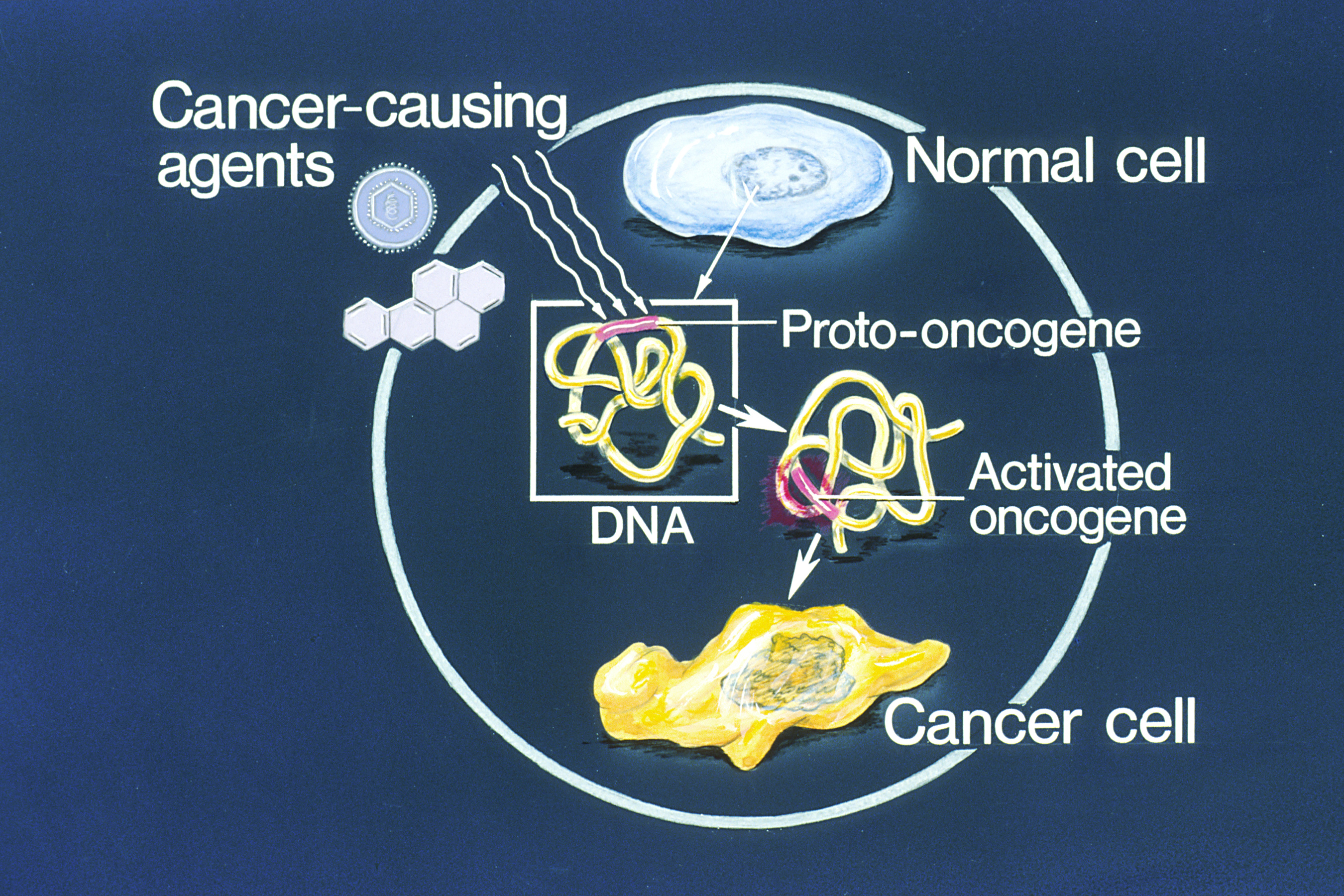
An oncogene is a gene that has the potential to cause cancer. In tumor cells, these genes are often mutated, or expressed at high levels.Kimball's Biology Pages. "Oncogenes" Free full text Most normal cells undergo a preprogrammed rapid cell death () if critical functions are altered and then malfunction. Activated oncogenes can cause those cells designated for apoptosis to survive and proliferate instead. Most oncogenes began as proto-oncogenes: normal genes involved in cell growth and proliferation or inhibition of apoptosis. If, through mutation, normal genes promoting cellular growth are up-regulated (gain-of-function mutation), they predispose the cel ... [...More Info...] [...Related Items...] OR: [Wikipedia] [Google] [Baidu] |
Chromosomes
A chromosome is a package of DNA containing part or all of the genetic material of an organism. In most chromosomes, the very long thin DNA fibers are coated with nucleosome-forming packaging proteins; in eukaryotic cells, the most important of these proteins are the histones. Aided by chaperone proteins, the histones bind to and condense the DNA molecule to maintain its integrity. These eukaryotic chromosomes display a complex three-dimensional structure that has a significant role in transcriptional regulation. Normally, chromosomes are visible under a light microscope only during the metaphase of cell division, where all chromosomes are aligned in the center of the cell in their condensed form. Before this stage occurs, each chromosome is duplicated ( S phase), and the two copies are joined by a centromere—resulting in either an X-shaped structure if the centromere is located equatorially, or a two-armed structure if the centromere is located distally; the joined ... [...More Info...] [...Related Items...] OR: [Wikipedia] [Google] [Baidu] |
Cytogenetics
Cytogenetics is essentially a branch of genetics, but is also a part of cell biology/cytology (a subdivision of human anatomy), that is concerned with how the chromosomes relate to cell behaviour, particularly to their behaviour during mitosis and meiosis. Techniques used include Karyotype, karyotyping, analysis of G banding, G-banded chromosomes, other cytogenetic banding techniques, as well as molecular cytogenetics such as Fluorescence in situ hybridization, fluorescence ''in situ'' hybridization (FISH) and comparative genomic hybridization (CGH). History Beginnings Chromosomes were first observed in plant cells by Carl Nägeli in 1842. Their behavior in animal (salamander) cells was described by Walther Flemming, the discoverer of mitosis, in 1882. The name was coined by another German anatomist, Heinrich Wilhelm Gottfried von Waldeyer-Hartz, von Waldeyer in 1888. The next stage took place after the development of genetics in the early 20th century, when it was appreciated ... [...More Info...] [...Related Items...] OR: [Wikipedia] [Google] [Baidu] |
Genetic Counseling
Genetic counseling is the process of investigating individuals and families affected by or at risk of genetic disorders to help them understand and adapt to the medical, psychological and familial implications of genetic contributions to disease. This field is considered necessary for the implementation of genomic medicine. The process integrates: * Interpretation of family and medical histories to assess the chance of disease occurrence or recurrence * Education about inheritance, testing, management, prevention, resources * Counseling to promote informed choices, adaptation to the risk or condition and support in reaching out to relatives that are also at risk History The practice of advising people about inherited traits began around the turn of the 20th century, shortly after William Bateson suggested that the new medical and biological study of heredity be called "genetics". Heredity became intertwined with social reforms when the field of modern eugenics took form. Although ... [...More Info...] [...Related Items...] OR: [Wikipedia] [Google] [Baidu] |
Chromosome
A chromosome is a package of DNA containing part or all of the genetic material of an organism. In most chromosomes, the very long thin DNA fibers are coated with nucleosome-forming packaging proteins; in eukaryotic cells, the most important of these proteins are the histones. Aided by chaperone proteins, the histones bind to and condense the DNA molecule to maintain its integrity. These eukaryotic chromosomes display a complex three-dimensional structure that has a significant role in transcriptional regulation. Normally, chromosomes are visible under a light microscope only during the metaphase of cell division, where all chromosomes are aligned in the center of the cell in their condensed form. Before this stage occurs, each chromosome is duplicated ( S phase), and the two copies are joined by a centromere—resulting in either an X-shaped structure if the centromere is located equatorially, or a two-armed structure if the centromere is located distally; the jo ... [...More Info...] [...Related Items...] OR: [Wikipedia] [Google] [Baidu] |
Philadelphia Chromosome
The Philadelphia chromosome or Philadelphia translocation (Ph) is an abnormal version of chromosome 22 where a part of the ''ABL (gene), Abelson murine leukemia'' 1 (''ABL1'') gene on chromosome 9 breaks off and attaches to the ''BCR (gene), breakpoint cluster region'' (''BCR'') gene in chromosome 22. The balanced reciprocal Translocation (genetics), translocation between the long arms of 9 and 22 chromosomes [t (9; 22) (q34; q11)] results in the fusion gene ''BCR::ABL1''. The Oncogene, oncogenic protein with persistently enhanced tyrosine kinase (TK) activity transcribed by the ''BCR''::''ABL1'' fusion gene can lead to rapid, uncontrolled growth of immature white blood cells that accumulates in the blood and bone marrow. The Philadelphia chromosome is present in the bone marrow cells of a vast majority ''chronic myelogenous leukemia'' (CML) patients. The expression patterns off different BCR-ABL1 transcripts vary during the progression of CML. Each variant is present in a distinct ... [...More Info...] [...Related Items...] OR: [Wikipedia] [Google] [Baidu] |


