|
Terminologia Anatomica
''Terminologia Anatomica'' (commonly abbreviated TA) is the international standard for human anatomy, human anatomical terminology. It is developed by the Federative International Programme on Anatomical Terminology (FIPAT) a program of the International Federation of Associations of Anatomists (IFAA). History The sixth edition of the previous standard, ''Nomina Anatomica'', was released in 1989. The first edition of ''Terminologia Anatomica'', superseding Nomina Anatomica, was developed by the Federative Committee on Anatomical Terminology (FCAT) and the International Federation of Associations of Anatomists (IFAA) and released in 1998. In April 2011, this edition was published online by the Federative International Programme on Anatomical Terminologies (FIPAT), the successor of FCAT. The first edition contained 7635 Latin items. The second edition was released online by FIPAT in 2019 and approved and adopted by the IFAA General Assembly in 2020. The latest errata is dated Au ... [...More Info...] [...Related Items...] OR: [Wikipedia] [Google] [Baidu] |
Rembrandt - The Anatomy Lesson Of Dr Nicolaes Tulp
Rembrandt Harmenszoon van Rijn (; ; 15 July 1606 – 4 October 1669), mononymously known as Rembrandt was a Dutch Golden Age painter, printmaker, and draughtsman. He is generally considered one of the greatest visual artists in the history of Western art.Gombrich, p. 420. It is estimated that Rembrandt's surviving works amount to about three hundred paintings, three hundred etchings and several hundred drawings. Unlike most Dutch painters of the 17th century, Rembrandt's works depict a wide range of styles and subject matter, from portraits and self-portraits to landscapes, genre scenes, allegorical and historical scenes, biblical and mythological subjects and animal studies. His contributions to art came in a period that historians call the Dutch Golden Age. Rembrandt never went abroad but was considerably influenced by the work of the Italian Old Masters and Dutch and Flemish artists who had studied in Italy. After he achieved youthful success as a portrait ... [...More Info...] [...Related Items...] OR: [Wikipedia] [Google] [Baidu] |
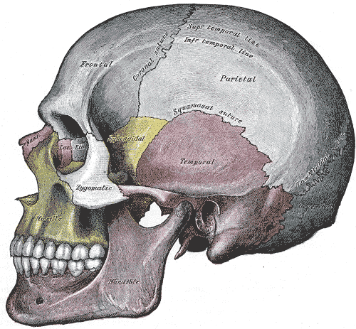
Human Cranium
The skull, or cranium, is typically a bony enclosure around the brain of a vertebrate. In some fish, and amphibians, the skull is of cartilage. The skull is at the head end of the vertebrate. In the human, the skull comprises two prominent parts: the neurocranium and the facial skeleton, which evolved from the first pharyngeal arch. The skull forms the frontmost portion of the axial skeleton and is a product of cephalization and vesicular enlargement of the brain, with several special senses structures such as the eyes, ears, nose, tongue and, in fish, specialized tactile organs such as barbels near the mouth. The skull is composed of three types of bone: cranial bones, facial bones and ossicles, which is made up of a number of fused flat and irregular bones. The cranial bones are joined at firm fibrous junctions called sutures and contains many foramina, fossae, processes, and sinuses. In zoology, the openings in the skull are called fenestrae, the most p ... [...More Info...] [...Related Items...] OR: [Wikipedia] [Google] [Baidu] |
Vertebral Joint
The spinal column, also known as the vertebral column, spine or backbone, is the core part of the axial skeleton in vertebrates. The vertebral column is the defining and eponymous characteristic of the vertebrate. The spinal column is a segmented column of vertebrae that surrounds and protects the spinal cord. The vertebrae are separated by intervertebral discs in a series of cartilaginous joints. The dorsal portion of the spinal column houses the spinal canal, an elongated cavity formed by the alignment of the vertebral neural arches that encloses and protects the spinal cord, with spinal nerves exiting via the intervertebral foramina to innervate each body segment. There are around 50,000 species of animals that have a vertebral column. The human spine is one of the most-studied examples, as the general structure of human vertebrae is fairly typical of that found in other mammals, reptiles, and birds. The shape of the vertebral body does, however, vary somewhat between diffe ... [...More Info...] [...Related Items...] OR: [Wikipedia] [Google] [Baidu] |
Suture (joint)
In anatomy, fibrous joints are joints connected by fibrous tissue, consisting mainly of collagen. These are fixed joints where bones are united by a layer of white fibrous tissue of varying thickness. In the skull, the joints between the bones are called sutures. Such immovable joints are also referred to as synarthroses. Types Most fibrous joints are also called "fixed" or "immovable". These joints have no joint cavity and are connected via fibrous connective tissue. * Sutures: The skull bones are connected by fibrous joints called '' sutures''. In fetal skulls, the sutures are wide to allow slight movement during birth. They later become rigid ( synarthrodial). * Syndesmosis: Some of the long bones in the body such as the radius and ulna in the forearm are joined by a '' syndesmosis'' (along the interosseous membrane). Syndemoses are slightly moveable ( amphiarthrodial). The distal tibiofibular joint is another example. * A '' gomphosis'' is a joint between the roo ... [...More Info...] [...Related Items...] OR: [Wikipedia] [Google] [Baidu] |
Joint
A joint or articulation (or articular surface) is the connection made between bones, ossicles, or other hard structures in the body which link an animal's skeletal system into a functional whole.Saladin, Ken. Anatomy & Physiology. 7th ed. McGraw-Hill Connect. Webp.274/ref> They are constructed to allow for different degrees and types of movement. Some joints, such as the knee, elbow, and shoulder, are self-lubricating, almost frictionless, and are able to withstand compression and maintain heavy loads while still executing smooth and precise movements. Other joints such as suture (joint), sutures between the bones of the skull permit very little movement (only during birth) in order to protect the brain and the sense organs. The connection between a tooth and the jawbone is also called a joint, and is described as a fibrous joint known as a gomphosis. Joints are classified both structurally and functionally. Joints play a vital role in the human body, contributing to movement, sta ... [...More Info...] [...Related Items...] OR: [Wikipedia] [Google] [Baidu] |
Lower Limb
Lower may refer to: *Lower (album), ''Lower'' (album), 2025 album by Benjamin Booker *Lower (surname) *Lower Township, New Jersey *Lower Receiver (firearms) *Lower Wick Gloucestershire, England See also *Nizhny {{Disambiguation ... [...More Info...] [...Related Items...] OR: [Wikipedia] [Google] [Baidu] |
Pelvis
The pelvis (: pelves or pelvises) is the lower part of an Anatomy, anatomical Trunk (anatomy), trunk, between the human abdomen, abdomen and the thighs (sometimes also called pelvic region), together with its embedded skeleton (sometimes also called bony pelvis or pelvic skeleton). The pelvic region of the trunk includes the bony pelvis, the pelvic cavity (the space enclosed by the bony pelvis), the pelvic floor, below the pelvic cavity, and the perineum, below the pelvic floor. The pelvic skeleton is formed in the area of the back, by the sacrum and the coccyx and anteriorly and to the left and right sides, by a pair of hip bones. The two hip bones connect the spine with the lower limbs. They are attached to the sacrum posteriorly, connected to each other anteriorly, and joined with the two femurs at the hip joints. The gap enclosed by the bony pelvis, called the pelvic cavity, is the section of the body underneath the abdomen and mainly consists of the reproductive organs and ... [...More Info...] [...Related Items...] OR: [Wikipedia] [Google] [Baidu] |
Upper Limb
The upper Limb (anatomy), limbs or upper extremities are the forelimbs of an upright posture, upright-postured tetrapod vertebrate, extending from the scapulae and clavicles down to and including the digit (anatomy), digits, including all the musculatures and ligaments involved with the shoulder, elbow, wrist and knuckle joints. In humans, each upper limb is divided into the shoulder, arm, elbow, forearm, wrist and hand, and is primarily used for climbing, manual handling of loads, lifting and dexterity, manipulating objects. In anatomy, just as arm refers to the upper arm, leg refers to the lower leg. Definition In formal usage, the term "arm" only refers to the structures from the shoulder to the elbow, explicitly excluding the forearm, and thus "upper limb" and "arm" are not synonymous. However, in casual usage, the terms are often used interchangeably. The term "upper arm" is redundant in anatomy, but in informal usage is used to distinguish between the two terms. Structure I ... [...More Info...] [...Related Items...] OR: [Wikipedia] [Google] [Baidu] |
Thorax
The thorax (: thoraces or thoraxes) or chest is a part of the anatomy of mammals and other tetrapod animals located between the neck and the abdomen. In insects, crustaceans, and the extinct trilobites, the thorax is one of the three main divisions of the body, each in turn composed of multiple segments. The human thorax includes the thoracic cavity and the thoracic wall. It contains organs including the heart, lungs, and thymus gland, as well as muscles and various other internal structures. The chest may be affected by many diseases, of which the most common symptom is chest pain. Etymology The word thorax comes from the Greek θώραξ ''thṓrax'' " breastplate, cuirass, corslet" via . Humans Structure In humans and other hominids, the thorax is the chest region of the body between the neck and the abdomen, along with its internal organs and other contents. It is mostly protected and supported by the rib cage, spine, and shoulder girdle. Contents The ... [...More Info...] [...Related Items...] OR: [Wikipedia] [Google] [Baidu] |
Vertebral Column
The spinal column, also known as the vertebral column, spine or backbone, is the core part of the axial skeleton in vertebrates. The vertebral column is the defining and eponymous characteristic of the vertebrate. The spinal column is a segmented column of vertebrae that surrounds and protects the spinal cord. The vertebrae are separated by intervertebral discs in a series of cartilaginous joints. The dorsal portion of the spinal column houses the spinal canal, an elongated body cavity, cavity formed by the alignment of the vertebral neural arches that encloses and protects the spinal cord, with spinal nerves exiting via the intervertebral foramina to innervate each body segment. There are around 50,000 species of animals that have a vertebral column. The human spine is one of the most-studied examples, as the general structure of human vertebrae is fairly homology (biology), typical of that found in other mammals, reptiles, and birds. The shape of the vertebral body does, howev ... [...More Info...] [...Related Items...] OR: [Wikipedia] [Google] [Baidu] |
Laryngeal Cartilages
Laryngeal cartilages are cartilages which surround and protect the larynx. They form during embryonic development from pharyngeal arches. There are a total of nine laryngeal skeleton in humans: * Thyroid cartilage - unpaired * Cricoid cartilage - unpaired * Epiglottis - unpaired * Arytenoid cartilages - paired * Corniculate cartilages The corniculate cartilages (cartilages of Santorini) are two small conical nodules in the larynx, consisting of elastic cartilage, which articulate with the summits of the arytenoid cartilages and serve to prolong them posteriorly and medially. T ... - paired * Cuneiform cartilages - paired Skeletal system ... [...More Info...] [...Related Items...] OR: [Wikipedia] [Google] [Baidu] |
Auricle (anatomy)
The auricle or auricula is the visible part of the ear that is outside the head. It is also called the pinna (Latin for 'wing' or ' fin', : pinnae), a term that is used more in zoology. Structure The diagram shows the shape and location of most of these components: * ''antihelix'' forms a 'Y' shape where the upper parts are: ** ''Superior crus'' (to the left of the ''fossa triangularis'' in the diagram) ** ''Inferior crus'' (to the right of the ''fossa triangularis'' in the diagram) * ''Antitragus'' is below the ''tragus'' * ''Aperture'' is the entrance to the ear canal * ''Auricular sulcus'' is the depression behind the ear next to the head * ''Concha'' is the hollow next to the ear canal * Conchal angle is the angle that the back of the ''concha'' makes with the side of the head * ''Crus'' of the helix is just above the ''tragus'' * ''Cymba conchae'' is the narrowest end of the ''concha'' * External auditory meatus is the ear canal * ''Fossa triangularis'' is the depression ... [...More Info...] [...Related Items...] OR: [Wikipedia] [Google] [Baidu] |


