|
Synovial Sarcoma
A synovial sarcoma (also known as malignant synovioma) is a rare form of cancer which occurs primarily in the extremities of the arms or legs, often in proximity to joint capsules and tendon sheaths. It is a type of soft-tissue sarcoma. The name "synovial sarcoma" was coined early in the 20th century, as some researchers thought that the microscopic similarity of some tumors to synovium, and its propensity to arise adjacent to joints, indicated a synovial origin; however, the actual cells from which the tumor develops are unknown and not necessarily synovial. Primary synovial sarcomas are most common in the soft tissue near the large joints of the arm and leg but have been documented in most human tissues and organs, including the brain, prostate, and heart. Synovial sarcoma occurs in about 1–2 per 1,000,000 people a year. They occur most commonly in the third decade of life, with males being affected more often than females (ratio around 1.2:1). Signs and symptoms Synovial ... [...More Info...] [...Related Items...] OR: [Wikipedia] [Google] [Baidu] |
Micrograph
A micrograph or photomicrograph is a photograph or digital image taken through a microscope or similar device to show a magnified image of an object. This is opposed to a macrograph or photomacrograph, an image which is also taken on a microscope but is only slightly magnified, usually less than 10 times. Micrography is the practice or art of using microscopes to make photographs. A micrograph contains extensive details of microstructure. A wealth of information can be obtained from a simple micrograph like behavior of the material under different conditions, the phases found in the system, failure analysis, grain size estimation, elemental analysis and so on. Micrographs are widely used in all fields of microscopy. Types Photomicrograph A light micrograph or photomicrograph is a micrograph prepared using an optical microscope, a process referred to as ''photomicroscopy''. At a basic level, photomicroscopy may be performed simply by connecting a camera to a microscope ... [...More Info...] [...Related Items...] OR: [Wikipedia] [Google] [Baidu] |
Epithelium
Epithelium or epithelial tissue is one of the four basic types of animal tissue, along with connective tissue, muscle tissue and nervous tissue. It is a thin, continuous, protective layer of compactly packed cells with a little intercellular matrix. Epithelial tissues line the outer surfaces of organs and blood vessels throughout the body, as well as the inner surfaces of cavities in many internal organs. An example is the epidermis, the outermost layer of the skin. There are three principal shapes of epithelial cell: squamous (scaly), columnar, and cuboidal. These can be arranged in a singular layer of cells as simple epithelium, either squamous, columnar, or cuboidal, or in layers of two or more cells deep as stratified (layered), or ''compound'', either squamous, columnar or cuboidal. In some tissues, a layer of columnar cells may appear to be stratified due to the placement of the nuclei. This sort of tissue is called pseudostratified. All glands are made up of epit ... [...More Info...] [...Related Items...] OR: [Wikipedia] [Google] [Baidu] |
Chromosomal Translocation
In genetics, chromosome translocation is a phenomenon that results in unusual rearrangement of chromosomes. This includes balanced and unbalanced translocation, with two main types: reciprocal-, and Robertsonian translocation. Reciprocal translocation is a chromosome abnormality caused by exchange of parts between non-homologous chromosomes. Two detached fragments of two different chromosomes are switched. Robertsonian translocation occurs when two non-homologous chromosomes get attached, meaning that given two healthy pairs of chromosomes, one of each pair "sticks" and blends together homogeneously. A gene fusion may be created when the translocation joins two otherwise-separated genes. It is detected on cytogenetics or a karyotype of affected cells. Translocations can be balanced (in an even exchange of material with no genetic information extra or missing, and ideally full functionality) or unbalanced (where the exchange of chromosome material is unequal resulting in extr ... [...More Info...] [...Related Items...] OR: [Wikipedia] [Google] [Baidu] |
CD99
CD99 antigen (Cluster of differentiation 99), also known as MIC2 or single-chain type-1 glycoprotein, is a heavily O-glycosylated transmembrane protein that is encoded by the ''CD99'' gene in humans. The protein has a mass of 32 kD. Unusually for a gene present on the X chromosome, the CD99 gene does not undergo X inactivation, and it was the first such pseudoautosomal gene to be discovered in humans. Expression It is expressed on all leukocytes but highest on thymocytes and is believed to augment T-cell adhesion and apoptosis of double positive T cells. It has been found in endothelial cells and in the periodontium, including gingival fibroblasts and gingival epithelial cells. It also participates in migration and activation. There is also experimental evidence that it binds to cyclophilin A. It is found on the cell surface of Ewing's sarcoma tumors and is positive in granulosa cell tumors. It is more expressed in malignant gliomas than in the brain, and such overexpression ... [...More Info...] [...Related Items...] OR: [Wikipedia] [Google] [Baidu] |
Bcl-2
Bcl-2 (B-cell lymphoma 2), encoded in humans by the ''BCL2'' gene, is the founding member of the Bcl-2 family of regulator proteins that regulate cell death (apoptosis), by either inhibiting (anti-apoptotic) or inducing (pro-apoptotic) apoptosis. It was the first apoptosis regulator identified in any organism. Bcl-2 derives its name from ''B-cell lymphoma 2'', as it is the second member of a range of proteins initially described in chromosomal translocations involving chromosomes 14 and 18 in follicular lymphomas. Orthologs (such as ''Bcl2'' in mice) have been identified in numerous mammals for which complete genome data are available. Like BCL3, BCL5, BCL6, BCL7A, BCL9, and BCL10, it has clinical significance in lymphoma. Isoforms The two isoforms of Bcl-2, Isoform 1, and Isoform 2, exhibit a similar fold. However, results in the ability of these isoforms to bind to the BAD and BAK proteins, as well as in the structural topology and electrostatic potential of the bind ... [...More Info...] [...Related Items...] OR: [Wikipedia] [Google] [Baidu] |
TLE1
Transducin-like enhancer protein 1 is a protein that in humans is encoded by the ''TLE1'' gene. Interactions TLE1 has been shown to interact Advocates for Informed Choice, doing business as, dba interACT or interACT Advocates for Intersex Youth, is a 501(c)(3) nonprofit organization using innovative strategies to advocate for the legal and human rights of children with intersex trai ... with: * Glycoprotein 130, * HES6, * RUNX1, * RUNX3, * SIX3 * TLE2, and * UTY. References Further reading * * * * * * * * * * * * * * * * * {{PDB Gallery, geneid=7088 ... [...More Info...] [...Related Items...] OR: [Wikipedia] [Google] [Baidu] |
Cytokeratin
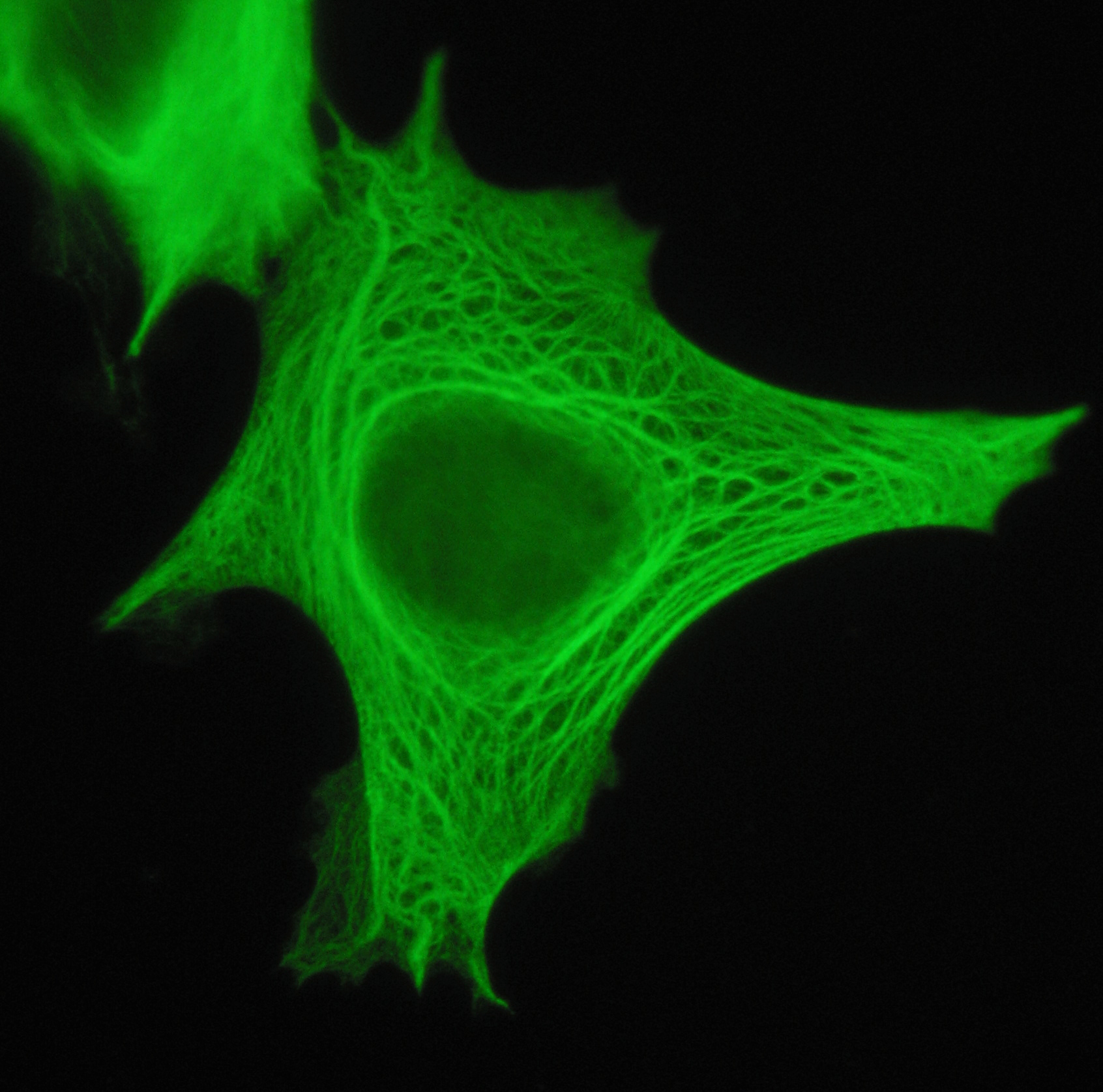
Cytokeratins are keratin proteins found in the intracytoplasmic cytoskeleton of epithelial tissue. They are an important component of intermediate filaments, which help cells resist mechanical stress. Expression of these cytokeratins within epithelial cells is largely specific to particular organs or tissues. Thus they are used clinically to identify the cell of origin of various human tumors. Naming The term ''cytokeratin'' began to be used in the late 1970s, when the protein subunits of keratin intermediate filaments inside cells were first being identified and characterized. In 2006 a new systematic nomenclature for mammalian keratins was created, and the proteins previously called ''cytokeratins'' are simply called ''keratins'' (human epithelial category). For example, cytokeratin-4 (CK-4) has been renamed keratin-4 (K4). However, they are still commonly referred to as cytokeratins in clinical practice. Types There are two categories of cytokeratins: the acidic type I cy ... [...More Info...] [...Related Items...] OR: [Wikipedia] [Google] [Baidu] |
The American Journal Of Surgical Pathology
''The American Journal of Surgical Pathology'' is a peer-reviewed medical journal covering surgical pathology. It was established in 1977. Its first editor-in-chief was Stephen Sternberg ( Memorial Sloan-Kettering Cancer Center); the current editor-in-chief is Stacey Mills (University of Virginia). According to the ''Journal Citation Reports'', the journal has a 2018 impact factor The impact factor (IF) or journal impact factor (JIF) of an academic journal is a scientometric index calculated by Clarivate that reflects the yearly mean number of citations of articles published in the last two years in a given journal, as ... of 6.155. References External links * Publications established in 1977 English-language journals Surgery journals Lippincott Williams & Wilkins academic journals Monthly journals {{surgery-journal-stub ... [...More Info...] [...Related Items...] OR: [Wikipedia] [Google] [Baidu] |
Histopathology (journal)
''Histopathology'' is a monthly peer-reviewed medical journal covering diagnostic, research, and surgical pathology. It was established in 1977 and is published by Wiley-Blackwell. The editor-in-chief An editor-in-chief (EIC), also known as lead editor or chief editor, is a publication's editorial leader who has final responsibility for its operations and policies. The highest-ranking editor of a publication may also be titled editor, managing ... is Daniel Berney. Publications established in 1977 Wiley-Blackwell academic journals Quarterly journals Pathology journals English-language journals Histopathology {{Med-journal-stub ... [...More Info...] [...Related Items...] OR: [Wikipedia] [Google] [Baidu] |
Mitotic Index
Mitotic index is defined as the ratio between the number of a population's cells undergoing mitosis to its total number of cells. Purpose The mitotic index is a measure of cellular proliferation. It is defined as the percentage of cells undergoing mitosis in a given population of cells. Mitosis is the division of somatic cells into two daughter cells. Durations of the cell cycle and mitosis vary in different cell types. An elevated mitotic index indicates more cells are dividing. In cancer cells, the mitotic index may be elevated compared to normal growth of tissues or cellular repair of the site of an injury. The mitotic index is therefore an important prognostic factor predicting both overall survival and response to chemotherapy in most types of cancer. It may lose much of its predictive value for elderly populations. For example, a low mitotic index loses any prognostic value for women over 70 years old with breast cancer. Calculation The mitotic index is the number of c ... [...More Info...] [...Related Items...] OR: [Wikipedia] [Google] [Baidu] |
National Cancer Institute
The National Cancer Institute (NCI) coordinates the United States National Cancer Program and is part of the National Institutes of Health (NIH), which is one of eleven agencies that are part of the U.S. Department of Health and Human Services. The NCI conducts and supports research, training, health information dissemination, and other activities related to the causes, prevention, diagnosis, and treatment of cancer; the supportive care of cancer patients and their families; and cancer survivorship. NCI is the oldest and has the largest budget and research program of the 27 institutes and centers of the NIH ($6.9 billion in 2020). It fulfills the majority of its mission via an extramural program that provides grants for cancer research. Additionally, the National Cancer Institute has intramural research programs in Bethesda, Maryland, and at the Frederick National Laboratory for Cancer Research at Fort Detrick in Frederick, Maryland. The NCI receives more than in funding ea ... [...More Info...] [...Related Items...] OR: [Wikipedia] [Google] [Baidu] |
Archives Of Pathology & Laboratory Medicine
The ''Archives of Pathology & Laboratory Medicine'' (Arch Pathol Lab Med) is a core clinical medical journal published by the College of American Pathologists and the American Medical Association The American Medical Association (AMA) is a professional association and lobbying group of physicians and medical students. Founded in 1847, it is headquartered in Chicago, Illinois. Membership was approximately 240,000 in 2016. The AMA's sta .... It continues in series publications entitled the Archives of Pathology and Laboratory Medicine (1926–28), the Archives of Pathology (1928–50), the A.M.A. Archives of Pathology (1950–60), and the Archives of Pathology (1960–75). References External links * Laboratory medicine journals Pathology journals Publications established in 1976 Monthly journals Academic journals published by learned and professional societies of the United States English-language journals Allen Press academic journals {{med-journal-stub ... [...More Info...] [...Related Items...] OR: [Wikipedia] [Google] [Baidu] |


