|
Schneiderian Membrane
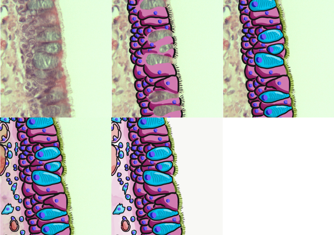
In anatomy, the Schneiderian membrane is the membranous lining of the maxillary sinus cavity. Microscopically there is a bilaminar membrane with pseudostratified ciliated columnar epithelial cells on the internal (or cavernous) side and periosteum The periosteum is a membrane that covers the outer surface of all bones, except at the articular surfaces (i.e. the parts within a joint space) of long bones. Endosteum lines the inner surface of the medullary cavity of all long bones. Structu ... on the Osseous tissue, osseous side. The size of the sinuses varies in different Skull, skulls, and even on the two sides of the same skull. This membrane is present in the nasal chamber which helps to smell (olfactory receptor) References {{reflist Human head and neck ... [...More Info...] [...Related Items...] OR: [Wikipedia] [Google] [Baidu] |
Lateral Window Sinus Lift
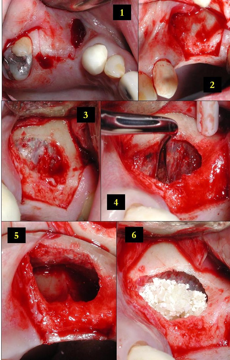
Lateral is a geometric term of location which may refer to: Healthcare *Lateral (anatomy), an anatomical direction *Lateral cricoarytenoid muscle *Lateral release (surgery), a surgical procedure on the side of a kneecap Phonetics *Lateral consonant, an l-like consonant in which air flows along the sides of the tongue **Lateral release (phonetics), the release of a plosive consonant into a lateral consonant Other uses *''Lateral'', journal of the Cultural Studies Association *Lateral canal, a canal built beside another stream *Lateral hiring, recruiting that targets employees of another organization *Lateral mark, a sea mark used in maritime pilotage to indicate the edge of a channel * Lateral stability of aircraft during flight *Lateral pass, a type of pass in American and Canadian football *Lateral support (other), various meanings *Lateral thinking, the solution of problems through an indirect and creative approach *Lateral number, a proposed alternate term for imagi ... [...More Info...] [...Related Items...] OR: [Wikipedia] [Google] [Baidu] |
Anatomy
Anatomy () is the branch of biology concerned with the study of the structure of organisms and their parts. Anatomy is a branch of natural science that deals with the structural organization of living things. It is an old science, having its beginnings in prehistoric times. Anatomy is inherently tied to developmental biology, embryology, comparative anatomy, evolutionary biology, and phylogeny, as these are the processes by which anatomy is generated, both over immediate and long-term timescales. Anatomy and physiology, which study the structure and function of organisms and their parts respectively, make a natural pair of related disciplines, and are often studied together. Human anatomy is one of the essential basic sciences that are applied in medicine. The discipline of anatomy is divided into macroscopic and microscopic. Macroscopic anatomy, or gross anatomy, is the examination of an animal's body parts using unaided eyesight. Gross anatomy also includes the br ... [...More Info...] [...Related Items...] OR: [Wikipedia] [Google] [Baidu] |
Mucous Membrane
A mucous membrane or mucosa is a membrane that lines various cavities in the body of an organism and covers the surface of internal organs. It consists of one or more layers of epithelial cells overlying a layer of loose connective tissue. It is mostly of endodermal origin and is continuous with the skin at body openings such as the eyes, eyelids, ears, inside the nose, inside the mouth, lips, the genital areas, the urethral opening and the anus. Some mucous membranes secrete mucus, a thick protective fluid. The function of the membrane is to stop pathogens and dirt from entering the body and to prevent bodily tissues from becoming dehydrated. Structure The mucosa is composed of one or more layers of epithelial cells that secrete mucus, and an underlying lamina propria of loose connective tissue. The type of cells and type of mucus secreted vary from organ to organ and each can differ along a given tract. Mucous membranes line the digestive, respiratory and reproductive ... [...More Info...] [...Related Items...] OR: [Wikipedia] [Google] [Baidu] |
Maxillary Sinus
The pyramid-shaped maxillary sinus (or antrum of Highmore) is the largest of the paranasal sinuses, and drains into the middle meatus of the nose through the osteomeatal complex.Human Anatomy, Jacobs, Elsevier, 2008, page 209-210 Structure It is the largest air sinus in the body. Found in the body of the maxilla, this sinus has three recesses: an alveolar recess pointed inferiorly, bounded by the alveolar process of the maxilla; a zygomatic recess pointed laterally, bounded by the zygomatic bone; and an infraorbital recess pointed superiorly, bounded by the inferior orbital surface of the maxilla. The medial wall is composed primarily of cartilage. The ostia for drainage are located high on the medial wall and open into the semilunar hiatus of the lateral nasal cavity; because of the position of the ostia, gravity cannot drain the maxillary sinus contents when the head is erect (see pathology). The ostium of the maxillary sinus is high up on the medial wall and on average is ... [...More Info...] [...Related Items...] OR: [Wikipedia] [Google] [Baidu] |
Pseudostratified Columnar Epithelium
A pseudostratified epithelium is a type of epithelium that, though comprising only a single layer of cells, has its cell nuclei positioned in a manner suggestive of stratified epithelia. As it rarely occurs as squamous or cuboidal epithelia, it is usually considered synonymous with the term pseudostratified columnar epithelium. The term ''pseudostratified'' is derived from the appearance of this epithelium in the section which conveys the erroneous (''pseudo'' means almost or approaching) impression that there is more than one layer of cells, when in fact this is a true simple epithelium since all the cells rest on the basement membrane. The nuclei of these cells, however, are disposed at different levels, thus creating the illusion of cellular stratification. All cells are not of equal size and not all cells extend to the luminal/apical surface; such cells are capable of cell division providing replacements for cells lost or damaged. Pseudostratified epithelia function in ... [...More Info...] [...Related Items...] OR: [Wikipedia] [Google] [Baidu] |
Periosteum
The periosteum is a membrane that covers the outer surface of all bones, except at the articular surfaces (i.e. the parts within a joint space) of long bones. Endosteum lines the inner surface of the medullary cavity of all long bones. Structure The periosteum consists of an outer fibrous layer, and an inner cambium layer (or osteogenic layer). The fibrous layer is of dense irregular connective tissue, containing fibroblasts, while the cambium layer is highly cellular containing progenitor cells that develop into osteoblasts. These osteoblasts are responsible for increasing the width of a long bone and the overall size of the other bone types. After a bone fracture, the progenitor cells develop into osteoblasts and chondroblasts, which are essential to the healing process. The outer fibrous layer and the inner cambium layer is differentiated under electron micrography. As opposed to osseous tissue, the periosteum has nociceptors, sensory neurons that make it very sensitiv ... [...More Info...] [...Related Items...] OR: [Wikipedia] [Google] [Baidu] |
Osseous Tissue
A bone is a rigid organ that constitutes part of the skeleton in most vertebrate animals. Bones protect the various other organs of the body, produce red and white blood cells, store minerals, provide structure and support for the body, and enable mobility. Bones come in a variety of shapes and sizes and have complex internal and external structures. They are lightweight yet strong and hard and serve multiple functions. Bone tissue (osseous tissue), which is also called bone in the uncountable sense of that word, is hard tissue, a type of specialized connective tissue. It has a honeycomb-like matrix internally, which helps to give the bone rigidity. Bone tissue is made up of different types of bone cells. Osteoblasts and osteocytes are involved in the formation and mineralization of bone; osteoclasts are involved in the resorption of bone tissue. Modified (flattened) osteoblasts become the lining cells that form a protective layer on the bone surface. The minera ... [...More Info...] [...Related Items...] OR: [Wikipedia] [Google] [Baidu] |
Skull
The skull is a bone protective cavity for the brain. The skull is composed of four types of bone i.e., cranial bones, facial bones, ear ossicles and hyoid bone. However two parts are more prominent: the cranium and the mandible. In humans, these two parts are the neurocranium and the viscerocranium ( facial skeleton) that includes the mandible as its largest bone. The skull forms the anterior-most portion of the skeleton and is a product of cephalisation—housing the brain, and several sensory structures such as the eyes, ears, nose, and mouth. In humans these sensory structures are part of the facial skeleton. Functions of the skull include protection of the brain, fixing the distance between the eyes to allow stereoscopic vision, and fixing the position of the ears to enable sound localisation of the direction and distance of sounds. In some animals, such as horned ungulates (mammals with hooves), the skull also has a defensive function by providing the mount (on the ... [...More Info...] [...Related Items...] OR: [Wikipedia] [Google] [Baidu] |


