|
Spinal Needle
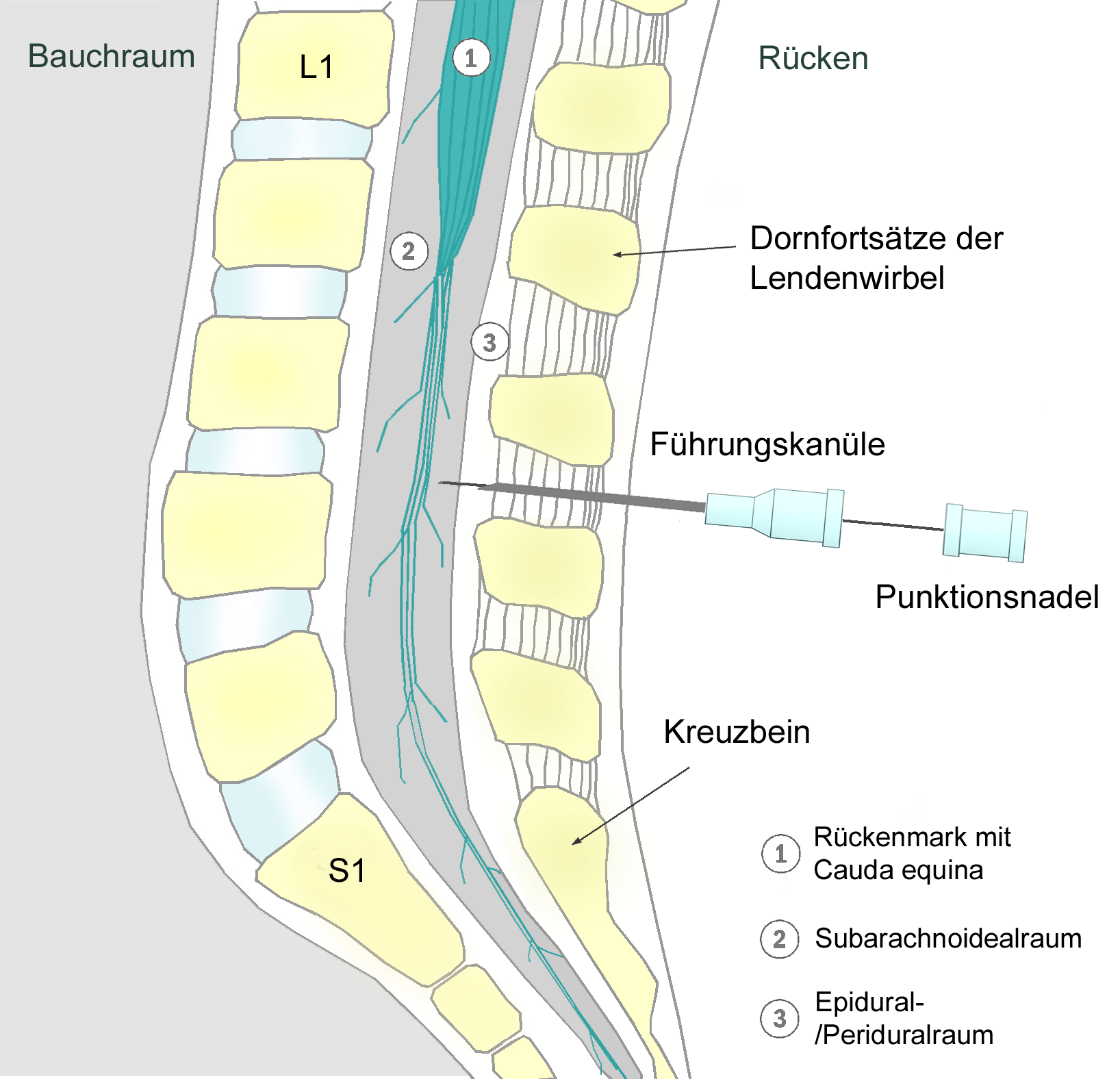
Spinal anaesthesia (or spinal anesthesia), also called spinal block, subarachnoid block, intradural block and intrathecal block, is a form of neuraxial regional anaesthesia involving the injection of a local anaesthetic with or without an opioid into the subarachnoid space. Usually a single-shot dose is administrered through a fine needle, alternatively continuous spinal anaesthesia through a intrathecal catheter can be performed. It is a safe and effective form of anesthesia usually performed by anesthesiologists and CRNAs that can be used as an alternative to general anesthesia commonly in surgeries involving the lower extremities and surgeries below the umbilicus. The local anesthetic with or without an opioid injected into the cerebrospinal fluid provides locoregional anaesthesia: true anaesthesia, motor, sensory and autonomic (sympathetic) blockade. Administering analgesics (opioid, alpha2-adrenoreceptor agonist) in the cerebrospinal fluid without a local anaesthetic prod ... [...More Info...] [...Related Items...] OR: [Wikipedia] [Google] [Baidu] |
Cerebrospinal Fluid
Cerebrospinal fluid (CSF) is a clear, colorless Extracellular fluid#Transcellular fluid, transcellular body fluid found within the meninges, meningeal tissue that surrounds the vertebrate brain and spinal cord, and in the ventricular system, ventricles of the brain. CSF is mostly produced by specialized Ependyma, ependymal cells in the choroid plexuses of the ventricles of the brain, and absorbed in the arachnoid granulations. It is also produced by ependymal cells in the lining of the ventricles. In humans, there is about 125 mL of CSF at any one time, and about 500 mL is generated every day. CSF acts as a shock absorber, cushion or buffer, providing basic mechanical and immune system, immunological protection to the brain inside the Human skull, skull. CSF also serves a vital function in the cerebral autoregulation of cerebral blood flow. CSF occupies the subarachnoid space (between the arachnoid mater and the pia mater) and the ventricular system around and inside t ... [...More Info...] [...Related Items...] OR: [Wikipedia] [Google] [Baidu] |
Sedation
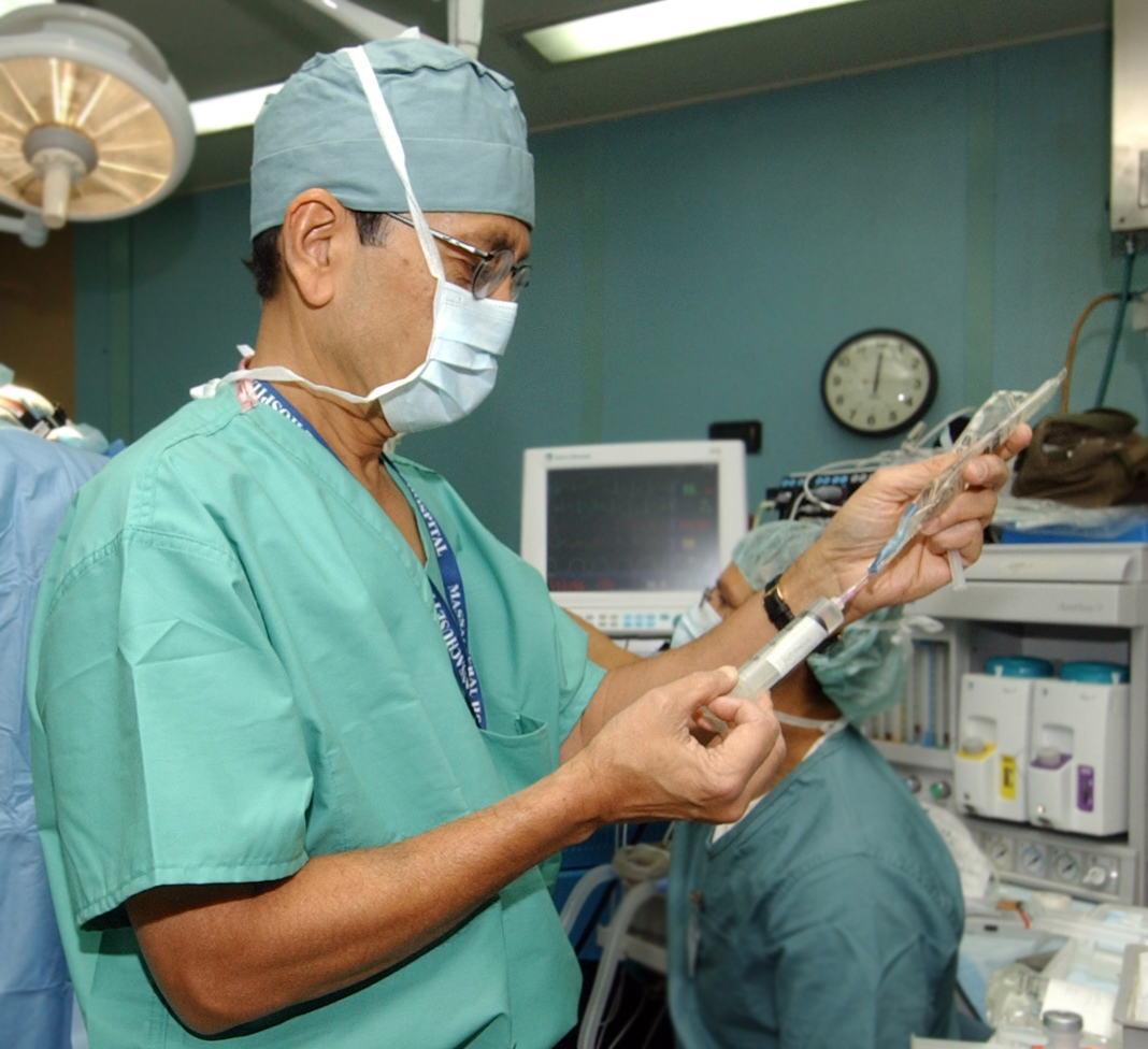
Sedation is the reduction of irritability or agitation by administration of sedative drugs, generally to facilitate a medical procedure or diagnostic procedure. Examples of drugs which can be used for sedation include isoflurane, diethyl ether, propofol, etomidate, ketamine, pentobarbital, lorazepam and midazolam. Medical uses Sedation is typically used in minor surgical procedures such as endoscopy, vasectomy, or dentistry and for reconstructive surgery, some cosmetic surgeries, removal of wisdom teeth, or for high-anxiety patients. Sedation methods in dentistry include inhalation sedation (using nitrous oxide), oral sedation, and intravenous (IV) sedation. Inhalation sedation is also sometimes referred to as "relative analgesia". Sedation is also used extensively in the intensive care unit so that patients who are being mechanical ventilation, ventilated tolerate having an endotracheal tube in their vertebrate trachea, trachea. It can also be used during a long term brain EEG ... [...More Info...] [...Related Items...] OR: [Wikipedia] [Google] [Baidu] |
Hernias
A hernia (: hernias or herniae, from Latin, meaning 'rupture') is the abnormal exit of tissue or an organ, such as the bowel, through the wall of the cavity in which it normally resides. The term is also used for the normal development of the intestinal tract, referring to the retraction of the intestine from the extra-embryonal navel coelom into the abdomen in the healthy embryo at about 7 weeks. Various types of hernias can occur, most commonly involving the abdomen, and specifically the groin. Groin hernias are most commonly inguinal hernias but may also be femoral hernias. Other types of hernias include hiatus, incisional, and umbilical hernias. Symptoms are present in about 66% of people with groin hernias. This may include pain or discomfort in the lower abdomen, especially with coughing, exercise, or urinating or defecating. Often, it gets worse throughout the day and improves when lying down. A bulge may appear at the site of hernia, that becomes larger when bend ... [...More Info...] [...Related Items...] OR: [Wikipedia] [Google] [Baidu] |
Endovascular Aneurysm Repair
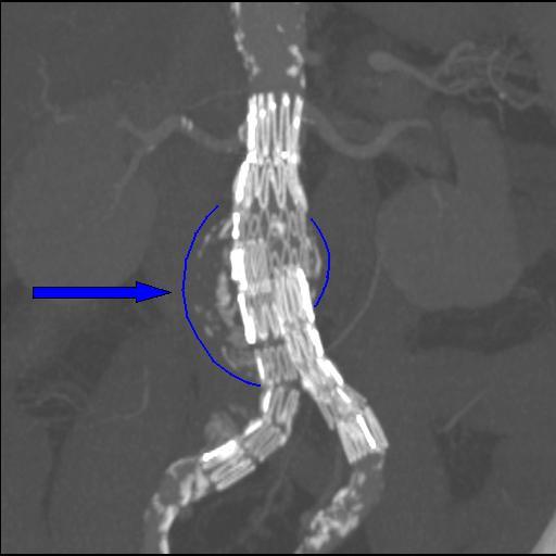
Endovascular aneurysm repair (EVAR) is a type of minimally-invasive endovascular surgery used to treat pathology of the aorta, most commonly an abdominal aortic aneurysm (AAA). When used to treat thoracic aortic disease, the procedure is then specifically termed TEVAR for "thoracic endovascular aortic/aneurysm repair." EVAR involves the placement of an expandable stent graft within the aorta to treat aortic disease without operating directly on the aorta. In 2003, EVAR surpassed open aortic surgery as the most common technique for repair of AAA, and in 2010, EVAR accounted for 78% of all intact AAA repair in the United States. Medical uses Standard EVAR is appropriate for aneurysms that begin below the renal arteries, where there exists an adequate length of normal aorta (the ''"proximal aortic neck"'') for reliable attachment of the endograft without leakage of blood around the device ("'' endoleak''"). If the proximal aortic neck is also involved with the aneurysm, the pati ... [...More Info...] [...Related Items...] OR: [Wikipedia] [Google] [Baidu] |
Human Leg
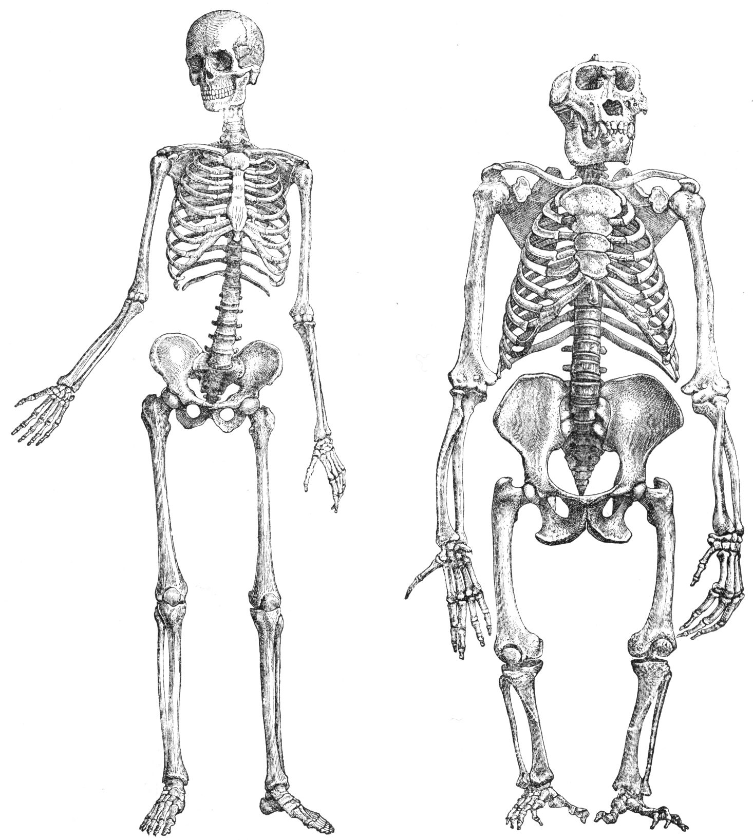
The leg is the entire lower limb (anatomy), limb of the human body, including the foot, thigh or sometimes even the hip or Gluteal muscles, buttock region. The major bones of the leg are the femur (thigh bone), tibia (shin bone), and adjacent fibula. There are 30 bones in each leg. The thigh is located in between the hip and knee. The calf (leg), calf (rear) and Tibia#Structure, shin (front), or shank, are located between the knee and ankle. Legs are used for standing, many forms of human movement, recreation such as dancing, and constitute a significant portion of a person's mass. Evolution has led to the human leg's development into a mechanism specifically adapted for efficient bipedalism, bipedal gait. While the capacity to walk upright is not unique to humans, other primates can only achieve this for short periods and at a great expenditure of energy. In humans, female legs generally have greater hip anteversion and tibiofemoral angles, while male legs have longer femur a ... [...More Info...] [...Related Items...] OR: [Wikipedia] [Google] [Baidu] |
Vascular Surgery
Vascular surgery is a surgical subspecialty in which vascular diseases involving the arteries, veins, or lymphatic vessels, are managed by medical therapy, minimally-invasive catheter procedures and surgical reconstruction. The specialty evolved from general surgery, general and cardiac surgery, cardiovascular surgery where it refined the management of just the vessels, no longer treating the heart or other organs. Modern vascular surgery includes open surgery techniques, endovascular (minimally invasive) techniques and medical management of vascular diseases - unlike the parent specialities. The vascular surgeon is trained in the diagnosis and management of diseases affecting all parts of the vascular system excluding the coronaries and intracranial vasculature. Vascular surgeons also are called to assist other physicians to carry out surgery near vessels, or to salvage vascular injuries that include hemorrhage control, dissection, occlusion or simply for safe exposure of vascul ... [...More Info...] [...Related Items...] OR: [Wikipedia] [Google] [Baidu] |
Joint Replacement
Joint replacement is a procedure of orthopedic surgery known also as arthroplasty, in which an arthritic or dysfunctional joint surface is replaced with an orthopedic prosthesis. Joint replacement is considered as a treatment when severe joint pain or dysfunction is not alleviated by less-invasive therapies. Joint replacement surgery is often indicated from various Arthropathy, joint diseases, including osteoarthritis and rheumatoid arthritis. Joint replacement has become more common, mostly with knee replacement, knee and hip replacements. About 773,000 Americans had a hip or knee replaced in 2009.Joint Replacement Surgery and You. (April, 2009) In ''Arthritis, Musculoskeletal and Skin Disease online''. Retrieved from http://www.niams.nih.gov/#. Uses Shoulder For shoulder replacement, there are a few major approaches to access the shoulder joint. The first is the deltopectoral approach, which saves the deltoid, but requires the supraspinatus to be cut. The second is the transde ... [...More Info...] [...Related Items...] OR: [Wikipedia] [Google] [Baidu] |
Arthroplasty
Arthroplasty (literally " e-orming of joint") is an orthopedic surgical procedure where the articular surface of a musculoskeletal joint is replaced, remodeled, or realigned by osteotomy or some other procedure. It is an elective procedure that is done to relieve pain and restore function to the joint after damage by arthritis or some other type of trauma. Types For the last 45 years, the most successful and common form of arthroplasty is the surgical replacement of arthritic or destructive or necrotic joint or joint surface with a prosthesis. For example, a hip joint that is affected by osteoarthritis may be replaced entirely ( total hip arthroplasty) with a prosthetic hip. This would involve replacing both the acetabulum (hip socket) and the head and neck of the femur. The purpose of this procedure is to relieve pain, to restore range of motion and to improve walking ability, thus leading to the improvement of muscle strength. Other types of arthroplasty * Interposit ... [...More Info...] [...Related Items...] OR: [Wikipedia] [Google] [Baidu] |
Ankle
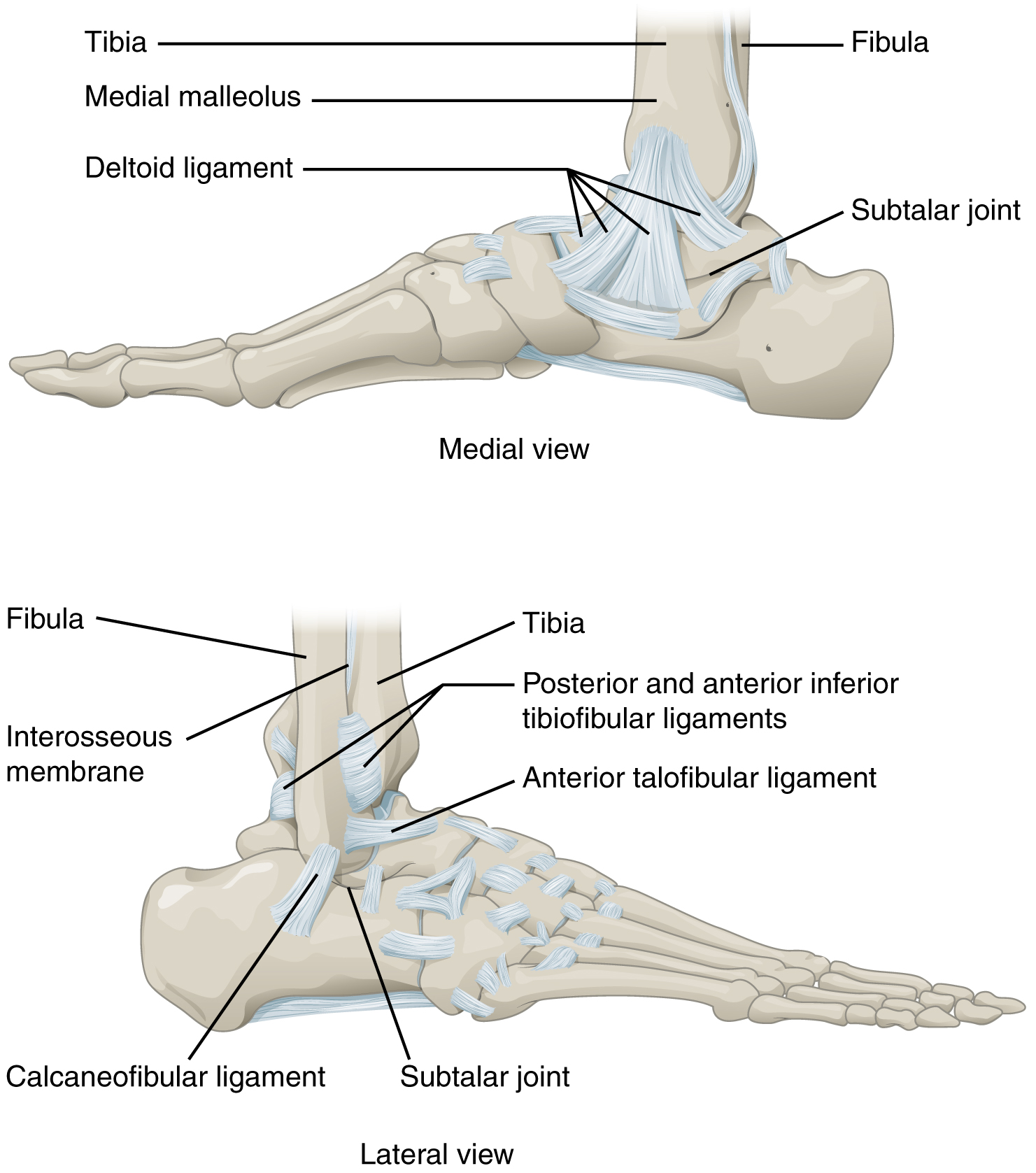
The ankle, the talocrural region or the jumping bone (informal) is the area where the foot and the leg meet. The ankle includes three joints: the ankle joint proper or talocrural joint, the subtalar joint, and the inferior tibiofibular joint. The movements produced at this joint are dorsiflexion and plantarflexion of the foot. In common usage, the term ankle refers exclusively to the ankle region. In medical terminology, "ankle" (without qualifiers) can refer broadly to the region or specifically to the talocrural joint. The main bones of the ankle region are the talus bone, talus (in the foot), the tibia, and fibula (both in the leg). The talocrural joint is a Synovial joint, synovial hinge joint that connects the distal ends of the tibia and fibula in the lower limb with the proximal end of the talus. The articulation between the tibia and the talus bears more weight than that between the smaller fibula and the talus. Structure Region The ankle region is found at the junction ... [...More Info...] [...Related Items...] OR: [Wikipedia] [Google] [Baidu] |
Tibia
The tibia (; : tibiae or tibias), also known as the shinbone or shankbone, is the larger, stronger, and anterior (frontal) of the two Leg bones, bones in the leg below the knee in vertebrates (the other being the fibula, behind and to the outside of the tibia); it connects the knee with the ankle bones, ankle. The tibia is found on the anatomical terms of location#Medial, medial side of the leg next to the fibula and closer to the median plane. The tibia is connected to the fibula by the interosseous membrane of leg, forming a type of fibrous joint called a syndesmosis with very little movement. The tibia is named for the flute ''aulos, tibia''. It is the second largest bone in the human body, after the femur. The leg bones are the strongest long bones as they support the rest of the body. Structure In human anatomy, the tibia is the second largest bone next to the femur. As in other vertebrates the tibia is one of two bones in the lower leg, the other being the fibula, and is a ... [...More Info...] [...Related Items...] OR: [Wikipedia] [Google] [Baidu] |
Knee
In humans and other primates, the knee joins the thigh with the leg and consists of two joints: one between the femur and tibia (tibiofemoral joint), and one between the femur and patella (patellofemoral joint). It is the largest joint in the human body. The knee is a modified hinge joint, which permits flexion and extension (kinesiology), extension as well as slight internal and external rotation. The knee is vulnerable to injury and to the development of osteoarthritis. It is often termed a ''compound joint'' having tibiofemoral and patellofemoral components. (The fibular collateral ligament is often considered with tibiofemoral components.) Structure The knee is a modified hinge joint, a type of synovial joint, which is composed of three functional compartments: the patellofemoral articulation, consisting of the patella, or "kneecap", and the patellar groove on the front of the femur through which it slides; and the medial and lateral tibiofemoral articulations linking the ... [...More Info...] [...Related Items...] OR: [Wikipedia] [Google] [Baidu] |
Femur
The femur (; : femurs or femora ), or thigh bone is the only long bone, bone in the thigh — the region of the lower limb between the hip and the knee. In many quadrupeds, four-legged animals the femur is the upper bone of the hindleg. The Femoral head, top of the femur fits into a socket in the pelvis called the hip joint, and the bottom of the femur connects to the shinbone (tibia) and kneecap (patella) to form the knee. In humans the femur is the largest and thickest bone in the body. Structure The femur is the only bone in the upper Human leg, leg. The two femurs converge Anatomical terms of location, medially toward the knees, where they articulate with the Anatomical terms of location, proximal ends of the tibiae. The angle at which the femora converge is an important factor in determining the femoral-tibial angle. In females, thicker pelvic bones cause the femora to converge more than in males. In the condition genu valgum, ''genu valgum'' (knock knee), the femurs conve ... [...More Info...] [...Related Items...] OR: [Wikipedia] [Google] [Baidu] |
