|
Rectus Capitis Lateralis Muscle
The rectus capitis lateralis, a short, flat muscle, arises from the upper surface of the transverse process of the atlas, and is inserted into the under surface of the jugular process of the occipital bone. Additional images File:Rectus capitis lateralis muscle - animation01.gif, Position of rectus capitis lateralis muscle (shown in red). Animation. File:Rectus capitis lateralis muscle - animation05.gif, Close up. Skull has been removed (except occipital bone). File:Rectus capitis lateralis muscle03.png, Lateral view. Still image. File:Gray129.png, Occipital bone. Outer surface. File:Gray187.png, Base of skull. Inferior surface. See also * Atlanto-occipital joint * Rectus capitis posterior major muscle * Rectus capitis posterior minor muscle * Rectus capitis anterior muscle The rectus capitis anterior (rectus capitis anticus minor) is a short, flat muscle, situated immediately behind the upper part of the Longus capitis. It arises from the anterior surface of the lateral ... [...More Info...] [...Related Items...] OR: [Wikipedia] [Google] [Baidu] |
Transverse Process
The spinal column, a defining synapomorphy shared by nearly all vertebrates,Hagfish are believed to have secondarily lost their spinal column is a moderately flexible series of vertebrae (singular vertebra), each constituting a characteristic irregular bone whose complex structure is composed primarily of bone, and secondarily of hyaline cartilage. They show variation in the proportion contributed by these two tissue types; such variations correlate on one hand with the cerebral/caudal rank (i.e., location within the vertebral column, backbone), and on the other with phylogenetic differences among the vertebrate taxon, taxa. The basic configuration of a vertebra varies, but the bone is its ''body'', with the central part of the body constituting the ''centrum''. The upper (closer to) and lower (further from), respectively, the cranium and its central nervous system surfaces of the vertebra body support attachment to the intervertebral discs. The posterior part of a vertebra fo ... [...More Info...] [...Related Items...] OR: [Wikipedia] [Google] [Baidu] |
Atlas (anatomy)
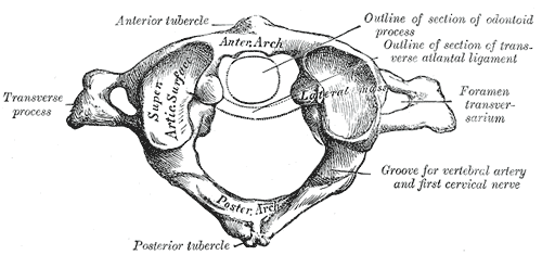
In anatomy, the atlas (C1) is the most superior (first) cervical vertebra of the spine and is located in the neck. It is named for Atlas of Greek mythology because, just as Atlas supported the globe, it supports the entire head. The atlas is the topmost vertebra and, with the axis (the vertebra below it), forms the joint connecting the skull and spine. The atlas and axis are specialized to allow a greater range of motion than normal vertebrae. They are responsible for the nodding and rotation movements of the head. The atlanto-occipital joint allows the head to nod up and down on the vertebral column. The dens acts as a pivot that allows the atlas and attached head to rotate on the axis, side to side. The atlas's chief peculiarity is that it has no body. It is ring-like and consists of an anterior and a posterior arch and two lateral masses. The atlas and axis are important neurologically because the brainstem extends down to the axis. Structure Anterior arch The anterio ... [...More Info...] [...Related Items...] OR: [Wikipedia] [Google] [Baidu] |
Jugular Process
The jugular process is a quadrilateral or triangular bony plate projecting lateralward from the posterior half of the occipital condyle; it is a part of the lateral part of the occipital bone. The jugular process is excavated in front by the jugular notch of occipital bone (which forms the posterior part of the jugular foramen). The posterolateral side of the jugular formanen is divided from the anteromedial side by the intrajugular process of occipital bone. The jugular process serves as the insertion of the rectus capitis lateralis The rectus capitis lateralis, a short, flat muscle, arises from the upper surface of the transverse process of the atlas, and is inserted into the under surface of the jugular process of the occipital bone. Additional images File:Rectus capitis .... References External links * Bones of the head and neck {{musculoskeletal-stub ... [...More Info...] [...Related Items...] OR: [Wikipedia] [Google] [Baidu] |
Occipital Bone
The occipital bone () is a neurocranium, cranial dermal bone and the main bone of the occiput (back and lower part of the skull). It is trapezoidal in shape and curved on itself like a shallow dish. The occipital bone overlies the occipital lobes of the cerebrum. At the base of skull in the occipital bone, there is a large oval opening called the foramen magnum, which allows the passage of the spinal cord. Like the other cranial bones, it is classed as a flat bone. Due to its many attachments and features, the occipital bone is described in terms of separate parts. From its front to the back is the basilar part of occipital bone, basilar part, also called the basioccipital, at the sides of the foramen magnum are the lateral parts of occipital bone, lateral parts, also called the exoccipitals, and the back is named as the squamous part of occipital bone, squamous part. The basilar part is a thick, somewhat quadrilateral piece in front of the foramen magnum and directed towards the ... [...More Info...] [...Related Items...] OR: [Wikipedia] [Google] [Baidu] |
Cervical Spinal Nerve 1
The cervical spinal nerve 1 (C1) is a spinal nerve of the cervical segment. from spinalcordinjuryzone.com. Published February 23, 2004 Archived Dec 23, 2011. Retrieved June 12, 2018. C1 carries predominantly motor fibres, but also a small meningeal branch that supplies sensation to parts of the dura around the foramen magnum (via dorsal rami). It originates from the spinal column from above the (C1). The dorsal root and gang ... [...More Info...] [...Related Items...] OR: [Wikipedia] [Google] [Baidu] |
Cervical Spinal Nerve 2
The cervical spinal nerve 2 (C2) is a spinal nerve of the cervical segment. Nervous System -- Groups of Nerves It is a part of the along with the C1 and C3 nerves sometimes forming part of Superior root of the ansa cervicalis. it also connects into the with the C3. It originates from the s ... [...More Info...] [...Related Items...] OR: [Wikipedia] [Google] [Baidu] |
Atlanto-occipital Joint
The atlanto-occipital joint (''Capsula articularis atlantooccipitalis'') is an articulation between the atlas bone and the occipital bone. It consists of a pair of condyloid joints. It is a synovial joint. Structure The atlanto-occipital joint is an articulation between the atlas bone and the occipital bone. It consists of a pair of condyloid joints. It is a synovial joint. Ligaments The ligaments connecting the bones are: * Two articular capsules * Posterior atlanto-occipital membrane * Anterior atlanto-occipital membrane Capsule The capsules of the atlantooccipital articulation surround the condyles of the occipital bone, and connect them with the articular processes of the atlas: they are thin and loose. Function The movements permitted in this joint are: * (a) flexion and extension around the mediolateral axis, which give rise to the ordinary forward and backward nodding of the head. * (b) slight lateral motion, lateroflexion, to one or other side around the anteroposter ... [...More Info...] [...Related Items...] OR: [Wikipedia] [Google] [Baidu] |
Human Skull
The skull is a bone protective cavity for the brain. The skull is composed of four types of bone i.e., cranial bones, facial bones, ear ossicles and hyoid bone. However two parts are more prominent: the cranium and the mandible. In humans, these two parts are the neurocranium and the viscerocranium ( facial skeleton) that includes the mandible as its largest bone. The skull forms the anterior-most portion of the skeleton and is a product of cephalisation—housing the brain, and several sensory structures such as the eyes, ears, nose, and mouth. In humans these sensory structures are part of the facial skeleton. Functions of the skull include protection of the brain, fixing the distance between the eyes to allow stereoscopic vision, and fixing the position of the ears to enable sound localisation of the direction and distance of sounds. In some animals, such as horned ungulates (mammals with hooves), the skull also has a defensive function by providing the mount (on the front ... [...More Info...] [...Related Items...] OR: [Wikipedia] [Google] [Baidu] |
Atlanto-occipital Joint
The atlanto-occipital joint (''Capsula articularis atlantooccipitalis'') is an articulation between the atlas bone and the occipital bone. It consists of a pair of condyloid joints. It is a synovial joint. Structure The atlanto-occipital joint is an articulation between the atlas bone and the occipital bone. It consists of a pair of condyloid joints. It is a synovial joint. Ligaments The ligaments connecting the bones are: * Two articular capsules * Posterior atlanto-occipital membrane * Anterior atlanto-occipital membrane Capsule The capsules of the atlantooccipital articulation surround the condyles of the occipital bone, and connect them with the articular processes of the atlas: they are thin and loose. Function The movements permitted in this joint are: * (a) flexion and extension around the mediolateral axis, which give rise to the ordinary forward and backward nodding of the head. * (b) slight lateral motion, lateroflexion, to one or other side around the anteroposter ... [...More Info...] [...Related Items...] OR: [Wikipedia] [Google] [Baidu] |
Rectus Capitis Posterior Major Muscle
The rectus capitis posterior major (or rectus capitis posticus major, both being Latin for ''larger posterior straight muscle of the head'') arises by a pointed tendon from the spinous process of the axis, and, becoming broader as it ascends, is inserted into the lateral part of the inferior nuchal line of the occipital bone and the surface of the bone immediately below the line. A soft tissue connection bridging from the rectus capitis posterior major to the cervical dura mater was described in 2011. Various clinical manifestations may be linked to this anatomical relationship. It has also been postulated that this connection serves as a monitor of dural tension along with the rectus capitis posterior minor and the obliquus capitis inferior. As the muscles of the two sides pass upward and lateralward, they leave between them a triangular space, in which the rectus capitis posterior minor is seen. Its main actions are to extend and rotate the atlanto-occipital joint. See also * ... [...More Info...] [...Related Items...] OR: [Wikipedia] [Google] [Baidu] |
Rectus Capitis Posterior Minor Muscle
The rectus capitis posterior minor (or rectus capitis posticus minor, both being Latin for ''lesser posterior straight muscle of the head'') arises by a narrow pointed tendon from the tubercle on the posterior arch of the atlas, and, widening as it ascends, is inserted into the medial part of the inferior nuchal line of the occipital bone and the surface between it and the foramen magnum, and also takes some attachment to the spinal dura mater. The synergists are the rectus capitis posterior major and the obliquus capitis superior. Connective tissue bridges were noted at the atlanto-occipital joint between the rectus capitis posterior minor (RCPm) muscle and the dorsal spinal dura. Similar connective tissue connections of the rectus capitis posterior major have been reported recently as well. The perpendicular arrangement of these fibers appears to restrict dural movement toward the spinal cord. The ligamentum nuchae was found to be continuous with the posterior cervical spinal ... [...More Info...] [...Related Items...] OR: [Wikipedia] [Google] [Baidu] |
Rectus Capitis Anterior Muscle
The rectus capitis anterior (rectus capitis anticus minor) is a short, flat muscle, situated immediately behind the upper part of the Longus capitis. It arises from the anterior surface of the lateral mass of the atlas, and from the root of its transverse process, and passing obliquely upward and medialward, is inserted into the inferior surface of the basilar part of the occipital bone immediately in front of the foramen magnum. action: aids in flexion of the head and the neck; nerve supply: C1, C2. Additional images File:Rectus capitis anterior muscle - animation01.gif, Animation. Position of rectus capitis anterior muscle. Some bones around the muscle are shown in semi-transparent. File:Rectus capitis anterior muscle - animation02.gif, Skull has been removed (except for occipital bone The occipital bone () is a neurocranium, cranial dermal bone and the main bone of the occiput (back and lower part of the skull). It is trapezoidal in shape and curved on itself like a sh ... [...More Info...] [...Related Items...] OR: [Wikipedia] [Google] [Baidu] |


