|
Radial Recurrent Artery
The radial recurrent artery arises from the radial artery immediately below the elbow. It ascends between the branches of the radial nerve, lying on the supinator muscle and then between the brachioradialis muscle and the brachialis muscle, supplying these muscles and the elbow-joint, and anastomosing with the terminal part of the profunda brachii The deep artery of arm (also known as arteria profunda brachii and the deep brachial artery) is a large vessel which arises from the lateral and posterior part of the brachial artery, just below the lower border of the teres major. Structure It .... Additional images File:Slide1MMMM.JPG, Radial recurrent artery File:Slide4PPPP.JPG, Radial recurrent artery File:Slide9PPPP.JPG, Radial recurrent artery References External links * * Arteries of the upper limb {{circulatory-stub ... [...More Info...] [...Related Items...] OR: [Wikipedia] [Google] [Baidu] |
Anastomosis
An anastomosis (, plural anastomoses) is a connection or opening between two things (especially cavities or passages) that are normally diverging or branching, such as between blood vessels, leaf veins, or streams. Such a connection may be normal (such as the foramen ovale in a fetus's heart) or abnormal (such as the patent foramen ovale in an adult's heart); it may be acquired (such as an arteriovenous fistula) or innate (such as the arteriovenous shunt of a metarteriole); and it may be natural (such as the aforementioned examples) or artificial (such as a surgical anastomosis). The reestablishment of an anastomosis that had become blocked is called a reanastomosis. Anastomoses that are abnormal, whether congenital or acquired, are often called fistulas. The term is used in medicine, biology, mycology, geology, and geography. Etymology Anastomosis: medical or Modern Latin, from Greek ἀναστόμωσις, anastomosis, "outlet, opening", Gr ana- "up, on, upon", stoma " ... [...More Info...] [...Related Items...] OR: [Wikipedia] [Google] [Baidu] |
Elbow-joint
The elbow is the region between the arm and the forearm that surrounds the elbow joint. The elbow includes prominent landmarks such as the olecranon, the cubital fossa (also called the chelidon, or the elbow pit), and the lateral and the medial epicondyles of the humerus. The elbow joint is a hinge joint between the arm and the forearm; more specifically between the humerus in the upper arm and the radius and ulna in the forearm which allows the forearm and hand to be moved towards and away from the body. The term ''elbow'' is specifically used for humans and other primates, and in other vertebrates forelimb plus joint is used. The name for the elbow in Latin is ''cubitus'', and so the word cubital is used in some elbow-related terms, as in ''cubital nodes'' for example. Structure Joint The elbow joint has three different portions surrounded by a common joint capsule. These are joints between the three bones of the elbow, the humerus of the upper arm, and the radius a ... [...More Info...] [...Related Items...] OR: [Wikipedia] [Google] [Baidu] |
Radial Artery
In human anatomy, the radial artery is the main artery of the lateral aspect of the forearm. Structure The radial artery arises from the bifurcation of the brachial artery in the antecubital fossa. It runs distally on the anterior part of the forearm. There, it serves as a landmark for the division between the anterior and posterior compartments of the forearm, with the posterior compartment beginning just lateral to the artery. The artery winds laterally around the wrist, passing through the anatomical snuff box and between the heads of the first dorsal interosseous muscle. It passes anteriorly between the heads of the adductor pollicis, and becomes the deep palmar arch, which joins with the deep branch of the ulnar artery. Along its course, it is accompanied by a similarly named vein, the radial vein. Branches The named branches of the radial artery may be divided into three groups, corresponding with the three regions in which the vessel is situated. In the fore ... [...More Info...] [...Related Items...] OR: [Wikipedia] [Google] [Baidu] |
Elbow-joint
The elbow is the region between the arm and the forearm that surrounds the elbow joint. The elbow includes prominent landmarks such as the olecranon, the cubital fossa (also called the chelidon, or the elbow pit), and the lateral and the medial epicondyles of the humerus. The elbow joint is a hinge joint between the arm and the forearm; more specifically between the humerus in the upper arm and the radius and ulna in the forearm which allows the forearm and hand to be moved towards and away from the body. The term ''elbow'' is specifically used for humans and other primates, and in other vertebrates forelimb plus joint is used. The name for the elbow in Latin is ''cubitus'', and so the word cubital is used in some elbow-related terms, as in ''cubital nodes'' for example. Structure Joint The elbow joint has three different portions surrounded by a common joint capsule. These are joints between the three bones of the elbow, the humerus of the upper arm, and the radius a ... [...More Info...] [...Related Items...] OR: [Wikipedia] [Google] [Baidu] |
Radial Nerve
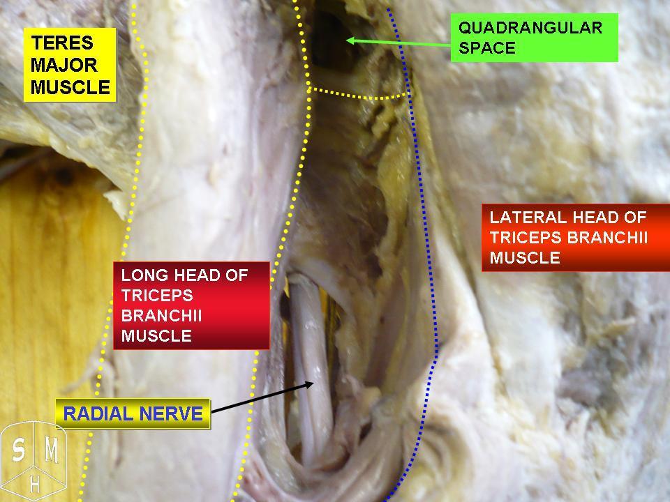
The radial nerve is a nerve in the human body that supplies the posterior portion of the upper limb. It innervates the medial and lateral heads of the triceps brachii muscle of the arm, as well as all 12 muscles in the posterior osteofascial compartment of the forearm and the associated joints and overlying skin. It originates from the brachial plexus, carrying fibers from the ventral roots of spinal nerves C5, C6, C7, C8 & T1. The radial nerve and its branches provide motor innervation to the dorsal arm muscles (the triceps brachii and the anconeus) and the extrinsic extensors of the wrists and hands; it also provides cutaneous sensory innervation to most of the back of the hand, except for the back of the little finger and adjacent half of the ring finger (which are innervated by the ulnar nerve). The radial nerve divides into a deep branch, which becomes the posterior interosseous nerve, and a superficial branch, which goes on to innervate the dorsum (back) of the hand ... [...More Info...] [...Related Items...] OR: [Wikipedia] [Google] [Baidu] |
Supinator Muscle
In human anatomy, the supinator is a broad muscle in the posterior compartment of the forearm, curved around the upper third of the radius. Its function is to supinate the forearm. Structure Supinator consists of two planes of fibers, between which the deep branch of the radial nerve ls. The two planes arise in common — the superficial one by tendinous (the initial portion of the muscle is actually just tendon) and the deeper by muscular fibers —''Gray's Anatomy'' (1918), see infobox from the supinator crest of the ulna, the lateral epicondyle of humerus, the radial collateral ligament, and the annular radial ligament. The superficial fibers (''pars superficialis'') surround the upper part of the radius, and are inserted into the lateral edge of the radial tuberosity and the oblique line of the radius, as low down as the insertion of the pronator teres. The upper fibers (''pars profunda'') of the deeper plane form a sling-like fasciculus, which encircles the ne ... [...More Info...] [...Related Items...] OR: [Wikipedia] [Google] [Baidu] |
Brachioradialis Muscle
The brachioradialis is a muscle of the forearm that flexes the forearm at the elbow. It is also capable of both pronation and supination, depending on the position of the forearm. It is attached to the distal styloid process of the radius by way of the brachioradialis tendon, and to the lateral supracondylar ridge of the humerus. Structure The brachioradialis is a superficial, fusiform muscle on the lateral side of the forearm. It originates proximally on the lateral supracondylar ridge of the humerus. It inserts distally on the radius, at the base of its styloid process. Near the elbow, it forms the lateral limit of the cubital fossa, or elbow pit. Nerve supply Despite the bulk of the muscle body being visible from the anterior aspect of the forearm, the brachioradialis is a posterior compartment muscle and consequently is innervated by the radial nerve. Of the muscles that receive innervation from the radial nerve, it is one of only four that receive input directly fr ... [...More Info...] [...Related Items...] OR: [Wikipedia] [Google] [Baidu] |
Brachialis Muscle
The brachialis (brachialis anticus), also known as the Teichmann muscle, is a muscle in the upper arm that flexes the elbow. It lies deeper than the biceps brachii, and makes up part of the floor of the region known as the cubital fossa (elbow pit). The brachialis is the prime mover of elbow flexion generating about 50% more power than the biceps.Saladin, Kenneth S, Stephen J. Sullivan, and Christina A. Gan. Anatomy & Physiology: The Unity of Form and Function. 2015. Print. Structure The brachialis originates from the anterior surface of the distal half of the humerus, near the insertion of the deltoid muscle, which it embraces by two angular processes. Its origin extends below to within 2.5 cm of the margin of the articular surface of the humerus at the elbow joint. Its fibers converge to a thick tendon, which is inserted into the tuberosity of the ulna and the rough depression on the anterior surface of the coronoid process of the ulna. Blood supply The brachialis is su ... [...More Info...] [...Related Items...] OR: [Wikipedia] [Google] [Baidu] |
Profunda Brachii
The deep artery of arm (also known as arteria profunda brachii and the deep brachial artery) is a large vessel which arises from the lateral and posterior part of the brachial artery, just below the lower border of the teres major. Structure It follows closely the radial nerve, running at first backward between the long and medial heads of the triceps brachii, then along the groove for the radial nerve (the radial sulcus), where it is covered by the lateral head of the triceps brachii, to the lateral side of the arm; there it pierces the lateral intermuscular septum, and, descending between the brachioradialis and the brachialis to the front of the lateral epicondyle of the humerus, ends by anastomosing with the radial recurrent artery. Branches and anastomoses It gives branches to the deltoid muscle (which, however, primarily is supplied by the posterior circumflex humeral artery) and to the muscles between which it lies; it supplies an occasional nutrient artery which enters ... [...More Info...] [...Related Items...] OR: [Wikipedia] [Google] [Baidu] |

