|
Relay Neuron
Interneurons (also called internuncial neurons, association neurons, connector neurons, or intermediate neurons) are neurons that are not specifically motor neurons or sensory neurons. Interneurons are the central nodes of neural circuits, enabling communication between sensory or motor neurons and the central nervous system (CNS). They play vital roles in reflexes, neuronal oscillations, and neurogenesis in the adult mammalian brain. Interneurons can be further broken down into two groups: local interneurons and relay interneurons. Local interneurons have short axons and form circuits with nearby neurons to analyze small pieces of information. Relay interneurons have long axons and connect circuits of neurons in one region of the brain with those in other regions. However, interneurons are generally considered to operate mainly within local brain areas. The interaction between interneurons allows the brain to perform complex functions such as learning and decision-making. Str ... [...More Info...] [...Related Items...] OR: [Wikipedia] [Google] [Baidu] |
Spinal Interneuron
A spinal interneuron, found in the spinal cord, relays signals between (afferent) sensory neurons, and (efferent) motor neurons. Different classes of spinal interneurons are involved in the process of sensory-motor integration. Most interneurons are found in the grey column, a region of grey matter in the spinal cord. Structure The grey column of the spinal cord appears to have groups of small neurons, often referred to as spinal interneurons, that are neither primary sensory cells nor motor neurons. The versatile properties of these spinal interneurons cover a wide range of activities. Their functions include the processing of sensory input, the modulation of motor neuron activity, the coordination of activity at different spinal levels, and the relay of sensory or proprioceptive data to the brain. There has been extensive research on the identification and characterization of the spinal cord interneurons based on factors such as location, size, structure, connectivity, ... [...More Info...] [...Related Items...] OR: [Wikipedia] [Google] [Baidu] |
Cholinergic
Cholinergic agents are compounds which mimic the action of acetylcholine and/or butyrylcholine. In general, the word " choline" describes the various quaternary ammonium salts containing the ''N'',''N'',''N''-trimethylethanolammonium cation. Found in most animal tissues, choline is a primary component of the neurotransmitter acetylcholine and functions with inositol as a basic constituent of lecithin. Choline also prevents fat deposits in the liver and facilitates the movement of fats into cells. The parasympathetic nervous system, which uses acetylcholine almost exclusively to send its messages, is said to be almost entirely cholinergic. Neuromuscular junctions, preganglionic neurons of the sympathetic nervous system, the basal forebrain, and brain stem complexes are also cholinergic, as are the receptor for the merocrine sweat glands. In neuroscience and related fields, the term cholinergic is used in these related contexts: * A substance (or ligand) is cholinergic if ... [...More Info...] [...Related Items...] OR: [Wikipedia] [Google] [Baidu] |
Unipolar Brush Cell
Unipolar brush cells (UBCs) are a class of excitatory glutamatergic interneuron found in the granular layer of the cerebellar cortex and also in the granule cell domain of the cochlear nucleus. Structure The UBC has a round or oval cell body with usually a single short dendrite that ends in a brush-like tuft of short dendrioles (dendrites unique to UBCs). These brush dendrioles form very large synaptic junctions. The dendritic brush and the large endings of the axonal branches are involved in the formation of cerebellar glomeruli. The UBC has one short dendrite where the granule cell has four or five. The brush dendrioles emit numerous, thin evaginations called filopodia, unique to UBCs. The filopodia emanate from all over the neuron, even including the dendritic stem and the cell body in some cells. Although UBC filopodia do not bear synaptic junctions, they are nevertheless involved in cell signaling. Function UBCs are intrinsically firing neurons and considered as a ... [...More Info...] [...Related Items...] OR: [Wikipedia] [Google] [Baidu] |
Lugaro Cell
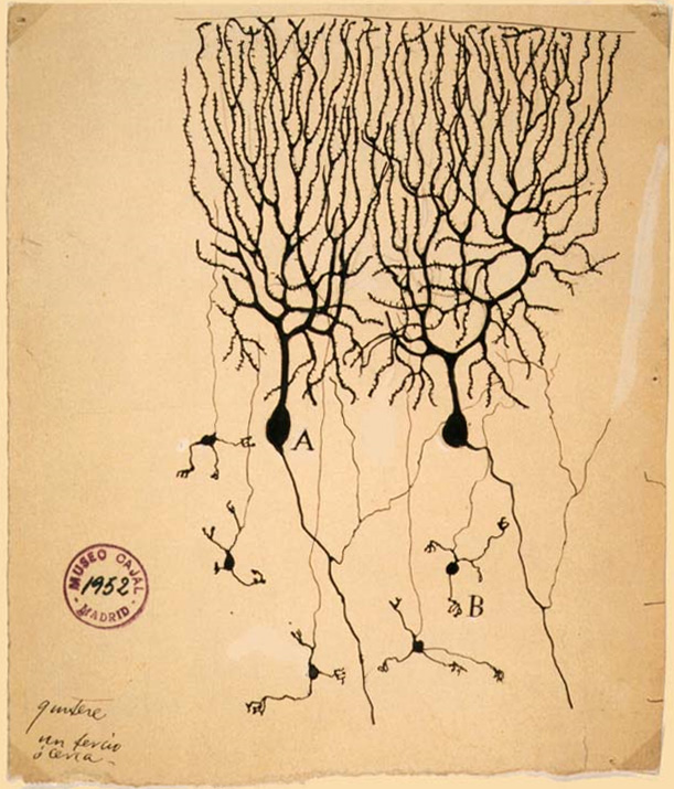
Lugaro cells are primary sensory interneurons of the cerebellum, that have an inhibitory function. They are fusiform, having a spindle shape that tapers at each end. They were first described by Ernesto Lugaro in the early 20th century. Lugaro cells are found just beneath the layer of Purkinje cells between the molecular layer and the granular layer. They have thick principal dendrites coming from opposite poles of their bodies. These dendrites are very long and travel along the boundary between the Purkinje layer and the granular layer. They seem to contact from 5 to 15 Purkinje cells in a horizontal direction. They can also sense stimuli close to the Purkinje cells and their dendrites form a large receptive area that monitors the environment near to the Purkinje cells. Whilst their dendrites make contact with the Purkinje cells they also receive inputs from branches of the Purkinje axons by which they seem to have a sampling and integration role.Lainé J, Axelrad H. Lugaro cells ... [...More Info...] [...Related Items...] OR: [Wikipedia] [Google] [Baidu] |
Granule Cell
The name granule cell has been used for a number of different types of neurons whose only common feature is that they all have very small cell bodies. Granule cells are found within the granular layer of the cerebellum, the dentate gyrus of the hippocampus, the superficial layer of the dorsal cochlear nucleus, the olfactory bulb, and the cerebral cortex. Cerebellar granule cells account for the majority of neurons in the human brain. These granule cells receive excitatory input from mossy fibers originating from pontine nuclei. Cerebellar granule cells project up through the Purkinje layer into the molecular layer where they branch out into parallel fibers that spread through Purkinje cell dendritic arbors. These parallel fibers form thousands of excitatory granule-cell–Purkinje-cell synapses onto the intermediate and distal dendrites of Purkinje cells using glutamate as a neurotransmitter. Layer 4 granule cells of the cerebral cortex receive inputs from the thala ... [...More Info...] [...Related Items...] OR: [Wikipedia] [Google] [Baidu] |
Golgi Cell
In neuroscience, Golgi cells are the most abundant inhibitory interneurons found within the granular layer of the cerebellum. Golgi cells can be found in the granular layer at various layers. The Golgi cell is essential for controlling the activity of the granular layer. They were first identified as inhibitory in 1964. It was also the first example of an inhibitory feedback network in which the inhibitory interneuron was identified anatomically. Golgi cells produce a wide lateral inhibition that reaches beyond the afferent synaptic field and inhibit granule cells via feedforward and feedback inhibitory loops. These cells synapse onto the dendrite of granule cells and unipolar brush cells. They receive excitatory input from mossy fibres, also synapsing on granule cells, and parallel fibers, which are long granule cell axons. Thereby this circuitry allows for feed-forward and feed-back inhibition of granule cells. Connections The cerebellar network contains a large number of ... [...More Info...] [...Related Items...] OR: [Wikipedia] [Google] [Baidu] |
Stellate Cell
Stellate cells are neurons in the central nervous system, named for their star-like shape formed by dendritic processes radiating from the cell body. These cells play significant roles in various brain functions, including inhibition in the cerebellum and excitation in the cortex, and are involved in synaptic plasticity and neurovascular coupling. Morphology Stellate cells are characterized by their star-shaped dendritic trees. Dendrites can vary between neurons, with stellate cells being either spiny or aspinous. In contrast, pyramidal cells, which are also found in the cerebral cortex, are always spiny and pyramid-shaped. The classification of neurons often depends on the presence or absence of dendritic spines: those with spines are classified as spiny, while those without are classified as aspinous. Types and locations Cerebellar Many stellate cells are GABAergic and are located in the molecular layer of the cerebellum. Most common stellate cells are the inhibito ... [...More Info...] [...Related Items...] OR: [Wikipedia] [Google] [Baidu] |
Basket Cell
Basket cells are inhibitory GABAergic interneurons of the brain, found throughout different regions of the cortex and cerebellum. Anatomy and physiology Basket cells are multipolar GABAergic interneurons that function to make inhibitory synapses and control the overall potentials of target cells. In general, dendrites of basket cells are free branching, contain smooth spines, and extend from 3 to 9 mm. Axons are highly branched, ranging in total from 20 to 50mm in total length. The branched axonal arborizations give rise to the name as they appear as baskets surrounding the soma of the target cell. Basket cells form axo-somatic synapses, meaning their synapses target somas of other cells. By controlling the somas of other neurons, basket cells can directly control the action potential discharge rate of target cells. Basket cells can be found throughout the brain, in among other the cortex, hippocampus, amygdala, basal ganglia, and the cerebellum. Cortex In the cortex, basket cel ... [...More Info...] [...Related Items...] OR: [Wikipedia] [Google] [Baidu] |
Cerebellum
The cerebellum (: cerebella or cerebellums; Latin for 'little brain') is a major feature of the hindbrain of all vertebrates. Although usually smaller than the cerebrum, in some animals such as the mormyrid fishes it may be as large as it or even larger. In humans, the cerebellum plays an important role in motor control and cognition, cognitive functions such as attention and language as well as emotion, emotional control such as regulating fear and pleasure responses, but its movement-related functions are the most solidly established. The human cerebellum does not initiate movement, but contributes to motor coordination, coordination, precision, and accurate timing: it receives input from sensory systems of the spinal cord and from other parts of the brain, and integrates these inputs to fine-tune motor activity. Cerebellar damage produces disorders in fine motor skill, fine movement, sense of balance, equilibrium, list of human positions, posture, and motor learning in humans. ... [...More Info...] [...Related Items...] OR: [Wikipedia] [Google] [Baidu] |
Parvalbumin
Parvalbumin (PV) is a calcium-binding protein with low molecular weight (typically 9–11 kDa). In humans, it is encoded by the ''PVALB'' gene. It is a member of the albumin family; it is named for its size (''parv-'', from Latin ' which means "small") and its ability to coagulate. It has three EF hand motifs and is structurally related to calmodulin and troponin C. Parvalbumin is found in fast-contracting muscles, where its levels are highest, as well as in the brain and some endocrine tissues. Parvalbumin is a small, stable protein containing EF-hand type calcium binding sites. It is involved in calcium signaling. Typically, this protein is broken into three domains, domains AB, CD and EF, each individually containing a helix-loop-helix motif. The AB domain houses a two amino-acid deletion in the loop region, whereas domains CD and EF contain the N-terminal and C-terminal, respectively. Calcium binding proteins like parvalbumin play a role in many physiological processes, nam ... [...More Info...] [...Related Items...] OR: [Wikipedia] [Google] [Baidu] |
Golgi Tendon Organ
The Golgi tendon organ (GTO) (also called Golgi organ, tendon organ, neurotendinous organ or neurotendinous spindle) is a proprioceptor – a type of sensory receptor that senses changes in muscle tension. It lies at the interface between a muscle and its tendon known as the musculotendinous junction also known as the myotendinous junction. It provides the sensory component of the Golgi tendon reflex. The Golgi tendon organ is one of several eponymous terms named after the Italian physician Camillo Golgi. Structure The body of the Golgi tendon organ is made up of braided strands of collagen (intrafusal fasciculi) that are less compact than elsewhere in the tendon and are encapsulated. The capsule is connected in series (along a single path) with a group of muscle fibers () at one end, and merge into the tendon proper at the other. Each capsule is about long, has a diameter of about , and is perforated by one or more afferent type Ib sensory nerve fibers ( Aɑ fiber), whic ... [...More Info...] [...Related Items...] OR: [Wikipedia] [Google] [Baidu] |


