|
Posterior Atlantooccipital Membrane
The posterior atlantooccipital membrane (posterior atlantooccipital ligament) is a broad but thin membrane. It is connected above to the posterior margin of the foramen magnum and below to the upper border of the posterior arch of the atlas. On each side of this membrane there is a defect above the groove for the vertebral artery which serves as an opening for the entrance of the artery. The suboccipital nerve also passes through this defect. The free border of the membrane arches over the artery and nerve and is sometimes ossified. The membrane is deep to the Recti capitis posteriores minores and Obliqui capitis superiores and is superficial to the dura mater of the vertebral canal to which it is closely associated. In 2015, Scali et al. revisited the anatomy of the posterior atlantooccipital membrane via plastination. Their findings revealed that the PAO membrane superiorly consisted of periosteum of the occiput, whereas inferiorly it formed part of the dura at the cerebrosp ... [...More Info...] [...Related Items...] OR: [Wikipedia] [Google] [Baidu] |
Foramen Magnum
The foramen magnum ( la, great hole) is a large, oval-shaped opening in the occipital bone of the skull. It is one of the several oval or circular openings (foramina) in the base of the skull. The spinal cord, an extension of the medulla oblongata, passes through the foramen magnum as it exits the cranial cavity. Apart from the transmission of the medulla oblongata and its membranes, the foramen magnum transmits the vertebral arteries, the anterior and posterior spinal arteries, the tectorial membranes and alar ligaments. It also transmits the accessory nerve into the skull. The foramen magnum is a very important feature in bipedal mammals. One of the attributes of a biped's foramen magnum is a forward shift of the anterior border of the cerebellar tentorium; this is caused by the shortening of the cranial base. Studies on the foramen magnum position have shown a connection to the functional influences of both posture and locomotion. The forward shift of the foramen magn ... [...More Info...] [...Related Items...] OR: [Wikipedia] [Google] [Baidu] |
Atlas (anatomy)
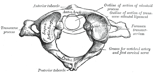
In anatomy, the atlas (C1) is the most superior (first) cervical vertebra of the spine and is located in the neck. It is named for Atlas of Greek mythology because, just as Atlas supported the globe, it supports the entire head. The atlas is the topmost vertebra and, with the axis (the vertebra below it), forms the joint connecting the skull and spine. The atlas and axis are specialized to allow a greater range of motion than normal vertebrae. They are responsible for the nodding and rotation movements of the head. The atlanto-occipital joint allows the head to nod up and down on the vertebral column. The dens acts as a pivot that allows the atlas and attached head to rotate on the axis, side to side. The atlas's chief peculiarity is that it has no body. It is ring-like and consists of an anterior and a posterior arch and two lateral masses. The atlas and axis are important neurologically because the brainstem extends down to the axis. Structure Anterior arch The anter ... [...More Info...] [...Related Items...] OR: [Wikipedia] [Google] [Baidu] |
Vertebral Artery
The vertebral arteries are major arteries of the neck. Typically, the vertebral arteries originate from the subclavian arteries. Each vessel courses superiorly along each side of the neck, merging within the skull to form the single, midline basilar artery. As the supplying component of the ''vertebrobasilar vascular system'', the vertebral arteries supply blood to the upper spinal cord, brainstem, cerebellum, and posterior part of brain. Structure The vertebral arteries usually arise from the posterosuperior aspect of the central subclavian arteries on each side of the body, then enter deep to the transverse process at the level of the 6th cervical vertebrae (C6), or occasionally (in 7.5% of cases) at the level of C7. They then proceed superiorly, in the transverse foramen of each cervical vertebra. Once they have passed through the transverse foramen of C1 (also known as the atlas), the vertebral arteries travel across the posterior arch of C1 and through the suboccipi ... [...More Info...] [...Related Items...] OR: [Wikipedia] [Google] [Baidu] |
Suboccipital Nerve
The suboccipital nerve (first cervical dorsal ramus) is the dorsal primary ramus of the first cervical nerve (C1). It exits the spinal cord between the skull and the first cervical vertebra, the atlas. It lies within the suboccipital triangle along with the vertebral artery, where the artery enters the foramen magnum. It supplies muscles of the suboccipital triangle, the rectus capitis posterior major, obliquus capitis superior, and obliquus capitis inferior. The suboccipital nerve also innervates rectus capitis posterior minor. See also * Vertebral artery The vertebral arteries are major arteries of the neck. Typically, the vertebral arteries originate from the subclavian arteries. Each vessel courses superiorly along each side of the neck, merging within the skull to form the single, midline ... Additional images File:Gray792.png, Upper part of medulla spinalis and hind- and mid-brains; posterior aspect, exposed in situ. File:Suboccipital_triangle.PNG, Subocci ... [...More Info...] [...Related Items...] OR: [Wikipedia] [Google] [Baidu] |
Ossified
Ossification (also called osteogenesis or bone mineralization) in bone remodeling is the process of laying down new bone material by cells named osteoblasts. It is synonymous with bone tissue formation. There are two processes resulting in the formation of normal, healthy bone tissue: Intramembranous ossification is the direct laying down of bone into the primitive connective tissue (mesenchyme), while endochondral ossification involves cartilage as a precursor. In fracture healing, endochondral osteogenesis is the most commonly occurring process, for example in fractures of long bones treated by plaster of Paris, whereas fractures treated by open reduction and internal fixation with metal plates, screws, pins, rods and nails may heal by intramembranous osteogenesis. Heterotopic ossification is a process resulting in the formation of bone tissue that is often atypical, at an extraskeletal location. Calcification is often confused with ossification. Calcification is syno ... [...More Info...] [...Related Items...] OR: [Wikipedia] [Google] [Baidu] |
Recti Capitis Posteriores Minores
The rectus capitis posterior minor (or rectus capitis posticus minor, both being Latin for ''lesser posterior straight muscle of the head'') arises by a narrow pointed tendon from the tubercle on the posterior arch of the atlas, and, widening as it ascends, is inserted into the medial part of the inferior nuchal line of the occipital bone and the surface between it and the foramen magnum, and also takes some attachment to the spinal dura mater. The synergists are the rectus capitis posterior major and the obliquus capitis superior. Connective tissue bridges were noted at the atlanto-occipital joint between the rectus capitis posterior minor (RCPm) muscle and the dorsal spinal dura. Similar connective tissue connections of the rectus capitis posterior major have been reported recently as well. The perpendicular arrangement of these fibers appears to restrict dural movement toward the spinal cord. The ligamentum nuchae was found to be continuous with the posterior cervical spi ... [...More Info...] [...Related Items...] OR: [Wikipedia] [Google] [Baidu] |
Obliqui Capitis Superiores
The obliquus capitis superior muscle () is a small muscle in the upper back part of the neck and is one of the suboccipital muscles and part of the suboccipital triangle. It arises from the lateral mass of the atlas bone. It passes superiorly and posteriorly to insert into the lateral half of the inferior nuchal line on the external surface of the occipital bone. The muscle is innervated by the suboccipital nerve, the dorsal ramus of the first spinal nerve. It acts at the atlanto-occipital joint to extend the head and flex the head to the ipsilateral side. Additional images File:Obliquus capitis superior muscle - animation04.gif, Position of obliquus capitis superior (shown in red). Animation. File:Obliquus capitis superior muscle05.png, Still image. Posterior view. File:Obliquus capitis superior.png, Deep muscles of the back (obliquus capitis superior labeled at upper left) File:Gray129.png, Occipital bone. Outer surface. Muscle attachments are shown as red circles. ... [...More Info...] [...Related Items...] OR: [Wikipedia] [Google] [Baidu] |
Vertebral Canal
The spinal canal (or vertebral canal or spinal cavity) is the canal that contains the spinal cord within the vertebral column. The spinal canal is formed by the vertebrae through which the spinal cord passes. It is a process of the dorsal body cavity. This canal is enclosed within the foramen of the vertebrae. In the intervertebral spaces, the canal is protected by the ligamentum flavum posteriorly and the posterior longitudinal ligament anteriorly. Structure The outermost layer of the meninges, the dura mater, is closely associated with the arachnoid mater which in turn is loosely connected to the innermost layer, the pia mater. The meninges divide the spinal canal into the epidural space and the subarachnoid space. The pia mater is closely attached to the spinal cord. A subdural space is generally only present due to trauma and/or pathological situations. The subarachnoid space is filled with cerebrospinal fluid and contains the vessels that supply the spinal cord, nam ... [...More Info...] [...Related Items...] OR: [Wikipedia] [Google] [Baidu] |
Plastination
Plastination is a technique or process used in anatomy to preserve bodies or body parts, first developed by Gunther von Hagens in 1977. The water and fat are replaced by certain plastics, yielding specimens that can be touched, do not smell or decay, and even retain most properties of the original sample. Process Four steps are used in the standard process of plastination: fixation, dehydration, forced impregnation in a vacuum, and hardening. Water and lipid tissues are replaced by curable polymers, which include silicone, epoxy, and polyester-copolymer. The first step of plastination, fixation, frequently uses a formaldehyde-based solution, and serves two functions. Dissecting the specimen to show specific anatomical elements can be time-consuming. Formaldehyde or other preserving solutions help prevent decomposition of the tissues. They may also confer a degree of rigidity. This can be beneficial in maintaining the shape or arrangement of a specimen. A stomach might be in ... [...More Info...] [...Related Items...] OR: [Wikipedia] [Google] [Baidu] |
Cerebrospinal Fluid
Cerebrospinal fluid (CSF) is a clear, colorless body fluid found within the tissue that surrounds the brain and spinal cord of all vertebrates. CSF is produced by specialised ependymal cells in the choroid plexus of the ventricles of the brain, and absorbed in the arachnoid granulations. There is about 125 mL of CSF at any one time, and about 500 mL is generated every day. CSF acts as a shock absorber, cushion or buffer, providing basic mechanical and immunological protection to the brain inside the skull. CSF also serves a vital function in the cerebral autoregulation of cerebral blood flow. CSF occupies the subarachnoid space (between the arachnoid mater and the pia mater) and the ventricular system around and inside the brain and spinal cord. It fills the ventricles of the brain, cisterns, and sulci, as well as the central canal of the spinal cord. There is also a connection from the subarachnoid space to the bony labyrinth of the inner ear via the ... [...More Info...] [...Related Items...] OR: [Wikipedia] [Google] [Baidu] |
Anterior Atlantooccipital Membrane
Standard anatomical terms of location are used to unambiguously describe the anatomy of animals, including humans. The terms, typically derived from Latin or Greek roots, describe something in its standard anatomical position. This position provides a definition of what is at the front ("anterior"), behind ("posterior") and so on. As part of defining and describing terms, the body is described through the use of anatomical planes and anatomical axes. The meaning of terms that are used can change depending on whether an organism is bipedal or quadrupedal. Additionally, for some animals such as invertebrates, some terms may not have any meaning at all; for example, an animal that is radially symmetrical will have no anterior surface, but can still have a description that a part is close to the middle ("proximal") or further from the middle ("distal"). International organisations have determined vocabularies that are often used as standard vocabularies for subdisciplines of anatom ... [...More Info...] [...Related Items...] OR: [Wikipedia] [Google] [Baidu] |


.jpg)

