|
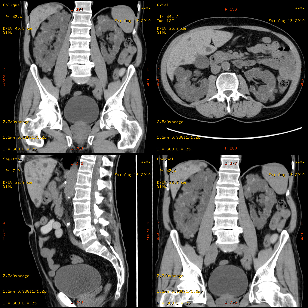
Polycystic Liver Disease
Polycystic liver disease (PLD) usually describes the presence of multiple cysts scattered throughout normal liver tissue. PLD is commonly seen in association with autosomal-dominant polycystic kidney disease, with a prevalence of 1 in 400 to 1000, and accounts for 8–10% of all cases of end-stage renal disease. The much rarer autosomal-dominant polycystic liver disease will progress without any kidney involvement. Presentation Pathophysiology Associations with ''PRKCSH'' and ''SEC63'' have been described. Polycystic liver disease comes in two forms: autosomal dominant polycystic kidney disease (with kidney cysts) and autosomal dominant polycystic liver disease (liver cysts only). Diagnosis Most patients with PLD are asymptomatic with simple cysts found following routine investigations. After confirming the presence of cysts in the liver, laboratory tests may be ordered to check for liver function including bilirubin, alkaline phosphatase, alanine aminotransferase, and prothrombin ... [...More Info...] [...Related Items...] OR: [Wikipedia] [Google] [Baidu] |
Micrograph
A micrograph or photomicrograph is a photograph or digital image taken through a microscope or similar device to show a magnified image of an object. This is opposed to a macrograph or photomacrograph, an image which is also taken on a microscope but is only slightly magnified, usually less than 10 times. Micrography is the practice or art of using microscopes to make photographs. A micrograph contains extensive details of microstructure. A wealth of information can be obtained from a simple micrograph like behavior of the material under different conditions, the phases found in the system, failure analysis, grain size estimation, elemental analysis and so on. Micrographs are widely used in all fields of microscopy. Types Photomicrograph A light micrograph or photomicrograph is a micrograph prepared using an optical microscope, a process referred to as ''photomicroscopy''. At a basic level, photomicroscopy may be performed simply by connecting a camera to a microscope, ... [...More Info...] [...Related Items...] OR: [Wikipedia] [Google] [Baidu] |
Von Meyenburg Complex
A bile duct hamartoma or biliary hamartoma, is a benign tumour-like malformation of the liver. They are classically associated with polycystic liver disease, as may be seen in the context of polycystic kidney disease, and represent a malformation of the liver plate. Cause It is a developmental anomaly in which abnormal tissues are present at normal site. Due to failure of regression of embryonic biliary duct. Diagnosis File:Histopathology of a bile duct hamartoma, high magnification.jpg, Histopathology of a bile duct hamartoma, high magnification, H&E stain. It shows typical features of bile duct hamartoma: Topic Completed: 23 November 2020. Minor changes: 23 November 2020 - Small to medium sized, irregularly shaped bile ducts lined by bland cuboidal epithelium (may also be flattened).- Prominent intervening collagenous stroma. - Bile ducts containing eosinophilic debris (may also contain inspissated bile) File:Von Meyenburg complex cropped.tif, von Meyenburg Complex in ultras ... [...More Info...] [...Related Items...] OR: [Wikipedia] [Google] [Baidu] |
Bile Duct
A bile duct is any of a number of long tube-like structures that carry bile, and is present in most vertebrates. Bile is required for the digestion of food and is secreted by the liver into passages that carry bile toward the hepatic duct. It joins the cystic duct (carrying bile to and from the gallbladder) to form the common bile duct which then opens into the intestine. Structure The top half of the common bile duct is associated with the liver, while the bottom half of the common bile duct is associated with the pancreas, through which it passes on its way to the intestine. It opens into the part of the intestine called the duodenum via the ampulla of Vater. Segments The biliary tree (see below) is the whole network of various sized ducts branching through the liver. The path is as follows: Bile canaliculi → Canals of Hering → interlobular bile ducts → intrahepatic bile ducts → left and right hepatic ducts ''merge to form'' → common hepatic duct ''exits ... [...More Info...] [...Related Items...] OR: [Wikipedia] [Google] [Baidu] |
Hamartoma
A hamartoma is a mostly benign, local malformation of cells that resembles a neoplasm of local tissue but is usually due to an overgrowth of multiple aberrant cells, with a basis in a systemic genetic condition, rather than a growth descended from a single mutated cell ( monoclonality), as would typically define a benign neoplasm/tumor. Despite this, many hamartomas are found to have clonal chromosomal aberrations that are acquired through somatic mutations, and on this basis the term ''hamartoma'' is sometimes considered synonymous with neoplasm. Hamartomas are by definition benign, slow-growing or self-limiting, though the underlying condition may still predispose the individual towards malignancies. Hamartomas are usually caused by a genetic syndrome that affects the development cycle of all or at least multiple cells. Many of these conditions are classified as overgrowth syndromes or cancer syndromes. Hamartomas occur in many different parts of the body and are most often ... [...More Info...] [...Related Items...] OR: [Wikipedia] [Google] [Baidu] |
Trichrome Stain
Trichrome staining is a histological staining method that uses two or more acid dyes in conjunction with a polyacid. Staining differentiates tissues by tinting them in contrasting colours. It increases the contrast of microscopic features in cells and tissues, which makes them easier to see when viewed through a microscope. The word ''trichrome'' means "three colours". The first staining protocol that was described as "trichrome" was Mallory's trichrome stain, which differentially stained erythrocytes to a red colour, muscle tissue to a red colour, and collagen to a blue colour. Some other trichrome staining protocols are the Masson's trichrome stain, Lillie's trichrome, and the Gömöri trichrome stain. Purpose Without trichrome staining, discerning one feature from another can be extremely difficult. Smooth muscle tissue, for example, is hard to differentiate from collagen. A trichrome stain can colour the muscle tissue red, and the collagen fibres green or blue. Liver bio ... [...More Info...] [...Related Items...] OR: [Wikipedia] [Google] [Baidu] |
Cyst
A cyst is a closed sac, having a distinct envelope and division compared with the nearby tissue. Hence, it is a cluster of cells that have grouped together to form a sac (like the manner in which water molecules group together to form a bubble); however, the distinguishing aspect of a cyst is that the cells forming the "shell" of such a sac are distinctly abnormal (in both appearance and behaviour) when compared with all surrounding cells for that given location. A cyst may contain air, fluids, or semi-solid material. A collection of pus is called an abscess, not a cyst. Once formed, a cyst may resolve on its own. When a cyst fails to resolve, it may need to be removed surgically, but that would depend upon its type and location. Cancer-related cysts are formed as a defense mechanism for the body following the development of mutations that lead to an uncontrolled cellular division. Once that mutation has occurred, the affected cells divide incessantly and become cancerous, fo ... [...More Info...] [...Related Items...] OR: [Wikipedia] [Google] [Baidu] |
Liver
The liver is a major organ only found in vertebrates which performs many essential biological functions such as detoxification of the organism, and the synthesis of proteins and biochemicals necessary for digestion and growth. In humans, it is located in the right upper quadrant of the abdomen, below the diaphragm. Its other roles in metabolism include the regulation of glycogen storage, decomposition of red blood cells, and the production of hormones. The liver is an accessory digestive organ that produces bile, an alkaline fluid containing cholesterol and bile acids, which helps the breakdown of fat. The gallbladder, a small pouch that sits just under the liver, stores bile produced by the liver which is later moved to the small intestine to complete digestion. The liver's highly specialized tissue, consisting mostly of hepatocytes, regulates a wide variety of high-volume biochemical reactions, including the synthesis and breakdown of small and complex molecule ... [...More Info...] [...Related Items...] OR: [Wikipedia] [Google] [Baidu] |
Polycystic Kidney Disease
Polycystic kidney disease (PKD or PCKD, also known as polycystic kidney syndrome) is a genetic disorder in which the renal tubules become structurally abnormal, resulting in the development and growth of multiple cysts within the kidney. These cysts may begin to develop in utero, in infancy, in childhood, or in adulthood. Cysts are non-functioning tubules filled with fluid pumped into them, which range in size from microscopic to enormous, crushing adjacent normal tubules and eventually rendering them non-functional as well. PKD is caused by abnormal genes that produce a specific abnormal protein; this protein has an adverse effect on tubule development. PKD is a general term for two types, each having their own pathology and genetic cause: autosomal dominant polycystic kidney disease (ADPKD) and autosomal recessive polycystic kidney disease (ARPKD). The abnormal gene exists in all cells in the body; as a result, cysts may occur in the liver, seminal vesicles, and pancreas. Th ... [...More Info...] [...Related Items...] OR: [Wikipedia] [Google] [Baidu] |
End-stage Renal Disease
Chronic kidney disease (CKD) is a type of kidney disease in which a gradual loss of kidney function occurs over a period of months to years. Initially generally no symptoms are seen, but later symptoms may include leg swelling, feeling tired, vomiting, loss of appetite, and confusion. Complications can relate to hormonal dysfunction of the kidneys and include (in chronological order) high blood pressure (often related to activation of the renin–angiotensin system system), bone disease, and anemia. Additionally CKD patients have markedly increased cardiovascular complications with increased risks of death and hospitalization. Causes of chronic kidney disease include diabetes, high blood pressure, glomerulonephritis, and polycystic kidney disease. Risk factors include a family history of chronic kidney disease. Diagnosis is by blood tests to measure the estimated glomerular filtration rate (eGFR), and a urine test to measure albumin. Ultrasound or kidney biopsy may be p ... [...More Info...] [...Related Items...] OR: [Wikipedia] [Google] [Baidu] |
PRKCSH
Glucosidase 2 subunit beta is an enzyme that in humans is encoded by the ''PRKCSH'' gene. This gene encodes the beta-subunit of glucosidase II, an N-linked glycan-processing enzyme in the endoplasmic reticulum (ER). This protein is an acidic phospho-protein known to be a substrate for protein kinase C In cell biology, Protein kinase C, commonly abbreviated to PKC (EC 2.7.11.13), is a family of protein kinase enzymes that are involved in controlling the function of other proteins through the phosphorylation of hydroxyl groups of serine and .... Mutations in this gene have been associated with the autosomal dominant polycystic liver disease (PCLD). Alternatively spliced transcript variants encoding distinct isoforms have been observed. References Further reading * * * * * * * * * * * * * * * * * * * EF-hand-containing proteins {{gene-19-stub ... [...More Info...] [...Related Items...] OR: [Wikipedia] [Google] [Baidu] |
SEC63
Translocation protein SEC63 homolog is a protein that in humans is encoded by the ''SEC63'' gene. Function The Sec61 complex is the central component of the protein translocation apparatus of the endoplasmic reticulum (ER) membrane. The protein encoded by this gene and SEC62 protein are found to be associated with ribosome-free SEC61 complex. It is speculated that Sec61-Sec62-Sec63 may perform post-translational protein translocation into the ER. The Sec61-Sec62-Sec63 complex might also perform the backward transport of ER proteins that are subject to the ubiquitin-proteasome Proteasomes are protein complexes which degrade unneeded or damaged proteins by proteolysis, a chemical reaction that breaks peptide bonds. Enzymes that help such reactions are called proteases. Proteasomes are part of a major mechanism by whi ...-dependent degradation pathway. The encoded protein is an integral membrane protein located in the rough ER. Clinical significance Mutations of this gen ... [...More Info...] [...Related Items...] OR: [Wikipedia] [Google] [Baidu] |
Suction (medicine)
In medicine, devices are sometimes necessary to create suction. Suction may be used to clear the airway of blood, saliva, vomit, or other secretions so that a patient may breathe. Suctioning can prevent pulmonary aspiration, which can lead to lung infections. In pulmonary hygiene, suction is used to remove fluids from the airways, to facilitate breathing and prevent growth of microorganisms. Small suction-providing devices are often called aspirators. In surgery suction can be used to remove blood from the area being operated on to allow surgeons to view and work on the area. Suction may also be used to remove blood that has built up within the skull after an intracranial hemorrhage. Suction devices may be mechanical hand pumps or battery or electrically operated mechanisms. In many hospitals and other health facilities, suction is typically provided by suction regulators, connected to a central medical vacuum supply by way of a pipeline system. The plastic, rigid Yanka ... [...More Info...] [...Related Items...] OR: [Wikipedia] [Google] [Baidu] |


