|
Parietal Eye
A parietal eye, also known as a third eye or pineal eye, is a part of the epithalamus present in some vertebrates. The eye is located at the top of the head, is photoreceptive and is associated with the pineal gland, regulating circadian rhythmicity and hormone production for thermoregulation. The hole in the head which contains the eye is known as a pineal foramen or parietal foramen, since it is often enclosed by the parietal bones. Presence in various animals The parietal eye is found in the tuatara, most lizards, frogs, salamanders, certain bony fish, sharks, and lampreys. It is absent in mammals, but was present in their closest extinct relatives, the therapsids, suggesting it was lost during the course of the mammalian evolution due to it being useless in endothermic animals. It is also absent in the ancestrally endothermic ("warm-blooded") archosaurs such as birds. The parietal eye is also lost in ectothermic ("cold-blooded") archosaurs like crocodilians, and in turtles, ... [...More Info...] [...Related Items...] OR: [Wikipedia] [Google] [Baidu] |
Frog Parietal Eye
A frog is any member of a diverse and largely carnivorous group of short-bodied, tailless amphibians composing the order Anura (ανοὐρά, literally ''without tail'' in Ancient Greek). The oldest fossil "proto-frog" ''Triadobatrachus'' is known from the Early Triassic of Madagascar, but molecular clock dating suggests their split from other amphibians may extend further back to the Permian, 265 million years ago. Frogs are widely distributed, ranging from the tropics to subarctic regions, but the greatest concentration of species diversity is in tropical rainforest. Frogs account for around 88% of extant amphibian species. They are also one of the five most diverse vertebrate orders. Warty frog species tend to be called toads, but the distinction between frogs and toads is informal, not from taxonomy or evolutionary history. An adult frog has a stout body, protruding eyes, anteriorly-attached tongue, limbs folded underneath, and no tail (the tail of tailed frogs is an ext ... [...More Info...] [...Related Items...] OR: [Wikipedia] [Google] [Baidu] |
Therapsid
Therapsida is a major group of eupelycosaurian synapsids that includes mammals, their ancestors and relatives. Many of the traits today seen as unique to mammals had their origin within early therapsids, including limbs that were oriented more underneath the body, as opposed to the sprawling posture of many reptiles and salamanders. Therapsids evolved from " pelycosaurs", specifically within the Sphenacodontia, more than 279.5 million years ago. They replaced the "pelycosaurs" as the dominant large land animals in the Middle Permian through to the Early Triassic. In the aftermath of the Permian–Triassic extinction event, therapsids declined in relative importance to the rapidly diversifying reptiles during the Middle Triassic. The therapsids include the cynodonts, the group that gave rise to mammals (Mammaliaformes) in the Late Triassic, around 225 million years ago. Of the non-mammalian therapsids, only cynodonts survived beyond the end of the Triassic, with the only oth ... [...More Info...] [...Related Items...] OR: [Wikipedia] [Google] [Baidu] |
Ostracoderm
Ostracoderms () are the armored jawless fish of the Paleozoic Era. The term does not often appear in classifications today because it is paraphyletic (excluding jawed fishes) (may also be polyphyletic if anaspids are closer to cyclostomes) and thus does not correspond to one evolutionary lineage. However, the term is still used as an informal way of loosely grouping together the armored jawless fishes. An innovation of ostracoderms was the use of gills not for feeding, but exclusively for respiration. Earlier chordates with gill precursors used them for both respiration and feeding. Ostracoderms had separate pharyngeal gill pouches along the side of the head, which were permanently open with no protective operculum. Unlike invertebrates that use ciliated motion to move food, ostracoderms used their muscular pharynx to create a suction that pulled small and slow moving prey into their mouths. Swiss anatomist Louis Agassiz received some fossils of bony armored fish from S ... [...More Info...] [...Related Items...] OR: [Wikipedia] [Google] [Baidu] |
Cone Cell
Cone cells, or cones, are photoreceptor cells in the retinas of vertebrate eyes including the human eye. They respond differently to light of different wavelengths, and the combination of their responses is responsible for color vision. Cones function best in relatively bright light, called the photopic region, as opposed to rod cells, which work better in dim light, or the scotopic region. Cone cells are densely packed in the fovea centralis, a 0.3 mm diameter rod-free area with very thin, densely packed cones which quickly reduce in number towards the periphery of the retina. Conversely, they are absent from the optic disc, contributing to the blind spot. There are about six to seven million cones in a human eye (vs ~92 million rods), with the highest concentration being towards the macula. Cones are less sensitive to light than the rod cells in the retina (which support vision at low light levels), but allow the perception of color. They are also able to perceive ... [...More Info...] [...Related Items...] OR: [Wikipedia] [Google] [Baidu] |
Rod Cell
Rod cells are photoreceptor cells in the retina of the eye that can function in lower light better than the other type of visual photoreceptor, cone cells. Rods are usually found concentrated at the outer edges of the retina and are used in peripheral vision. On average, there are approximately 92 million rod cells (vs ~6 million cones) in the human retina. Rod cells are more sensitive than cone cells and are almost entirely responsible for night vision. However, rods have little role in color vision, which is the main reason why colors are much less apparent in dim light. Structure Rods are a little longer and leaner than cones but have the same basic structure. Opsin-containing disks lie at the end of the cell adjacent to the retinal pigment epithelium, which in turn is attached to the inside of the eye. The stacked-disc structure of the detector portion of the cell allows for very high efficiency. Rods are much more common than cones, with about 120 million rod cells comp ... [...More Info...] [...Related Items...] OR: [Wikipedia] [Google] [Baidu] |
Skull
The skull is a bone protective cavity for the brain. The skull is composed of four types of bone i.e., cranial bones, facial bones, ear ossicles and hyoid bone. However two parts are more prominent: the cranium and the mandible. In humans, these two parts are the neurocranium and the viscerocranium ( facial skeleton) that includes the mandible as its largest bone. The skull forms the anterior-most portion of the skeleton and is a product of cephalisation—housing the brain, and several sensory structures such as the eyes, ears, nose, and mouth. In humans these sensory structures are part of the facial skeleton. Functions of the skull include protection of the brain, fixing the distance between the eyes to allow stereoscopic vision, and fixing the position of the ears to enable sound localisation of the direction and distance of sounds. In some animals, such as horned ungulates (mammals with hooves), the skull also has a defensive function by providing the mount (on the ... [...More Info...] [...Related Items...] OR: [Wikipedia] [Google] [Baidu] |
Diencephalon
The diencephalon (or interbrain) is a division of the forebrain (embryonic ''prosencephalon''). It is situated between the telencephalon and the midbrain (embryonic ''mesencephalon''). The diencephalon has also been known as the 'tweenbrain in older literature. It consists of structures that are on either side of the third ventricle, including the thalamus, the hypothalamus, the epithalamus and the subthalamus. The diencephalon is one of the main vesicles of the brain formed during embryogenesis. During the third week of development a neural tube is created from the ectoderm, one of the three primary germ layers. The tube forms three main vesicles during the third week of development: the prosencephalon, the mesencephalon and the rhombencephalon. The prosencephalon gradually divides into the telencephalon and the diencephalon. Structure The diencephalon consists of the following structures: *Thalamus *Hypothalamus including the posterior pituitary * Epithalamus which cons ... [...More Info...] [...Related Items...] OR: [Wikipedia] [Google] [Baidu] |
Evagination
Endodermic evagination relates to the inner germ layers of cells of the very early embryo, from which is formed the lining of the digestive tract, of other internal organs, and of certain glands, implies the extension of a layer of body tissue to form a pouch, or the turning inside out (protrusion) of some body part or organ from its basic position, for example the para-nasal sinuses are believed to be formed in the fetus by 'ballooning' of the developing nasal canal, and the prostate or Skene's gland formed out of evaginations of the urethra The urethra (from Greek οὐρήθρα – ''ourḗthrā'') is a tube that connects the urinary bladder to the urinary meatus for the removal of urine from the body of both females and males. In human females and other primates, the urethra c .... See also * List of human cell types derived from the germ layers References Embryology Developmental biology {{developmental-biology-stub ... [...More Info...] [...Related Items...] OR: [Wikipedia] [Google] [Baidu] |
Pineal Organ
The pineal gland, conarium, or epiphysis cerebri, is a small endocrine gland in the brain of most vertebrates. The pineal gland produces melatonin, a serotonin-derived hormone which modulates sleep patterns in both circadian and seasonal cycles. The shape of the gland resembles a pine cone, which gives it its name. The pineal gland is located in the epithalamus, near the center of the brain, between the two hemispheres, tucked in a groove where the two halves of the thalamus join. The pineal gland is one of the neuroendocrine secretory circumventricular organs in which capillaries are mostly permeable to solutes in the blood. Nearly all vertebrate species possess a pineal gland. The most important exception is a primitive vertebrate, the hagfish. Even in the hagfish, however, there may be a "pineal equivalent" structure in the dorsal diencephalon. The lancelet ''Branchiostoma lanceolatum'', an early chordate which is a close relative to vertebrates, also lacks a recognizab ... [...More Info...] [...Related Items...] OR: [Wikipedia] [Google] [Baidu] |
Snake
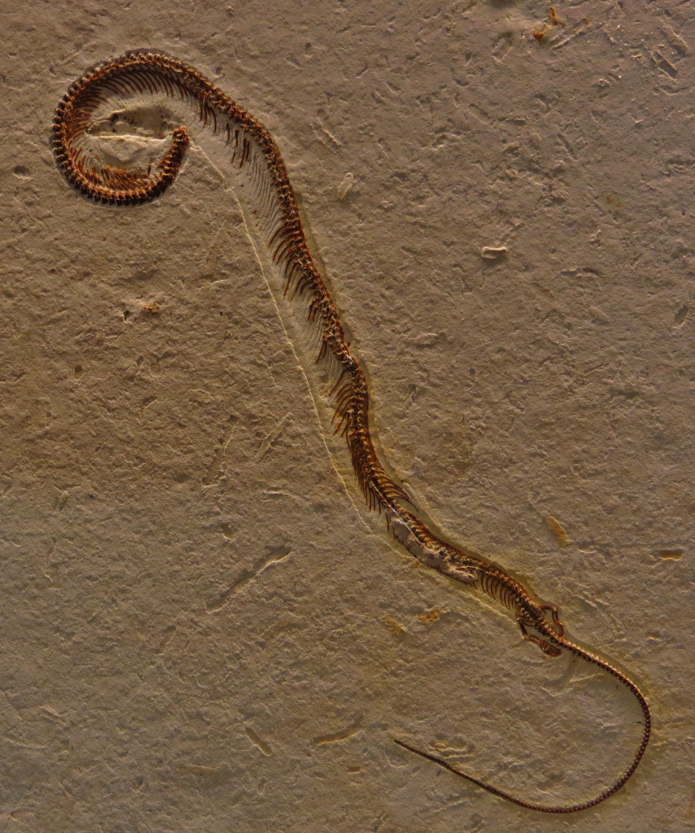
Snakes are elongated, limbless, carnivorous reptiles of the suborder Serpentes . Like all other squamates, snakes are ectothermic, amniote vertebrates covered in overlapping scales. Many species of snakes have skulls with several more joints than their lizard ancestors, enabling them to swallow prey much larger than their heads (cranial kinesis). To accommodate their narrow bodies, snakes' paired organs (such as kidneys) appear one in front of the other instead of side by side, and most have only one functional lung. Some species retain a pelvic girdle with a pair of vestigial claws on either side of the cloaca. Lizards have evolved elongate bodies without limbs or with greatly reduced limbs about twenty-five times independently via convergent evolution, leading to many lineages of legless lizards. These resemble snakes, but several common groups of legless lizards have eyelids and external ears, which snakes lack, although this rule is not universal (see Amphisbaen ... [...More Info...] [...Related Items...] OR: [Wikipedia] [Google] [Baidu] |
Lepidosauria
The Lepidosauria (, from Greek meaning ''scaled lizards'') is a subclass or superorder of reptiles, containing the orders Squamata and Rhynchocephalia. Squamata includes snakes, lizards, and amphisbaenians. Squamata contains over 9,000 species, making it by far the most species-rich and diverse order of reptiles in the present day. Rhynchocephalia was a formerly widespread and diverse group of reptiles in the Mesozoic Era. However, it is represented by only one living species: the tuatara (''Sphenodon punctatus),'' a superficially lizard-like reptile native to New Zealand. Lepidosauria is a monophyletic group (i.e. a clade), containing all descendants of the last common ancestor of squamates and rhynchocephalians. Lepidosaurs can be distinguished from other reptiles via several traits, such as large keratinous scales which may overlap one another. Purely in the context of modern taxa, Lepidosauria can be considered the sister taxon to Archosauria, which includes Aves (bird ... [...More Info...] [...Related Items...] OR: [Wikipedia] [Google] [Baidu] |
Archelosauria
Archelosauria is a clade grouping turtles and archosaurs (birds and crocodilians) and their fossil relatives, to the exclusion of lepidosaurs (the clade containing lizards, snakes and the tuatara). The majority of phylogenetic analyses based on molecular data (e.g. DNA and proteins) have supported a sister-group relationship between turtles and archosaurs. On the other hand, Archelosauria has not been supported by most morphological analyses, which have instead found turtles to either be descendants of parareptiles, early-diverging diapsids outside of Sauria, or close relatives of lepidosaurs within the clade Ankylopoda. Classification Multiple sequence alignments of DNA and protein sequences and phylogenetic inferences have shown that turtles are the closest living relatives to birds and crocodilians. There are about 1000 ultra-conserved elements in the genome that are unique to turtles and archosaurs, but which are not found in lepidosaurs. Other genome-wide analyses also sup ... [...More Info...] [...Related Items...] OR: [Wikipedia] [Google] [Baidu] |
_Ranomafana.jpg)


