|
Posterior Compartment Of Thigh
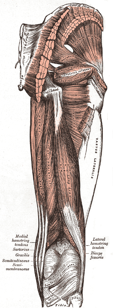
The posterior compartment of the thigh is one of the fascial compartments that contains the knee flexors and hip extensors known as the hamstring muscles, as well as vascular and nervous elements, particularly the sciatic nerve. Structure The posterior compartment is a fascial compartment bounded by fascia. It is separated from the anterior compartment by two folds of deep fascia, known as the medial intermuscular septum and the lateral intermuscular septum. The muscles of the posterior compartment of the thigh are the: * biceps femoris muscle, which consists of a short head and a long head. * semitendinosus muscle * semimembranosus muscle These muscles (or their tendons) apart from the short head of the biceps femoris, are commonly known as the hamstrings. The depression at the back of the knee, or ''kneepit'' is the popliteal fossa, colloquially called the ''ham''. The tendons of the above muscles can be felt as prominent cords on both sides of the fossa—the bicep ... [...More Info...] [...Related Items...] OR: [Wikipedia] [Google] [Baidu] |
Thigh
In anatomy, the thigh is the area between the hip (pelvis) and the knee. Anatomically, it is part of the lower limb. The single bone in the thigh is called the femur. This bone is very thick and strong (due to the high proportion of bone tissue), and forms a ball and socket joint at the hip, and a modified hinge joint at the knee. Structure Bones The femur is the only bone in the thigh and serves as an attachment site for all thigh muscles. The head of the femur articulates with the acetabulum in the pelvic bone forming the hip joint, while the distal part of the femur articulates with the tibia and patella forming the knee. By most measures, the femur is the strongest and longest bone in the body. The femur is categorised as a long bone and comprises a diaphysis, the shaft (or body) and two epiphyses, the lower extremity and the upper extremity of femur, that articulate with adjacent bones in the hip and knee. Muscular compartments In cross-section, the thigh is d ... [...More Info...] [...Related Items...] OR: [Wikipedia] [Google] [Baidu] |
Biceps Femoris Muscle
The biceps femoris () is a muscle of the thigh located to the posterior, or back. As its name implies, it consists of two heads; the long head is considered part of the hamstring muscle group, while the short head is sometimes excluded from this characterization, as it only causes knee flexion (but not hip extension) and is activated by a separate nerve (the peroneal, as opposed to the tibial branch of the sciatic nerve). Structure It has two heads of origin: *the ''long head'' arises from the lower and inner impression on the posterior part of the tuberosity of the ischium. This is a common tendon origin with the semitendinosus muscle, and from the lower part of the sacrotuberous ligament. *the ''short head'', arises from the lateral lip of the linea aspera, between the adductor magnus and vastus lateralis extending up almost as high as the insertion of the gluteus maximus, from the lateral prolongation of the linea aspera to within 5 cm. of the lateral condyle; and fr ... [...More Info...] [...Related Items...] OR: [Wikipedia] [Google] [Baidu] |
Dartmouth Medical School
The Geisel School of Medicine is the medical school of Dartmouth College located in Hanover, New Hampshire. The fourth oldest medical school in the United States, it was founded in 1797 by New England physician Nathan Smith. It is one of the seven Ivy League medical schools. Several milestones in medical care and research have taken place at Dartmouth, including the introduction of stethoscopes to U.S. medical education (1838), the first clinical X-ray (1896), and the first multispecialty intensive care unit (ICU) in the United States (1955). The Geisel School of Medicine grants the Doctor of Medicine (MD) and Doctor of Philosophy (PhD) degrees. The school has a student body of approximately 700 students and more than 2,300 faculty and researchers. Geisel organizes research through over a dozen research centers and institutes, attracting more than $140 million in grants annually, and is ranked as a top medical school by '' U.S. News & World Report'' for both primary care and ... [...More Info...] [...Related Items...] OR: [Wikipedia] [Google] [Baidu] |
Fascia Lata
The fascia lata is the deep fascia of the thigh. It encloses the thigh muscles and forms the outer limit of the fascial compartments of thigh, which are internally separated by the medial intermuscular septum and the lateral intermuscular septum. The fascia lata is thickened at its lateral side where it forms the iliotibial tract, a structure that runs to the tibia and serves as a site of muscle attachment. Structure The fascia lata is an investment for the whole of the thigh, but varies in thickness in different parts. It is thicker in the upper and lateral part of the thigh, where it receives a fibrous expansion from the gluteus maximus, and where the tensor fasciae latae is inserted between its layers; it is very thin behind and at the upper and medial part, where it covers the adductor muscles, and again becomes stronger around the knee, receiving fibrous expansions from the tendon of the biceps femoris laterally, from the sartorius medially, and from the quadriceps fem ... [...More Info...] [...Related Items...] OR: [Wikipedia] [Google] [Baidu] |
Compartment Syndrome
Compartment syndrome is a serious medical condition in which increased pressure within a Fascial compartment, body compartment compromises blood flow and tissue function, potentially leading to permanent damage if not promptly treated. There are two types: Acute (medicine), acute and Chronic condition, chronic. Acute compartment syndrome can lead to a loss of the affected limb due to tissue death. Symptoms of acute compartment syndrome (ACS) include severe pain, decreased blood flow, decreased movement, numbness, and a pale limb. It is most often due to Injury, physical trauma, like a bone fracture (up to 75% of cases) or a crush injury. It can also occur after Reperfusion injury, blood flow returns following a period of poor circulation. Diagnosis is Clinical diagnosis, clinical, based on symptoms, not a specific test. However, it may be supported by measuring the pressure inside the Fascial compartment, compartment. It is classically described by pain out of proportion to the in ... [...More Info...] [...Related Items...] OR: [Wikipedia] [Google] [Baidu] |
Popliteal Artery
The popliteal artery is a deeply placed continuation of the femoral artery opening in the distal portion of the adductor magnus muscle. It courses through the popliteal fossa and ends at the lower border of the popliteus muscle, where it branches into the anterior and posterior tibial arteries. The deepest (most anterior) structure in the fossa, the popliteal artery runs close to the joint capsule of the knee as it spans the intercondylar fossa. Five genicular branches of the popliteal artery supply the capsule and ligaments of the knee joint. The genicular arteries are the superior lateral, superior medial, middle, inferior lateral, and inferior medial genicular arteries. They participate in the formation of the periarticular genicular anastomosis, a network of vessels surrounding the knee that provides collateral circulation capable of maintaining blood supply to the leg during full knee flexion, which may kink the popliteal artery. Structure The popliteal artery is the con ... [...More Info...] [...Related Items...] OR: [Wikipedia] [Google] [Baidu] |
Adductor Hiatus
In human anatomy, the adductor hiatus also known as hiatus magnus is a hiatus (gap) between the adductor magnus muscle and the femur that allows the passage of the femoral vessels from the anterior thigh to the posterior thigh and then the popliteal fossa. It is the termination of the adductor canal and lies about superior to the adductor tubercle. Structure Kale et al. classified the adductor hiatus according to its shape and the structures surrounding. An adductor hiatus is described as oval or bridging depending on the shape of the upper boundary. It can also be described as muscular or fibrous depending on whether the structure surrounding is the muscular part or the tendinous part of the adductor magnus muscle. For example, the top drawing on the right shows an oval fibrous type of adductor hiatus, and the bottom one shows a bridging muscular adductor hiatus. Four structures are associated with the adductor hiatus. However, only two structures enter and then leave throug ... [...More Info...] [...Related Items...] OR: [Wikipedia] [Google] [Baidu] |
Inferior Gluteal Artery
The inferior gluteal artery (sciatic artery) is a terminal branch of the anterior trunk of the internal iliac artery. It exits the pelvis through the greater sciatic foramen. It is distributed chiefly to the buttock and the back of the thigh. Anatomy Origin It is the smaller of the two terminal branches of the anterior trunk of the internal iliac artery. Course It passes posterior-ward within parietal pelvic fascia. It travels in between the S1 nerve and S2 (or S2-S3) nerve(s). It descends upon the nerves of the sacral plexus and the piriformis muscle, posterior to the internal pudendal artery. It passes through the inferior part of the greater sciatic foramen. It exits the pelvis inferior to the piriformis muscle, between piriformis muscle and coccygeus muscle. It then descends in the interval between the greater trochanter of the femur and tuberosity of the ischium. It is accompanied by the sciatic nerve and the posterior femoral cutaneous nerves, and covered by the glu ... [...More Info...] [...Related Items...] OR: [Wikipedia] [Google] [Baidu] |
Common Fibular Nerve
The common fibular nerve (also known as the common peroneal nerve, external popliteal nerve, or lateral popliteal nerve) is a nerve in the lower leg that provides sensation over the posterolateral part of the leg and the knee joint. It divides at the knee into two terminal branches: the superficial fibular nerve and deep fibular nerve, which innervate the muscles of the lateral and anterior compartments of the leg respectively. When the common fibular nerve is damaged or compressed, foot drop can ensue. Structure The common fibular nerve is the smaller terminal branch of the sciatic nerve. The common fibular nerve has root values of L4, L5, S1, and S2. It arises from the superior angle of the popliteal fossa and extends to the lateral angle of the popliteal fossa, along the medial border of the biceps femoris. It then winds around the neck of the fibula to pierce the fibularis longus and divides into terminal branches of the superficial fibular nerve and the deep fibular nerve ... [...More Info...] [...Related Items...] OR: [Wikipedia] [Google] [Baidu] |
Tibial Nerve
The tibial nerve is a branch of the sciatic nerve. The tibial nerve passes through the popliteal fossa to pass below the arch of soleus. Structure Popliteal fossa The tibial nerve is the larger terminal branch of the sciatic nerve with root values of L4, L5, S1, S2, and S3. It lies superficial (or posterior) to the popliteal vessels, extending from the superior angle to the inferior angle of the popliteal fossa, crossing the popliteal vessels from lateral to medial side. It gives off branches as shown below: * Muscular branches - Muscular branches arise from the distal part of the popliteal fossa. It supplies the medial and lateral heads of gastrocnemius, soleus, plantaris and popliteus muscles. Nerve to popliteus crosses the popliteus muscle, runs downwards and laterally, winds around the lower border of the popliteus to supply the deep (or anterior) surface of the popliteus. This nerve also supplies the tibialis posterior muscle, superior tibiofibular joint, tibia bone, ... [...More Info...] [...Related Items...] OR: [Wikipedia] [Google] [Baidu] |
Popliteal Fossa
The popliteal fossa (also referred to as hough or kneepit in analogy to the cubital fossa) is a shallow depression located at the back of the knee joint. The bones of the popliteal fossa are the femur and the tibia. Like other flexion surfaces of large joints (groin, armpit, cubital fossa and essentially the anterior part of the neck), it is an area where blood vessels and nerves pass relatively superficially, and with an increased number of lymph nodes. Structure Boundaries The boundaries of the fossa are: Roof Moving from superficial to deep structures, the roof is formed by: # the skin. # the superficial fascia. This contains the small saphenous vein, the terminal branch of the posterior cutaneous nerve of the thigh, posterior division of the medial cutaneous nerve, lateral sural cutaneous nerve, and medial sural cutaneous nerve. # the popliteal fascia. Floor The floor is formed by: # the popliteal surface of the femur. # the capsule of the knee joint and the obli ... [...More Info...] [...Related Items...] OR: [Wikipedia] [Google] [Baidu] |
Hamstring
A hamstring () is any one of the three posterior thigh muscles in human anatomy between the hip and the knee: from medial to lateral, the semimembranosus, semitendinosus and biceps femoris. Etymology The word " ham" is derived from the Old English “ham” or “hom” meaning the hollow or bend of the knee, from a Germanic base where it meant "crooked". It gained the meaning of the leg of an animal around the 15th century. ''String'' refers to tendons, and thus the hamstrings' string-like tendons felt on either side of the back of the knee. Criteria The common criteria of any hamstring muscles are: # Muscles should originate from ischial tuberosity. # Muscles should be inserted over the knee joint, in the tibia or in the fibula. # Muscles will be innervated by the tibial branch of the sciatic nerve. # Muscle will participate in flexion of the knee joint and extension of the hip joint. Those muscles which fulfill all of the four criteria are called true hamstrings. ... [...More Info...] [...Related Items...] OR: [Wikipedia] [Google] [Baidu] |


