|
Plantaris Muscle
The plantaris is one of the superficial muscles of the superficial posterior compartment of the leg, one of the fascial compartments of the leg. It is composed of a thin muscle belly and a long thin tendon. While not as thick as the achilles tendon, the plantaris tendon (which tends to be between in length) is the longest tendon in the human body. Not including the tendon, the plantaris muscle is approximately long and is absent in 8-12% of the population. It is one of the plantar flexors in the posterior compartment of the leg, along with the gastrocnemius and soleus muscles. The plantaris is considered to have become an unimportant muscle when human ancestors switched from climbing trees to bipedalism and in anatomically modern humans it mainly acts with the gastrocnemius. Structure The plantaris muscle arises from the inferior part of the lateral supracondylar ridge of the femur at a position slightly superior to the origin of the lateral head of gastrocnemius. It pas ... [...More Info...] [...Related Items...] OR: [Wikipedia] [Google] [Baidu] |
Lateral Supracondylar Ridge
The lateral supracondylar ridge is a prominent, rough margin on the lower part of the lateral border of the humerus. It presents an anterior lip for the origin of forearm extensors, including the brachioradialis muscle above, and the extensor carpi radialis longus muscle below. It also presents a posterior lip for the triceps brachii, and an intermediate ridge for the attachment of the lateral intermuscular septum. Clinical significance The lateral supracondylar ridge may be broken in a supracondylar humerus fracture, common in children A child () is a human being between the stages of childbirth, birth and puberty, or between the Development of the human body, developmental period of infancy and puberty. The term may also refer to an unborn human being. In English-speaking .... References External links * Image at u-szeged.hu Humerus {{musculoskeletal-stub ... [...More Info...] [...Related Items...] OR: [Wikipedia] [Google] [Baidu] |
Anatomically Modern Humans
Early modern human (EMH), or anatomically modern human (AMH), are terms used to distinguish ''Homo sapiens'' ( sometimes ''Homo sapiens sapiens'') that are anatomically consistent with the range of phenotypes seen in contemporary humans, from extinct archaic human species (of which some are at times also identified with, but only one, prefix ''sapiens''). This distinction is useful especially for times and regions where anatomically modern and archaic humans co-existed, for example, in Paleolithic Europe. Among the oldest known remains of ''Homo sapiens'' are those found at the Omo-Kibish I archaeological site in south-western Ethiopia, dating to about 233,000 to 196,000 years ago, the Florisbad Skull founded at the Florisbad archaeological and paleontological site in South Africa, dating to about 259,000 years ago, and the Jebel Irhoud site in Morocco, dated about 315,000 years ago. Extinct species of the genus ''Homo'' include ''Homo erectus'' (extant from roughly 2 to ... [...More Info...] [...Related Items...] OR: [Wikipedia] [Google] [Baidu] |
Internal Capsule
The internal capsule is a paired white matter structure, as a two-way nerve tract, tract, carrying afferent nerve fiber, ascending and efferent nerve fiber, descending axon, fibers, to and from the cerebral cortex. The internal capsule is situated in the Anatomical terms of location#Medial and lateral, inferomedial part of each cerebral hemisphere of the brain. It carries information past the subcortical basal ganglia. As it courses it separates the caudate nucleus and the thalamus from the putamen and the globus pallidus. It also separates the caudate nucleus and the putamen in the dorsal striatum, a brain region involved in motor and reward pathways. The internal capsule is V-shaped in transection forming an anterior and posterior limb, with the angle between them called the genu. The corticospinal tract constitutes a large part of the internal capsule, carrying motor information from the primary motor cortex to the lower motor neurons in the spinal cord. Above the basal gangli ... [...More Info...] [...Related Items...] OR: [Wikipedia] [Google] [Baidu] |
Upper Motor Neuron
Upper motor neurons (UMNs) is a term introduced by William Gowers in 1886. They are found in the cerebral cortex and brainstem and carry information down to activate interneurons and lower motor neurons, which in turn directly signal muscles to contract or relax. UMNs represent the major origin point for voluntary somatic movement. Upper motor neurons represent the largest pyramidal cells in the motor regions of the cerebral cortex. The major cell type of the UMNs is the '' Betz cells'' residing in layer V of the primary motor cortex, located on the precentral gyrus in the posterior frontal lobe. The cell bodies of Betz cell neurons are the largest in the brain, approaching nearly 0.1 mm in diameter. The axons of the upper motor neurons project out of the precentral gyrus travelling through to the brainstem, where they will decussate (intersect) within the lower medulla oblongata to form the lateral corticospinal tract on each side of the spinal cord. The fibers that ... [...More Info...] [...Related Items...] OR: [Wikipedia] [Google] [Baidu] |
Primary Motor Cortex
The primary motor cortex ( Brodmann area 4) is a brain region that in humans is located in the dorsal portion of the frontal lobe. It is the primary region of the motor system and works in association with other motor areas including premotor cortex, the supplementary motor area, posterior parietal cortex, and several subcortical brain regions, to plan and execute voluntary movements. Primary motor cortex is defined anatomically as the region of cortex that contains large neurons known as Betz cells, which, along with other cortical neurons, send long axons down the spinal cord to synapse onto the interneuron circuitry of the spinal cord and also directly onto the alpha motor neurons in the spinal cord which connect to the muscles. At the primary motor cortex, motor representation is orderly arranged (in an inverted fashion) from the toe (at the top of the cerebral hemisphere) to mouth (at the bottom) along a fold in the cortex called the central sulcus. However, some body p ... [...More Info...] [...Related Items...] OR: [Wikipedia] [Google] [Baidu] |
Pre-central Gyrus
The precentral gyrus is a prominent gyrus on the surface of the posterior frontal lobe of the brain. It is the site of the primary motor cortex that in humans is cytoarchitecturally defined as Brodmann area 4. Structure The precentral gyrus lies in front of the postcentral gyrus - mostly on the lateral (convex) side of each cerebral hemisphere - from which it is separated by the central sulcus. Its anterior border is represented by the precentral sulcus, while inferiorly it borders to the lateral sulcus (Sylvian fissure). Medially, it is contiguous with the paracentral lobule. The internal pyramidal layer (layer V) of the precentral cortex contains giant (70-100 micrometers) pyramidal neurons called Betz cells, which send long axons to the contralateral motor nuclei of the cranial nerves and to the lower motor neurons in the ventral horn of the spinal cord. These axons form the corticospinal tract. The Betz cells along with their long axons are referred to as upper motor neuro ... [...More Info...] [...Related Items...] OR: [Wikipedia] [Google] [Baidu] |
Human Brain
The human brain is the central organ (anatomy), organ of the nervous system, and with the spinal cord, comprises the central nervous system. It consists of the cerebrum, the brainstem and the cerebellum. The brain controls most of the activities of the human body, body, processing, integrating, and coordinating the information it receives from the sensory nervous system. The brain integrates sensory information and coordinates instructions sent to the rest of the body. The cerebrum, the largest part of the human brain, consists of two cerebral hemispheres. Each hemisphere has an inner core composed of white matter, and an outer surface – the cerebral cortex – composed of grey matter. The cortex has an outer layer, the neocortex, and an inner allocortex. The neocortex is made up of six Cerebral cortex#Layers of neocortex, neuronal layers, while the allocortex has three or four. Each hemisphere is divided into four lobes of the brain, lobes – the frontal lobe, frontal, pa ... [...More Info...] [...Related Items...] OR: [Wikipedia] [Google] [Baidu] |
Frontal Lobe
The frontal lobe is the largest of the four major lobes of the brain in mammals, and is located at the front of each cerebral hemisphere (in front of the parietal lobe and the temporal lobe). It is parted from the parietal lobe by a Sulcus (neuroanatomy), groove between tissues called the central sulcus and from the temporal lobe by a deeper groove called the lateral sulcus (Sylvian fissure). The most anterior rounded part of the frontal lobe (though not well-defined) is known as the frontal pole, one of the three Cerebral hemisphere#Poles, poles of the cerebrum. The frontal lobe is covered by the frontal cortex. The frontal cortex includes the premotor cortex and the primary motor cortex – parts of the motor cortex. The front part of the frontal cortex is covered by the prefrontal cortex. The nonprimary motor cortex is a functionally defined portion of the frontal lobe. There are four principal Gyrus, gyri in the frontal lobe. The precentral gyrus is directly anterior to the ... [...More Info...] [...Related Items...] OR: [Wikipedia] [Google] [Baidu] |
Sacral Plexus
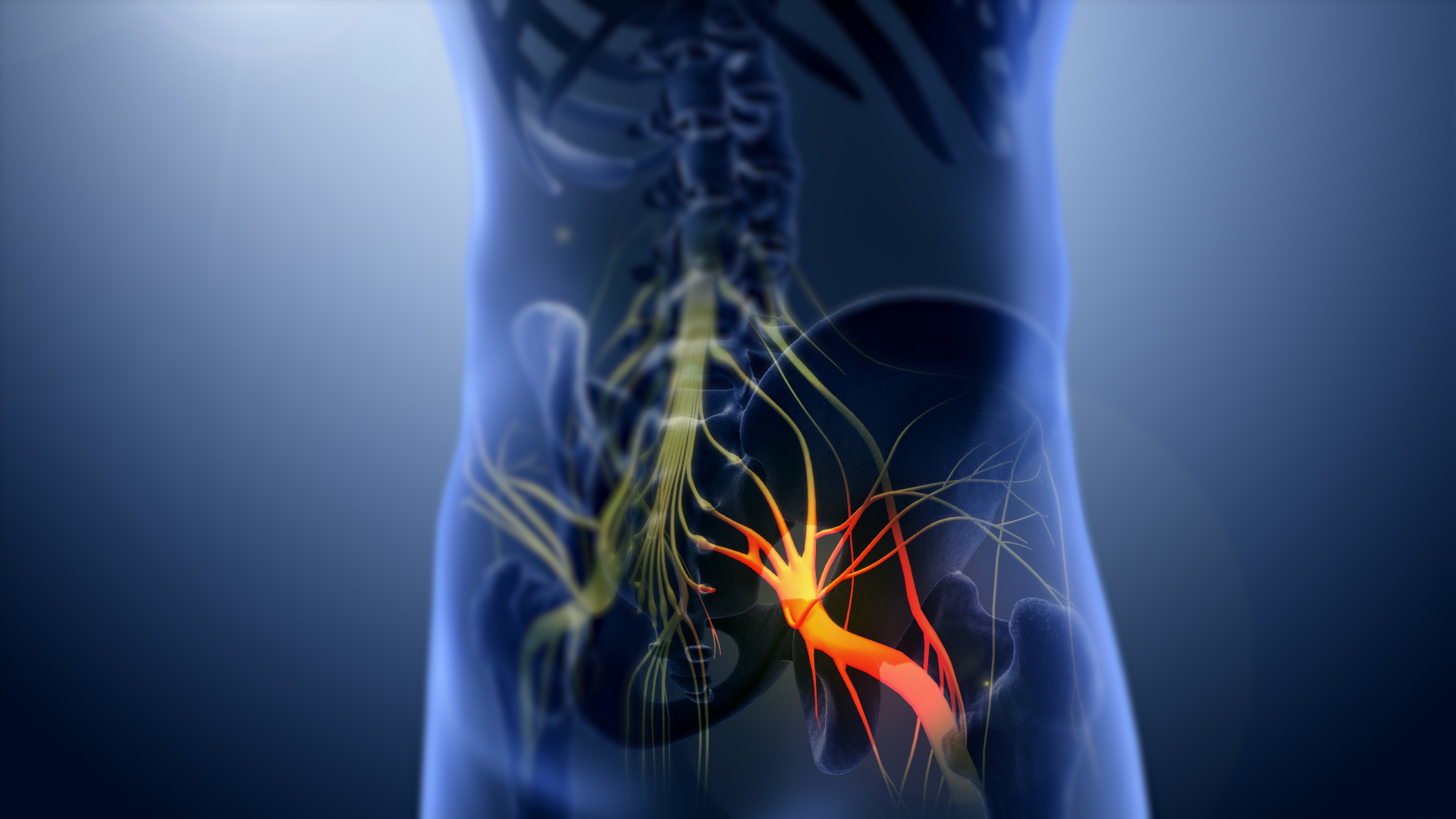
In human anatomy, the sacral plexus is a nerve plexus which provides motor and sensory nerves for the posterior thigh, most of the lower leg and foot, and part of the pelvis. It is part of the lumbosacral plexus and emerges from the lumbar vertebrae and sacral vertebrae (L4-S4).''Thieme Atlas of Anatomy'' (2006), pp 470-471 A sacral plexopathy is a disorder affecting the nerves of the sacral plexus, usually caused by trauma, nerve compression, vascular disease, or infection. Symptoms may include pain, loss of motor control, and sensory deficits. Structure The sacral plexus is formed by: * the lumbosacral trunk * the anterior division of the first sacral nerve * portions of the anterior divisions of the second and third sacral nerves The nerves forming the sacral plexus converge toward the lower part of the greater sciatic foramen, and unite to form a flattened band, from the anterior and posterior surfaces of which several branches arise. The band itself is continued as the ... [...More Info...] [...Related Items...] OR: [Wikipedia] [Google] [Baidu] |
Sciatic Nerve
The sciatic nerve, also called the ischiadic nerve, is a large nerve in humans and other vertebrate animals. It is the largest branch of the sacral plexus and runs alongside the hip joint and down the right lower limb. It is the longest and widest single nerve in the human body, going from the top of the leg to the foot on the posterior aspect. The sciatic nerve has no cutaneous branches for the thigh. This nerve provides the connection to the nervous system for the skin of the lateral leg and the whole foot, the muscles of the back of the thigh, and those of the leg and foot. It is derived from Spinal nerve, spinal nerves Lumbar spinal nerve 4, L4 to Sacral spinal nerve 3, S3. It contains Axon, fibres from both the anterior and posterior divisions of the lumbosacral plexus. Structure In humans, the sciatic nerve is formed from the L4 to S3 segments of the sacral plexus, a collection of nerve fibres that emerge from the Sacrum, sacral part of the spinal cord. The lumbosacral trunk ... [...More Info...] [...Related Items...] OR: [Wikipedia] [Google] [Baidu] |
Tibial Nerve
The tibial nerve is a branch of the sciatic nerve. The tibial nerve passes through the popliteal fossa to pass below the arch of soleus. Structure Popliteal fossa The tibial nerve is the larger terminal branch of the sciatic nerve with root values of L4, L5, S1, S2, and S3. It lies superficial (or posterior) to the popliteal vessels, extending from the superior angle to the inferior angle of the popliteal fossa, crossing the popliteal vessels from lateral to medial side. It gives off branches as shown below: * Muscular branches - Muscular branches arise from the distal part of the popliteal fossa. It supplies the medial and lateral heads of gastrocnemius, soleus, plantaris and popliteus muscles. Nerve to popliteus crosses the popliteus muscle, runs downwards and laterally, winds around the lower border of the popliteus to supply the deep (or anterior) surface of the popliteus. This nerve also supplies the tibialis posterior muscle, superior tibiofibular joint, tibia bone, ... [...More Info...] [...Related Items...] OR: [Wikipedia] [Google] [Baidu] |
Calcaneus
In humans and many other primates, the calcaneus (; from the Latin ''calcaneus'' or ''calcaneum'', meaning heel; : calcanei or calcanea) or heel bone is a bone of the Tarsus (skeleton), tarsus of the foot which constitutes the heel. In some other animals, it is the point of the hock (anatomy), hock. Structure In humans, the calcaneus is the largest of the tarsal bones and the largest bone of the foot. Its long axis is pointed forwards and laterally. The talus bone, calcaneus, and navicular bone are considered the proximal row of tarsal bones. In the calcaneus, several important structures can be distinguished:Platzer (2004), p 216 There is a large calcaneal tuberosity located posteriorly on plantar surface with medial and lateral tubercles on its surface. Besides, there is another peroneal tubercle on its lateral surface. On its lower edge on either side are its lateral and medial processes (serving as the origins of the Abductor hallucis muscle, abductor hallucis and Abductor di ... [...More Info...] [...Related Items...] OR: [Wikipedia] [Google] [Baidu] |


