|
Perisynaptic Schwann Cells
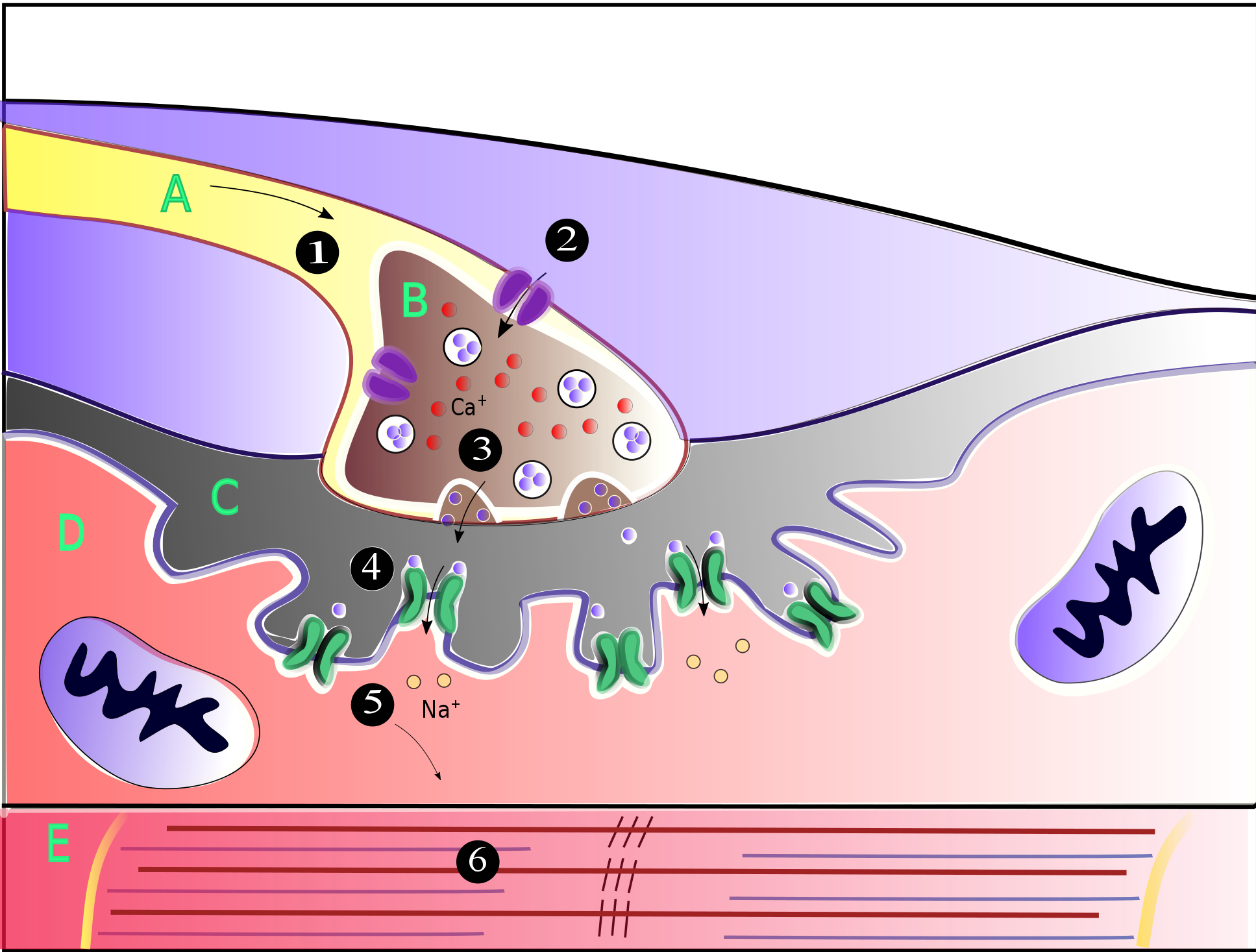
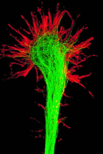
Perisynaptic schwann cells (also known as Terminal schwann cells or Teloglia) are neuroglia found at the Neuromuscular junction (NMJ) with known functions in synaptic transmission, synaptogenesis, and nerve regeneration. These cells share a common ancestor with both Myelinating and Non-Myelinating Schwann Cells called Neural Crest, Neural Crest cells. Perisynaptic Schwann Cells (PSCs) contribute to the tripartite synapse organization in combination with the pre-synaptic nerve and the post-synaptic muscle fiber. PSCs are considered to be the glial component of the Neuromuscular Junction (NMJ) and have a similar functionality to that of Astrocytes in the Central Nervous System. The characteristics of PSCs are based on both external synaptic properties and internal glial properties, where the internal characteristics of PSCs develop based on the associated synapse, for example: the PSCs of a fast-twitch muscle fiber differ from the PSCs of a slow-twitch muscle fiber even when removed ... [...More Info...] [...Related Items...] OR: [Wikipedia] [Google] [Baidu] |
Neuroglia
Glia, also called glial cells (gliocytes) or neuroglia, are non- neuronal cells in the central nervous system (the brain and the spinal cord) and in the peripheral nervous system that do not produce electrical impulses. The neuroglia make up more than one half the volume of neural tissue in the human body. They maintain homeostasis, form myelin, and provide support and protection for neurons. In the central nervous system, glial cells include oligodendrocytes (that produce myelin), astrocytes, ependymal cells and microglia, and in the peripheral nervous system they include Schwann cells (that produce myelin), and satellite cells. Function They have four main functions: * to surround neurons and hold them in place * to supply nutrients and oxygen to neurons * to insulate one neuron from another * to destroy pathogens and remove dead neurons. They also play a role in neurotransmission and synaptic connections, and in physiological processes such as breathing. While ... [...More Info...] [...Related Items...] OR: [Wikipedia] [Google] [Baidu] |
Neuromuscular Junction
A neuromuscular junction (or myoneural junction) is a chemical synapse between a motor neuron and a muscle fiber. It allows the motor neuron to transmit a signal to the muscle fiber, causing muscle contraction. Muscles require innervation to function—and even just to maintain muscle tone, avoiding atrophy. In the neuromuscular system, nerves from the central nervous system and the peripheral nervous system are linked and work together with muscles. Synaptic transmission at the neuromuscular junction begins when an action potential reaches the presynaptic terminal of a motor neuron, which activates voltage-gated calcium channels to allow calcium ions to enter the neuron. Calcium ions bind to sensor proteins (synaptotagmins) on synaptic vesicles, triggering vesicle fusion with the cell membrane and subsequent neurotransmitter release from the motor neuron into the synaptic cleft. In vertebrates, motor neurons release acetylcholine (ACh), a small molecule neurotransmitter, which ... [...More Info...] [...Related Items...] OR: [Wikipedia] [Google] [Baidu] |
Synaptogenesis
Synaptogenesis is the formation of synapses between neurons in the nervous system. Although it occurs throughout a healthy person's lifespan, an explosion of synapse formation occurs during early brain development, known as exuberant synaptogenesis. Synaptogenesis is particularly important during an individual's critical period, during which there is a certain degree of synaptic pruning due to competition for neural growth factors by neurons and synapses. Processes that are not used, or inhibited during their critical period will fail to develop normally later on in life. Exuberant synaptogenesis Brain growth and development begins during gestation and into the postnatal period. Brain development can be divided into stages including: neurogenesis, differentiation, proliferation, migration, synaptogenesis, gliogenesis and myelination, and apoptosis and synaptic pruning. Synaptogenesis occurs in the third trimester during gestation as well as the first two years postnatal. Dur ... [...More Info...] [...Related Items...] OR: [Wikipedia] [Google] [Baidu] |
Neural Crest
The neural crest is a ridge-like structure that is formed transiently between the epidermal ectoderm and neural plate during vertebrate development. Neural crest cells originate from this structure through the epithelial-mesenchymal transition, and in turn give rise to a diverse cell lineage—including melanocytes, craniofacial cartilage and bone, smooth muscle, dentin, peripheral and enteric neurons, adrenal medulla and glia. After gastrulation, the neural crest is specified at the border of the neural plate and the non-neural ectoderm. During neurulation, the borders of the neural plate, also known as the neural folds, converge at the dorsal midline to form the neural tube. Subsequently, neural crest cells from the roof plate of the neural tube undergo an epithelial to mesenchymal transition, delaminating from the neuroepithelium and migrating through the periphery, where they differentiate into varied cell types. The emergence of the neural crest was important in v ... [...More Info...] [...Related Items...] OR: [Wikipedia] [Google] [Baidu] |
Astrocyte
Astrocytes (from Ancient Greek , , "star" and , , "cavity", "cell"), also known collectively as astroglia, are characteristic star-shaped glial cells in the brain and spinal cord. They perform many functions, including biochemical control of endothelial cells that form the blood–brain barrier, provision of nutrients to the nervous tissue, maintenance of extracellular ion balance, regulation of cerebral blood flow, and a role in the repair and scarring process of the brain and spinal cord following infection and traumatic injuries. The proportion of astrocytes in the brain is not well defined; depending on the counting technique used, studies have found that the astrocyte proportion varies by region and ranges from 20% to around 40% of all glia. Another study reports that astrocytes are the most numerous cell type in the brain. Astrocytes are the major source of cholesterol in the central nervous system. Apolipoprotein E transports cholesterol from astrocytes to neurons and ot ... [...More Info...] [...Related Items...] OR: [Wikipedia] [Google] [Baidu] |
Central Nervous System
The central nervous system (CNS) is the part of the nervous system consisting primarily of the brain, spinal cord and retina. The CNS is so named because the brain integrates the received information and coordinates and influences the activity of all parts of the bodies of bilateria, bilaterally symmetric and triploblastic animals—that is, all multicellular animals except sponges and Coelenterata, diploblasts. It is a structure composed of nervous tissue positioned along the Anatomical_terms_of_location#Rostral,_cranial,_and_caudal, rostral (nose end) to caudal (tail end) axis of the body and may have an enlarged section at the rostral end which is a brain. Only arthropods, cephalopods and vertebrates have a true brain, though precursor structures exist in onychophorans, gastropods and lancelets. The rest of this article exclusively discusses the vertebrate central nervous system, which is radically distinct from all other animals. Overview In vertebrates, the brain and spinal ... [...More Info...] [...Related Items...] OR: [Wikipedia] [Google] [Baidu] |
Peripheral Nervous System
The peripheral nervous system (PNS) is one of two components that make up the nervous system of Bilateria, bilateral animals, with the other part being the central nervous system (CNS). The PNS consists of nerves and ganglia, which lie outside the brain and the spinal cord. The main function of the PNS is to connect the CNS to the Limb (anatomy), limbs and Organ (anatomy), organs, essentially serving as a relay between the brain and spinal cord and the rest of the body. Unlike the CNS, the PNS is not protected by the vertebral column and skull, or by the blood–brain barrier, which leaves it exposed to toxins. The peripheral nervous system can be divided into a somatic nervous system, somatic division and an autonomic nervous system, autonomic division. Each of these can further be differentiated into a sensory and a motor sector. In the somatic nervous system, the cranial nerves are part of the PNS with the exceptions of the olfactory nerve and epithelia and the optic nerve (c ... [...More Info...] [...Related Items...] OR: [Wikipedia] [Google] [Baidu] |
Louis-Antoine Ranvier
Louis-Antoine Ranvier (2 October 1835 – 22 March 1922) was a French physician, pathologist, anatomist and histologist, who discovered the nodes of Ranvier, regularly spaced discontinuities of the myelin sheath, occurring at varying intervals along the length of a nerve fiber. Career Ranvier was born and studied medicine at Lyon, graduating in 1865 from the Ecole Préparatoire de Médecine et de Pharmacie. He moved to Paris after receiving the internship of Parisian hospitals. Here he founded a small private research laboratory on Rue Christine along with fellow intern Victor André Cornil, and together they later offered a course in histology to medical students which involved the careful examination of tissues under a microscope. Their course was unique in the time as microscopy had not been viewed favourably in medicine especially by Henri Ducrotay de Blainville (1777-1850) and Auguste Comte (1798-1857). Their histology course material became an influential textbook o ... [...More Info...] [...Related Items...] OR: [Wikipedia] [Google] [Baidu] |
Schwann Cell Linage Flowchart
Schwann may refer to: *The Schwann cell * Henry Schwann, English cricketer *Theodor Schwann Theodor Schwann (; 7 December 181011 January 1882) was a German physician and physiology, physiologist. His most significant contribution to biology is considered to be the extension of cell theory to animals. Other contributions include the d ..., a German physiologist, histologist and cytologist * Schwann Records, a German record label * The Schwann catalog, a listing of in-print sound recordings in the United States, published from the late 1940s through 2001 See also * Schwan (other) {{disambig, surname German-language surnames ... [...More Info...] [...Related Items...] OR: [Wikipedia] [Google] [Baidu] |
Neural Tube
In the developing chordate (including vertebrates), the neural tube is the embryonic precursor to the central nervous system, which is made up of the brain and spinal cord. The neural groove gradually deepens as the neural folds become elevated, and ultimately the folds meet and coalesce in the middle line and convert the groove into the closed neural tube. In humans, neural tube closure usually occurs by the fourth week of pregnancy (the 28th day after conception). Development The neural tube develops in two ways: primary neurulation and secondary neurulation. Primary neurulation divides the ectoderm into three cell types: * The internally located neural tube * The externally located epidermis * The neural crest cells, which develop in the region between the neural tube and epidermis but then migrate to new locations # Primary neurulation begins after the neural plate forms. The edges of the neural plate start to thicken and lift upward, forming the neural folds. The center ... [...More Info...] [...Related Items...] OR: [Wikipedia] [Google] [Baidu] |
Enteric Nervous System
The enteric nervous system (ENS) is one of the three divisions of the autonomic nervous system (ANS), the others being the sympathetic nervous system (SNS) and parasympathetic nervous system (PSNS). It consists of a mesh-like system of neurons that governs the function of the gastrointestinal tract. The ENS is nicknamed the "second brain". It is derived from neural crest cells. The enteric nervous system is capable of operating independently of the brain and spinal cord, but is thought to rely on innervation from the vagus nerve and prevertebral ganglia in healthy subjects. However, studies have shown that the system is operable with a severed vagus nerve. The neurons of the enteric nervous system control the motor functions of the system, in addition to the secretion of gastrointestinal enzymes. These neurons communicate through many neurotransmitters similar to the CNS, including acetylcholine, dopamine, and serotonin. The large presence of serotonin and dopamine in the intest ... [...More Info...] [...Related Items...] OR: [Wikipedia] [Google] [Baidu] |
Transforming Growth Factor Beta
Transforming growth factor beta (TGF-β) is a multifunctional cytokine belonging to the transforming growth factor superfamily that includes three different mammalian isoforms (TGF-β 1 to 3, HGNC symbols TGFB1, TGFB2, TGFB3) and many other signaling proteins. TGFB proteins are produced by all white blood cell lineages. Activated TGF-β complexes with other factors to form a serine/threonine kinase complex that binds to TGF-β receptors. TGF-β receptors are composed of both type 1 and type 2 receptor subunits. After the binding of TGF-β, the type 2 receptor kinase phosphorylates and activates the type 1 receptor kinase that activates a signaling cascade. This leads to the activation of different downstream substrates and regulatory proteins, inducing transcription of different target genes that function in differentiation, chemotaxis, proliferation, and activation of many immune cells. TGF-β is secreted by many cell types, including macrophages, in a latent form in whic ... [...More Info...] [...Related Items...] OR: [Wikipedia] [Google] [Baidu] |


