|
Periodontology
Periodontology or periodontics (from Ancient Greek , – 'around'; and , – 'tooth', genitive , ) is the Specialty (dentistry), specialty of dentistry that studies supporting structures of Tooth, teeth, as well as diseases and conditions that affect them. The supporting tissues are known as the periodontium, which includes the gingiva (gums), Alveolar process, alveolar bone, cementum, and the periodontal ligament. A periodontist is a dentist that specializes in the prevention, diagnosis and treatment of periodontal disease and in the placement of dental implants. The periodontium The term ''periodontium'' is used to describe the group of structures that directly surround, support and protect the teeth. The periodontium is composed largely of the gingival tissue and the supporting bone. Gingivae Normal gingiva may range in color from light coral pink to heavily pigmented. The soft tissues and connective fibres that cover and protect the underlying cementum, periodontal lig ... [...More Info...] [...Related Items...] OR: [Wikipedia] [Google] [Baidu] |
Periodontal Disease
Periodontal disease, also known as gum disease, is a set of inflammatory conditions affecting the tissues surrounding the teeth. In its early stage, called gingivitis, the gums become swollen and red and may bleed. It is considered the main cause of tooth loss for adults worldwide. In its more serious form, called periodontitis, the gums can pull away from the tooth, bone can be lost, and the teeth may loosen or fall out. Halitosis (bad breath) may also occur. Periodontal disease typically arises from the development of plaque biofilm, which harbors harmful bacteria such as ''Porphyromonas gingivalis'' and ''Treponema denticola''. These bacteria infect the gum tissue surrounding the teeth, leading to inflammation and, if left untreated, progressive damage to the teeth and gum tissue. Recent meta-analysis have shown that the composition of the oral microbiota and its response to periodontal disease differ between men and women. These differences are particularly notable in t ... [...More Info...] [...Related Items...] OR: [Wikipedia] [Google] [Baidu] |
Dental Implant
A dental implant (also known as an endosseous implant or fixture) is a prosthesis that interfaces with the bone of the jaw or skull to support a dental prosthesis such as a crown (dentistry), crown, bridge (dentistry), bridge, dentures, denture, or facial prosthesis or to act as an Dental braces, orthodontic anchor. The basis for modern dental implants is a biological process called osseointegration, in which materials such as titanium or Zirconium dioxide, zirconia form an intimate bond to the bone. The implant fixture is first placed so that it is likely to osseointegrate, then a dental prosthetic is added. A variable amount of healing time is required for osseointegration before either the dental prosthetic (a tooth, bridge, or denture) is attached to the implant or an abutment (dentistry), abutment is placed which will hold a dental prosthetic or crown. Success or failure of implants depends primarily on the thickness and health of the bone and gingival tissues that surround ... [...More Info...] [...Related Items...] OR: [Wikipedia] [Google] [Baidu] |
Gingivitis
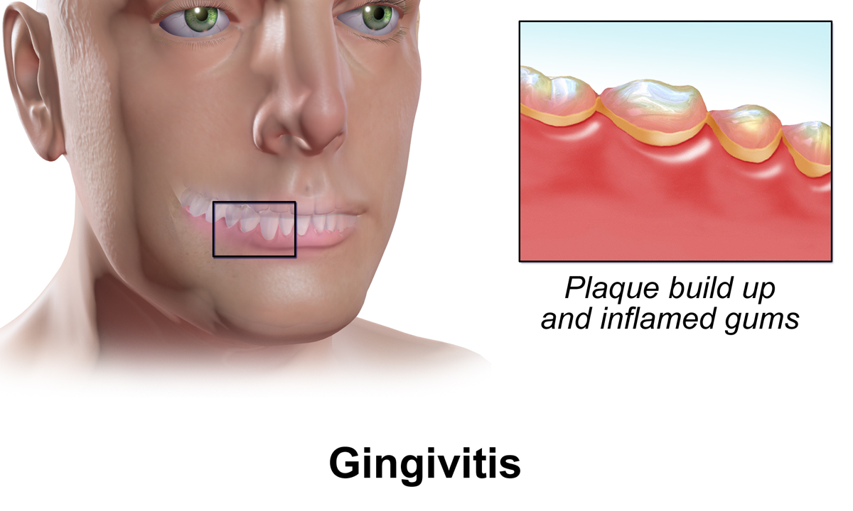
Gingivitis is a non-destructive disease that causes inflammation of the gums; ulitis is an alternative term. The most common form of gingivitis, and the most common form of periodontal disease overall, is in response to bacterial biofilms (also called plaque) that are attached to tooth surfaces, termed ''plaque-induced gingivitis''. Most forms of gingivitis are plaque-induced. While some cases of gingivitis never progress to periodontitis, periodontitis is always preceded by gingivitis. Gingivitis is reversible with good oral hygiene; however, without treatment, gingivitis can progress to periodontitis, in which the inflammation of the gums results in tissue destruction and bone resorption around the teeth. Periodontitis can ultimately lead to tooth loss. Signs and symptoms The symptoms of gingivitis are somewhat non-specific and manifest in the gum tissue as the classic signs of inflammation: *Swollen gums *Bright red gums *Gums that are tender or painful to the touch *B ... [...More Info...] [...Related Items...] OR: [Wikipedia] [Google] [Baidu] |
Dental Plaque
Dental plaque is a biofilm of microorganisms (mostly bacteria, but also fungi) that grows on surfaces within the mouth. It is a sticky colorless deposit at first, but when it forms Calculus (dental), tartar, it is often brown or pale yellow. It is commonly found between the teeth, on the front of teeth, behind teeth, on chewing surfaces, along the gums, gumline (supragingival), or below the gumline cervical margins (subgingival). Dental plaque is also known as microbial plaque, oral biofilm, dental biofilm, dental plaque biofilm or bacterial plaque biofilm. Bacterial plaque is one of the major causes for dental decay and gum disease. It has been observed that differences in the composition of dental plaque microbiota exist between men and women, particularly in the presence of periodontal disease, periodontitis. Progression and build-up of dental plaque can give rise to tooth decay – the localised destruction of the tissues of the tooth by acid produced from the bacterial degrad ... [...More Info...] [...Related Items...] OR: [Wikipedia] [Google] [Baidu] |
Periodontal Fiber
The periodontal ligament, commonly abbreviated as the PDL, are a group of specialized connective tissue fibers that essentially attach a tooth to the alveolar bone within which they sit. It inserts into root cementum on one side and onto alveolar bone on the other. Structure The PDL consists of principal fibers, loose connective tissue, blast and clast cells, oxytalan fibers and cell rest of Malassez. Alveolodental ligament The main principal fiber group is the alveolodental ligament, which consists of five fiber subgroups: alveolar crest, horizontal, oblique, apical, and interradicular on multirooted teeth. Principal fibers other than the alveolodental ligament are the transseptal fibers. All these fibers help the tooth withstand the naturally substantial compressive forces that occur during chewing and remain embedded in the bone. The ends of the principal fibers that are within either cementum or alveolar bone proper are considered Sharpey fibers. * Alveolar crest ... [...More Info...] [...Related Items...] OR: [Wikipedia] [Google] [Baidu] |
Periodontal Ligament
The periodontal ligament, commonly abbreviated as the PDL, are a group of specialized connective tissue fibers that essentially attach a tooth to the alveolar bone within which they sit. It inserts into root cementum on one side and onto alveolar bone on the other. Structure The PDL consists of principal fibers, loose connective tissue, blast and clast cells, oxytalan fibers and cell rest of Malassez. Alveolodental ligament The main principal fiber group is the alveolodental ligament, which consists of five fiber subgroups: alveolar crest, horizontal, oblique, apical, and interradicular on multirooted teeth. Principal fibers other than the alveolodental ligament are the transseptal fibers. All these fibers help the tooth withstand the naturally substantial compressive forces that occur during chewing and remain embedded in the bone. The ends of the principal fibers that are within either cementum or alveolar bone proper are considered Sharpey fibers. * Alveolar cre ... [...More Info...] [...Related Items...] OR: [Wikipedia] [Google] [Baidu] |
Gingival Sulcus
In dental anatomy, the gingival sulcus is an area of potential space between a tooth and the surrounding gingiva, gingival tissue and is lined by sulcular epithelium. The depth of the sulcus (Latin for ''groove'') is bounded by two entities: Commonly used terms of relationship and comparison in dentistry, apically by the gingival fibers of the connective tissue attachment and Commonly used terms of relationship and comparison in dentistry, coronally by the free gingival margin. A healthy sulcular depth is three millimeters or less, which is readily self-cleansable with a properly used toothbrush or the supplemental use of other oral hygiene aids. Anatomy The dentogingival tissues consist of many constituents, such as the enamel or cementum of the tooth and the connective tissue supporting epithelia like the junctional epithelium, the gingival epithelium and the sulcular epithelium. The junctional epithelium is developed during the eruption of teeth when the reduced enamel ... [...More Info...] [...Related Items...] OR: [Wikipedia] [Google] [Baidu] |
Mucogingival Junction
A mucogingival junction is an anatomical feature found on the intraoral mucosa. The mucosa of the cheeks and floor of the mouth are freely moveable and fragile, whereas the mucosa around the teeth and on the palate are firm and keratinized. Where the two tissue types meet is known as a mucogingival junction. There are three mucogingival junctions: on the facial of the maxilla and on both the facial and lingual of the mandible. The palatal gingiva of the maxilla is continuous with the tissue of the palate, which is bound down to the palatal bones. Because the palate is devoid of freely moveable alveolar mucosa, there is no mucogingival junction.Carranza's Clinical Periodontology, W.B. Saunders 2002, page 17. Clinical importance The clinical importance of the mucogingival junction is in measuring the width of attached gingiva. Attached gingiva is important because it is bound very tightly to the underlying alveolar bone and provides protection to the mucosa during functiona ... [...More Info...] [...Related Items...] OR: [Wikipedia] [Google] [Baidu] |
Sulcular Epithelium
In dental anatomy, the sulcular epithelium is that epithelium Epithelium or epithelial tissue is a thin, continuous, protective layer of cells with little extracellular matrix. An example is the epidermis, the outermost layer of the skin. Epithelial ( mesothelial) tissues line the outer surfaces of man ... which lines the gingival sulcus.Carranza's Clinical Periodontology, W.B. Saunders, 2002, page 23. It is apically bounded by the junctional epithelium and meets the epithelium of the oral cavity at the height of the free gingival margin. The sulcular epithelium is nonkeratinized. References Dental anatomy {{dentistry-stub ... [...More Info...] [...Related Items...] OR: [Wikipedia] [Google] [Baidu] |
Alveolar Process
The alveolar process () is the portion of bone containing the tooth sockets on the jaw bones (in humans, the maxilla and the mandible). The alveolar process is covered by gums within the mouth, terminating roughly along the line of the mandibular canal. Partially comprising compact bone, it is penetrated by many small openings for blood vessels and connective fibres. The bone is of clinical, phonetic and forensic significance. Terminology The term ''alveolar'' () ('hollow') refers to the cavities of the tooth sockets, known as dental alveoli. The alveolar process is also called the ''alveolar bone'' or ''alveolar ridge''. In phonetics, the term refers more specifically to the ridges on the inside of the mouth which can be felt with the tongue, either on roof of the mouth between the upper teeth and the hard palate or on the bottom of the mouth behind the lower teeth. The curved portion of the process is referred to as the alveolar arch. The alveolar bone proper, also ca ... [...More Info...] [...Related Items...] OR: [Wikipedia] [Google] [Baidu] |
Cementum
Cementum is a specialized calcified substance covering the root of a tooth. The cementum is the part of the periodontium that attaches the teeth to the alveolar bone by anchoring the periodontal ligament. Structure The cells of cementum are the entrapped cementoblasts, the cementocytes. Each cementocyte lies in its lacuna, similar to the pattern noted in bone. These lacunae also have canaliculi or canals. Unlike those in bone, however, these canals in cementum do not contain nerves, nor do they radiate outward. Instead, the canals are oriented toward the periodontal ligament and contain cementocytic processes that exist to diffuse nutrients from the ligament because it is vascularized. After the apposition of cementum in layers, the cementoblasts that do not become entrapped in cementum line up along the cemental surface along the length of the outer covering of the periodontal ligament. These cementoblasts can form subsequent layers of cementum if the tooth is injured. Sh ... [...More Info...] [...Related Items...] OR: [Wikipedia] [Google] [Baidu] |
Periodontium
The periodontium () is the specialized tissues that both surround and support the teeth, maintaining them in the maxillary and mandibular bones. Periodontics is the dental specialty that relates specifically to the care and maintenance of these tissues. It provides the support necessary to maintain teeth in function. It consists of four principal components, namely: * Gingiva (the gums) * Periodontal ligament (PDL) * Cementum * Alveolar bone proper Each of these components is distinct in location, architecture, and biochemical properties, which adapt during the life of the structure. For example, as teeth respond to forces or migrate medially, bone resorbs on the pressure side and is added on the tension side. Cementum similarly adapts to wear on the occlusal surfaces of the teeth by apical deposition. The periodontal ligament in itself is an area of high turnover that allows the tooth not only to be suspended in the alveolar bone but also to respond to the forces. Thus, ... [...More Info...] [...Related Items...] OR: [Wikipedia] [Google] [Baidu] |


