|
PET-CT
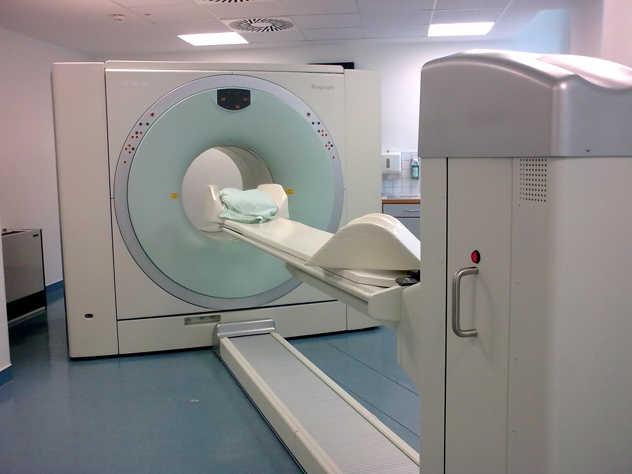
Positron emission tomography–computed tomography (better known as PET-CT or PET/CT) is a nuclear medicine technique which combines, in a single gantry, a positron emission tomography (PET) scanner and an x-ray computed tomography (CT) scanner, to acquire sequential images from both devices in the same session, which are combined into a single superposed ( co-registered) image. Thus, functional imaging obtained by PET, which depicts the spatial distribution of metabolic or biochemical activity in the body can be more precisely aligned or correlated with anatomic imaging obtained by CT scanning. Two- and three-dimensional image reconstruction may be rendered as a function of a common software and control system. PET-CT has revolutionized medical diagnosis in many fields, by adding precision of anatomic localization to functional imaging, which was previously lacking from pure PET imaging. For example, many diagnostic imaging procedures in oncology, surgical planning, radi ... [...More Info...] [...Related Items...] OR: [Wikipedia] [Google] [Baidu] |
PET-CT Siemens Biograph01
Positron emission tomography–computed tomography (better known as PET-CT or PET/CT) is a nuclear medicine technique which combines, in a single gantry, a positron emission tomography (PET) scanner and an x-ray computed tomography (CT) scanner, to acquire sequential images from both devices in the same session, which are combined into a single superposed ( co-registered) image. Thus, functional imaging obtained by PET, which depicts the spatial distribution of metabolic or biochemical activity in the body can be more precisely aligned or correlated with anatomic imaging obtained by CT scanning. Two- and three-dimensional image reconstruction may be rendered as a function of a common software and control system. PET-CT has revolutionized medical diagnosis in many fields, by adding precision of anatomic localization to functional imaging, which was previously lacking from pure PET imaging. For example, many diagnostic imaging procedures in oncology, surgical planning, radiation t ... [...More Info...] [...Related Items...] OR: [Wikipedia] [Google] [Baidu] |
Positron Emission Tomography
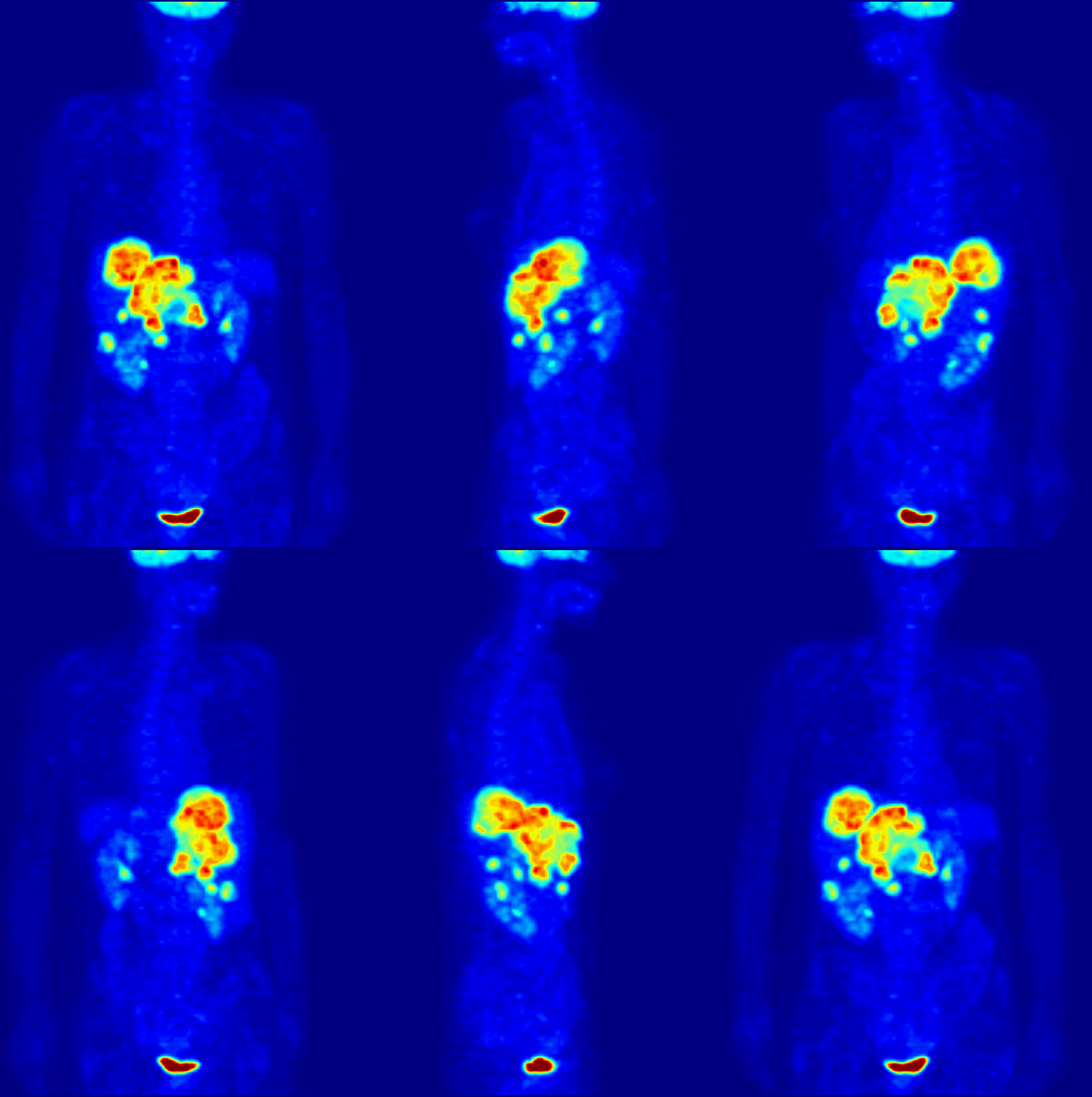
Positron emission tomography (PET) is a functional imaging technique that uses radioactive substances known as radiotracers to visualize and measure changes in metabolic processes, and in other physiological activities including blood flow, regional chemical composition, and absorption. Different tracers are used for various imaging purposes, depending on the target process within the body. For example: * Fluorodeoxyglucose ( 18F">sup>18FDG or FDG) is commonly used to detect cancer; * 18Fodium fluoride">sup>18Fodium fluoride (Na18F) is widely used for detecting bone formation; * Oxygen-15 (15O) is sometimes used to measure blood flow. PET is a common imaging technique, a medical scintillography technique used in nuclear medicine. A radiopharmaceutical – a radioisotope attached to a drug – is injected into the body as a radioactive tracer, tracer. When the radiopharmaceutical undergoes beta plus decay, a positron is emitted, and when the positron interacts with an or ... [...More Info...] [...Related Items...] OR: [Wikipedia] [Google] [Baidu] |
PET-MRI
Positron emission tomography–magnetic resonance imaging (PET–MRI) is a hybrid imaging technology that incorporates magnetic resonance imaging (MRI) soft tissue morphological imaging and positron emission tomography (PET) functional imaging. The combination of PET and MRI was mentioned in a 1991 Phd thesis by R. Raylman. Simultaneous PET/MR detection was first demonstrated in 1997, however it took another 13 years, and new detector technologies, for clinical systems to become commercially available. Applications Presently, the main clinical fields of PET-MRI are oncology, cardiology, neurology, and neuroscience. Research studies are actively conducted at the moment to understand benefits of the new PET-MRI diagnostic method. The technology combines the exquisite structural and functional characterization of tissue provided by MRI with the extreme sensitivity of PET imaging of metabolism and tracking of uniquely labeled cell types or cell receptors. Manufacturers Several compa ... [...More Info...] [...Related Items...] OR: [Wikipedia] [Google] [Baidu] |
CT Scan
A computed tomography scan (CT scan; formerly called computed axial tomography scan or CAT scan) is a medical imaging technique used to obtain detailed internal images of the body. The personnel that perform CT scans are called radiographers or radiology technologists. CT scanners use a rotating X-ray tube and a row of detectors placed in a gantry to measure X-ray attenuations by different tissues inside the body. The multiple X-ray measurements taken from different angles are then processed on a computer using tomographic reconstruction algorithms to produce tomographic (cross-sectional) images (virtual "slices") of a body. CT scans can be used in patients with metallic implants or pacemakers, for whom magnetic resonance imaging (MRI) is contraindicated. Since its development in the 1970s, CT scanning has proven to be a versatile imaging technique. While CT is most prominently used in medical diagnosis, it can also be used to form images of non-living objects. The 1979 N ... [...More Info...] [...Related Items...] OR: [Wikipedia] [Google] [Baidu] |
Nuclear Medicine
Nuclear medicine or nucleology is a medical specialty involving the application of radioactive substances in the diagnosis and treatment of disease. Nuclear imaging, in a sense, is "radiology done inside out" because it records radiation emitting from within the body rather than radiation that is generated by external sources like X-rays. In addition, nuclear medicine scans differ from radiology, as the emphasis is not on imaging anatomy, but on the function. For such reason, it is called a physiological imaging modality. Single photon emission computed tomography (SPECT) and positron emission tomography (PET) scans are the two most common imaging modalities in nuclear medicine. Diagnostic medical imaging Diagnostic In nuclear medicine imaging, radiopharmaceuticals are taken internally, for example, through inhalation, intravenously or orally. Then, external detectors ( gamma cameras) capture and form images from the radiation emitted by the radiopharmaceuticals. This ... [...More Info...] [...Related Items...] OR: [Wikipedia] [Google] [Baidu] |
Functional Imaging
Functional imaging (or physiological imaging) is a medical imaging technique of detecting or measuring changes in metabolism, blood flow, regional chemical composition, and absorption. As opposed to structural imaging, functional imaging centers on revealing physiological activities within a certain tissue or organ by employing medical image modalities that very often use radioactive tracers, tracers or probes to reflect spatial distribution of them within the body. These tracers are often structural analog, analogous to some chemical compounds, like glucose, within the body. To achieve this, isotopes are used because they have similar chemical and biological characteristics. By appropriate proportionality, the nuclear medicine physicians can determine the real intensity of certain substance within the body to evaluate the risk or danger of developing some diseases. Modalities * Positron emission tomography (PET) ** Fludeoxyglucose for Glucose metabolism ** O-15 as a flow tracer ... [...More Info...] [...Related Items...] OR: [Wikipedia] [Google] [Baidu] |
Cyclotron
A cyclotron is a type of particle accelerator invented by Ernest O. Lawrence in 1929–1930 at the University of California, Berkeley, and patented in 1932. Lawrence, Ernest O. ''Method and apparatus for the acceleration of ions'', filed: January 26, 1932, granted: February 20, 1934 A cyclotron accelerates charged particles outwards from the center of a flat cylindrical vacuum chamber along a spiral path. The particles are held to a spiral trajectory by a static magnetic field and accelerated by a rapidly varying electric field. Lawrence was awarded the 1939 Nobel Prize in Physics for this invention. The cyclotron was the first "cyclical" accelerator. The primary accelerators before the development of the cyclotron were electrostatic accelerators, such as the Cockcroft–Walton accelerator and Van de Graaff generator. In these accelerators, particles would cross an accelerating electric field only once. Thus, the energy gained by the particles was limited by the maximu ... [...More Info...] [...Related Items...] OR: [Wikipedia] [Google] [Baidu] |
Gallium-68 Generator
A germanium-68/gallium-68 generator is a device used to extract the positron-emitting isotope 68Ga of gallium from a source of decaying germanium-68. The parent isotope 68Ge has a half-life of 271 days and can be easily utilized for in-hospital production of generator produced 68Ga. Its decay product gallium-68 (with a half-life of only 68 minutes, inconvenient for transport) is extracted and used for certain positron emission tomography nuclear medicine diagnostic procedures, where the radioisotope's relatively short half-life and emission of positrons for creation of 3-dimensional PET scans, are useful. Parent isotope (68Ge) source The parent isotope germanium-68 is the longest-lived (271 days) of the radioisotopes of germanium. It has been produced by several methods. In the U.S., it is primarily produced in proton accelerators: At Los Alamos National Laboratory, it may be separated out as a product of proton capture, after proton irradiation of Nb-encapsulated gallium metal. ... [...More Info...] [...Related Items...] OR: [Wikipedia] [Google] [Baidu] |
National Cancer Institute
The National Cancer Institute (NCI) coordinates the United States National Cancer Program and is part of the National Institutes of Health (NIH), which is one of eleven agencies that are part of the U.S. Department of Health and Human Services. The NCI conducts and supports research, training, health information dissemination, and other activities related to the causes, prevention, diagnosis, and treatment of cancer; the supportive care of cancer patients and their families; and cancer survivorship. NCI is the oldest and has the largest budget and research program of the 27 institutes and centers of the NIH ($6.9 billion in 2020). It fulfills the majority of its mission via an extramural program that provides grants for cancer research. Additionally, the National Cancer Institute has intramural research programs in Bethesda, Maryland, and at the Frederick National Laboratory for Cancer Research at Fort Detrick in Frederick, Maryland. The NCI receives more than in funding eac ... [...More Info...] [...Related Items...] OR: [Wikipedia] [Google] [Baidu] |
University Of Geneva
The University of Geneva (French: ''Université de Genève'') is a public research university located in Geneva, Switzerland. It was founded in 1559 by John Calvin as a theological seminary. It remained focused on theology until the 17th century, when it became a center for enlightenment scholarship. Today, it is the third largest university in Switzerland by number of students. In 1873, it dropped its religious affiliations and became officially secular. In 2009, the University of Geneva celebrated the 450th anniversary of its founding. Almost 40% of the students come from foreign countries. The university holds and actively pursues teaching, research, and community service as its primary objectives. In 2016, it was ranked 53rd worldwide by the Shanghai Academic Ranking of World Universities, 89th by the QS World University Rankings, and 131st in the Times Higher Education World University Ranking. UNIGE is a member of the League of European Research Universities (includi ... [...More Info...] [...Related Items...] OR: [Wikipedia] [Google] [Baidu] |
Knoxville, TN
Knoxville is a city in and the county seat of Knox County in the U.S. state of Tennessee. As of the 2020 United States census, Knoxville's population was 190,740, making it the largest city in the East Tennessee Grand Division and the state's third largest city after Nashville and Memphis.U.S. Census Bureau2010 Census Interactive Population Search. Retrieved: December 20, 2011. Knoxville is the principal city of the Knoxville Metropolitan Statistical Area, which had an estimated population of 869,046 in 2019. First settled in 1786, Knoxville was the first capital of Tennessee. The city struggled with geographic isolation throughout the early 19th century. The arrival of the railroad in 1855 led to an economic boom. The city was bitterly divided over the secession issue during the American Civil War and was occupied alternately by Confederate and Union armies, culminating in the Battle of Fort Sanders in 1863. Following the war, Knoxville grew rapidly as a major whole ... [...More Info...] [...Related Items...] OR: [Wikipedia] [Google] [Baidu] |


