|
Occipital Horn Syndrome
Occipital horn syndrome (OHS), formerly considered a variant of Ehlers–Danlos syndrome, is an X-linked recessive mitochondrial and connective tissue disorder. It is caused by a deficiency in the transport of the essential mineral copper, associated with mutations in the ATP7A gene. Only about 2/3 of children with OHS are thought to have genetically inherited the disorder; the other 1/3 do not have the disease in their family history. Since the disorder is X-linked recessive the disease affects more males. This is because they do not have a second X chromosome, unlike females, so essentially are lacking the 'backup' copy with proper function. Females are much more likely to be carriers only. For a female to be affected they must carry two defective X chromosomes, not just one.Horn Syndro ... [...More Info...] [...Related Items...] OR: [Wikipedia] [Google] [Baidu] |
X-linked Recessive
X-linked recessive inheritance is a mode of inheritance in which a mutation in a gene on the X chromosome causes the phenotype to be always expressed in males (who are necessarily homozygous for the gene mutation because they have one X and one Y chromosome) and in females who are homozygous for the gene mutation, see zygosity. Females with one copy of the mutated gene are carriers. X-linked inheritance means that the gene causing the trait or the disorder is located on the X chromosome. Females have two X chromosomes while males have one X and one Y chromosome. Carrier females who have only one copy of the mutation do not usually express the phenotype, although differences in X-chromosome inactivation (known as skewed X-inactivation) can lead to varying degrees of clinical expression in carrier females, since some cells will express one X allele and some will express the other. The current estimate of sequenced X-linked genes is 499, and the total, including vaguely defined trait ... [...More Info...] [...Related Items...] OR: [Wikipedia] [Google] [Baidu] |
Elbow-joint
The elbow is the region between the arm and the forearm that surrounds the elbow joint. The elbow includes prominent landmarks such as the olecranon, the cubital fossa (also called the chelidon, or the elbow pit), and the lateral and the medial epicondyles of the humerus. The elbow joint is a hinge joint between the arm and the forearm; more specifically between the humerus in the upper arm and the radius and ulna in the forearm which allows the forearm and hand to be moved towards and away from the body. The term ''elbow'' is specifically used for humans and other primates, and in other vertebrates forelimb plus joint is used. The name for the elbow in Latin is ''cubitus'', and so the word cubital is used in some elbow-related terms, as in ''cubital nodes'' for example. Structure Joint The elbow joint has three different portions surrounded by a common joint capsule. These are joints between the three bones of the elbow, the humerus of the upper arm, and the radius a ... [...More Info...] [...Related Items...] OR: [Wikipedia] [Google] [Baidu] |
X-linked Recessive Disorders
Sex linked describes the sex-specific patterns of inheritance and presentation when a gene mutation (allele) is present on a sex chromosome (allosome) rather than a non-sex chromosome (autosome). In humans, these are termed X-linked recessive, X-linked dominant and Y-linked. The inheritance and presentation of all three differ depending on the sex of both the parent and the child. This makes them characteristically different from autosomal dominance and recessiveness. There are many more X-linked conditions than Y-linked conditions, since humans have several times as many genes on the X chromosome than the Y chromosome. Only females are able to be carriers for X-linked conditions; males will always be affected by any X-linked condition, since they have no second X chromosome with a healthy copy of the gene. As such, X-linked recessive conditions affect males much more commonly than females. In X-linked recessive inheritance, a son born to a carrier mother and an unaffected fat ... [...More Info...] [...Related Items...] OR: [Wikipedia] [Google] [Baidu] |
Inborn Errors Of Metal Metabolism
Inborn errors of metabolism form a large class of genetic diseases involving congenital disorders of enzyme activities. The majority are due to defects of single genes that code for enzymes that facilitate conversion of various substances ( substrates) into others (products). In most of the disorders, problems arise due to accumulation of substances which are toxic or interfere with normal function, or due to the effects of reduced ability to synthesize essential compounds. Inborn errors of metabolism are now often referred to as congenital metabolic diseases or inherited metabolic disorders. To this concept it's possible to include the new term of Enzymopathy. This term was created following the study of Biodynamic Enzymology, a science based on the study of the enzymes and their derivated products. Finally, ''inborn errors of metabolism'' were studied for the first time by British physician Archibald Garrod (1857–1936), in 1908. He is known for work that prefigured the "one ge ... [...More Info...] [...Related Items...] OR: [Wikipedia] [Google] [Baidu] |
List Of Cutaneous Conditions
Many skin conditions affect the human integumentary system—the organ system covering the entire surface of the body and composed of skin, hair, nails, and related muscle and glands. The major function of this system is as a barrier against the external environment. The skin weighs an average of four kilograms, covers an area of two square metres, and is made of three distinct layers: the epidermis, dermis, and subcutaneous tissue. The two main types of human skin are: glabrous skin, the hairless skin on the palms and soles (also referred to as the "palmoplantar" surfaces), and hair-bearing skin.Burns, Tony; ''et al''. (2006) ''Rook's Textbook of Dermatology CD-ROM''. Wiley-Blackwell. . Within the latter type, the hairs occur in structures called pilosebaceous units, each with hair follicle, sebaceous gland, and associated arrector pili muscle. In the embryo, the epidermis, hair, and glands form from the ectoderm, which is chemically influenced by the underlying meso ... [...More Info...] [...Related Items...] OR: [Wikipedia] [Google] [Baidu] |
Cutis Laxa
Cutis laxa or pachydermatocele is a group of rare connective tissue disorders in which the skin becomes inelastic and hangs loosely in folds. Signs and symptoms It is characterised by skin that is loose, hanging, wrinkled, and lacking in elasticity. The loose skin can be either generalised or localised. Biopsies have shown reduction and degeneration of dermal elastic fibres in the affected areas of skin. The loose skin is often most noticeable on the face, resulting in a prematurely aged appearance. The affected areas of skin may be thickened and dark. In addition, the joints may be loose ( hypermobile) because of lax ligaments and tendons. When cutis laxa is severe, it can also affect the internal organs. The lungs, heart (supravalvular pulmonary stenosis), intestines, or arteries may be affected with a variety of severe impairments. In some cases, hernias and outpouching of the bladder can be observed. Patients can also present with whites of the eyes that are blue. Causes I ... [...More Info...] [...Related Items...] OR: [Wikipedia] [Google] [Baidu] |
Adeno-associated Virus
Adeno-associated viruses (AAV) are small viruses that infect humans and some other primate species. They belong to the genus ''Dependoparvovirus'', which in turn belongs to the family ''Parvoviridae''. They are small (approximately 26 nm in diameter) replication-defective, nonenveloped viruses and have linear single-stranded DNA (ssDNA) genome of approximately 4.8 kilobases (kb). AAV are not currently known to cause disease. The viruses cause a very mild immune response. Several additional features make AAV an attractive candidate for creating viral vectors for gene therapy, and for the creation of isogenic human disease models. Gene therapy vectors using AAV can infect both dividing and quiescent cells and persist in an extrachromosomal state without integrating into the genome of the host cell. In the native virus, however, integration of virally carried genes into the host genome does occur. Integration can be important for certain applications, but can also have unwan ... [...More Info...] [...Related Items...] OR: [Wikipedia] [Google] [Baidu] |
Human Pelvis
The pelvis (plural pelves or pelvises) is the lower part of the trunk, between the abdomen and the thighs (sometimes also called pelvic region), together with its embedded skeleton (sometimes also called bony pelvis, or pelvic skeleton). The pelvic region of the trunk includes the bony pelvis, the pelvic cavity (the space enclosed by the bony pelvis), the pelvic floor, below the pelvic cavity, and the perineum, below the pelvic floor. The pelvic skeleton is formed in the area of the back, by the sacrum and the coccyx and anteriorly and to the left and right sides, by a pair of hip bones. The two hip bones connect the spine with the lower limbs. They are attached to the sacrum posteriorly, connected to each other anteriorly, and joined with the two femurs at the hip joints. The gap enclosed by the bony pelvis, called the pelvic cavity, is the section of the body underneath the abdomen and mainly consists of the reproductive organs (sex organs) and the rectum, while the pel ... [...More Info...] [...Related Items...] OR: [Wikipedia] [Google] [Baidu] |
Clavicle
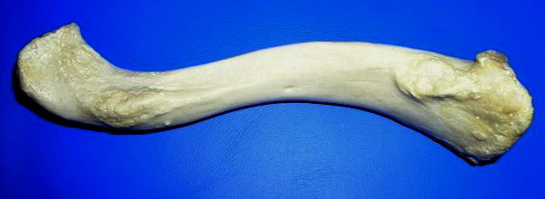
The clavicle, or collarbone, is a slender, S-shaped long bone approximately 6 inches (15 cm) long that serves as a strut between the shoulder blade and the sternum (breastbone). There are two clavicles, one on the left and one on the right. The clavicle is the only long bone in the body that lies horizontally. Together with the shoulder blade, it makes up the shoulder girdle. It is a palpable bone and, in people who have less fat in this region, the location of the bone is clearly visible. It receives its name from the Latin ''clavicula'' ("little key"), because the bone rotates along its axis like a key when the shoulder is abducted. The clavicle is the most commonly fractured bone. It can easily be fractured by impacts to the shoulder from the force of falling on outstretched arms or by a direct hit. Structure The collarbone is a thin doubly curved long bone that connects the arm to the trunk of the body. Located directly above the first rib, it acts as a strut t ... [...More Info...] [...Related Items...] OR: [Wikipedia] [Google] [Baidu] |
Radial Head
The head of the radius has a cylindrical form, and on its upper surface is a shallow cup or fovea for articulation with the capitulum of the humerus. The circumference of the head is smooth; it is broad medially where it articulates with the radial notch of the ulna, narrow in the rest of its extent, which is embraced by the annular ligament.'' Gray's Anatomy'' (1918), see infobox Articular surfaces The head of the radius is shaped to articulate with a complex of articular surfaces during both flexion-extension at the elbow and supination-pronation in the forearm: Humeroradial joint The head's proximal surface is concave and cup-shaped to correspond to the spherical surface of the capitulum of the humerus. The radius can thus glide on the capitulum during elbow flexion-extension while simultaneously rotate about its own main axis during supination-pronation. Between the capitulum and the trochlea of the humerus is the capitulotrochlear groove. A semi-lunar surface around th ... [...More Info...] [...Related Items...] OR: [Wikipedia] [Google] [Baidu] |
Occipital Bone
The occipital bone () is a cranial dermal bone and the main bone of the occiput (back and lower part of the skull). It is trapezoidal in shape and curved on itself like a shallow dish. The occipital bone overlies the occipital lobes of the cerebrum. At the base of skull in the occipital bone, there is a large oval opening called the foramen magnum, which allows the passage of the spinal cord. Like the other cranial bones, it is classed as a flat bone. Due to its many attachments and features, the occipital bone is described in terms of separate parts. From its front to the back is the basilar part, also called the basioccipital, at the sides of the foramen magnum are the lateral parts, also called the exoccipitals, and the back is named as the squamous part. The basilar part is a thick, somewhat quadrilateral piece in front of the foramen magnum and directed towards the pharynx. The squamous part is the curved, expanded plate behind the foramen magnum and is the largest par ... [...More Info...] [...Related Items...] OR: [Wikipedia] [Google] [Baidu] |


