|
OPN1LW
OPN1LW is a gene on the X chromosome that encodes for long wave sensitive (LWS) opsin, or red cone photopigment. The OPN1LW gene provides instructions for making an opsin pigment that is more sensitive to light in the yellow/orange part of the visible spectrum (long-wavelength light). The gene contains 6 exons with variability that induces shifts in the spectral range. OPN1LW is subject to homologous recombination with OPN1MW, as the two have very similar sequences. These recombinations can lead to various vision problems, such as red-green colourblindness and blue monochromacy. The protein encoded is a G-protein coupled receptor with embedded 11-''cis''-retinal, whose light excitation causes a cis-trans conformational change that begins the process of chemical signalling to the brain. It is responsible for perception of visible light in the yellow-green range on the visible spectrum (around 500-570nm). Gene OPN1LW produces red-sensitive opsin, while its counterparts, OPN1MW ... [...More Info...] [...Related Items...] OR: [Wikipedia] [Google] [Baidu] |
Opsin
Animal opsins are G-protein-coupled receptors and a group of proteins made light-sensitive via a chromophore, typically retinal. When bound to retinal, opsins become retinylidene proteins, but are usually still called opsins regardless. Most prominently, they are found in photoreceptor cells of the retina. Five classical groups of opsins are involved in Visual perception, vision, mediating the conversion of a photon of light into an electrochemical signal, the first step in the Visual phototransduction, visual transduction cascade. Another opsin found in the mammalian retina, melanopsin, is involved in circadian rhythms and Pupillary light reflex, pupillary reflex but not in vision. Humans have in total nine opsins. Beside vision and light perception, opsins may also sense temperature, sound, or chemicals. Structure and function Animal opsins detect light and are the molecules that allow us to see. Opsins are G-protein-coupled receptors (GPCRs), which are chemoreceptors and hav ... [...More Info...] [...Related Items...] OR: [Wikipedia] [Google] [Baidu] |
Cone Cell
Cone cells or cones are photoreceptor cells in the retina of the vertebrate eye. Cones are active in daylight conditions and enable photopic vision, as opposed to rod cells, which are active in dim light and enable scotopic vision. Most vertebrates (including humans) have several classes of cones, each sensitive to a different part of the visible spectrum of light. The comparison of the responses of different cone cell classes enables color vision. There are about six to seven million cones in a human eye (vs ~92 million rods), with the highest concentration occurring towards the macula and most densely packed in the fovea centralis, a diameter rod-free area with very thin, densely packed cones. Conversely, like rods, they are absent from the optic disc, contributing to the blind spot. Cones are less sensitive to light than the rod cells in the retina (which support vision at low light levels), but allow the perception of color. They are also able to perceive finer ... [...More Info...] [...Related Items...] OR: [Wikipedia] [Google] [Baidu] |
OPN1MW
Green-sensitive opsin is a protein that in humans is encoded by the ''OPN1MW'' gene. OPN1MW2 is a similar opsin. The OPN1MW gene provides instructions for making an opsin pigment that is more sensitive to light in the middle of the visible spectrum (yellow/green light). See also * Opsin * OPN1LW OPN1LW is a gene on the X chromosome that encodes for long wave sensitive (LWS) opsin, or red cone photopigment. The OPN1LW gene provides instructions for making an opsin pigment that is more sensitive to light in the yellow/orange part of the vis ... References Further reading * * * * * * * * * * * * * External links GeneReviews/NIH/NCBI/UW entry on Red-Green Color Vision Defects G protein-coupled receptors Color vision {{transmembranereceptor-stub ... [...More Info...] [...Related Items...] OR: [Wikipedia] [Google] [Baidu] |
11-cis Retinal
Retinal (also known as retinaldehyde) is a polyene chromophore. Retinal, bound to proteins called opsins, is the chemical basis of visual phototransduction, the light-detection stage of visual perception (vision). Some microorganisms use retinal to convert light into metabolic energy. One study suggests that approximately three billion years ago, most living organisms on Earth used retinal, rather than chlorophyll, to convert sunlight into energy. Because retinal absorbs mostly green light and transmits purple light, this gave rise to the Purple Earth hypothesis. Retinal itself is considered to be a form of vitamin A when eaten by an animal. There are many forms of vitamin A, all of which are converted to retinal, which cannot be made without them. The number of different molecules that can be converted to retinal varies from species to species. Retinal was originally called retinene, and was renamed after it was discovered to be vitamin A aldehyde. Vertebrate animals ingest ... [...More Info...] [...Related Items...] OR: [Wikipedia] [Google] [Baidu] |
OPN1SW
Blue-sensitive opsin is a protein that in humans is encoded by the ''OPN1SW'' gene. The OPN1SW gene provides instructions for making a protein that is essential for normal color vision. This protein is found in the retina, which is the light-sensitive tissue at the back of the eye. The OPN1SW gene provides instructions for making an opsin pigment that is more sensitive to light in the blue/violet part of the visible spectrum (short-wavelength light). Cones with this pigment are called short-wavelength-sensitive or S cones. In response to light, the photopigment triggers a series of chemical reactions within an S cone. These reactions ultimately alter the cell's electrical charge, generating a signal that is transmitted to the brain. The brain combines input from all three types of cones to produce normal color vision. See also * Opsin Animal opsins are G-protein-coupled receptors and a group of proteins made light-sensitive via a chromophore, typically retinal. When bound to r ... [...More Info...] [...Related Items...] OR: [Wikipedia] [Google] [Baidu] |
X Chromosome
The X chromosome is one of the two sex chromosomes in many organisms, including mammals, and is found in both males and females. It is a part of the XY sex-determination system and XO sex-determination system. The X chromosome was named for its unique properties by early researchers, which resulted in the naming of its counterpart Y chromosome, for the next letter in the alphabet, following its subsequent discovery. Discovery It was first noted that the X chromosome was special in 1890 by Hermann Henking in Leipzig. Henking was studying the testicles of '' Pyrrhocoris'' and noticed that one chromosome did not take part in meiosis. Chromosomes are so named because of their ability to take up staining (''chroma'' in Greek means ''color''). Although the X chromosome could be stained just as well as the others, Henking was unsure whether it was a different class of the object and consequently named it ''X element'', which later became X chromosome after it was established that it w ... [...More Info...] [...Related Items...] OR: [Wikipedia] [Google] [Baidu] |
Locus Control Region
A locus control region (LCR) is a long-range cis-regulatory element that enhances expression of linked genes at distal chromatin sites. It functions in a copy number-dependent manner and is tissue-specific, as seen in the selective expression of β-globin genes in erythroid cells. Expression levels of genes can be modified by the LCR and gene-proximal elements, such as promoters, enhancers, and silencers. The LCR functions by recruiting chromatin-modifying, coactivator, and transcription complexes. Its sequence is conserved in many vertebrates, and conservation of specific sites may suggest importance in function. It has been compared to a super-enhancer as both perform long-range ''cis'' regulation via recruitment of the transcription complex. History The β-globin LCR was identified over 20 years ago in studies of transgenic mice. These studies determined that the LCR was required for normal regulation of beta-globin gene expression. Evidence of the presence of this add ... [...More Info...] [...Related Items...] OR: [Wikipedia] [Google] [Baidu] |
Pi Bond
In chemistry, pi bonds (π bonds) are covalent chemical bonds, in each of which two lobes of an orbital on one atom overlap with two lobes of an orbital on another atom, and in which this overlap occurs laterally. Each of these atomic orbitals has an electron density of zero at a shared nodal plane that passes through the two bonded nuclei. This plane also is a nodal plane for the molecular orbital of the pi bond. Pi bonds can form in double and triple bonds but do not form in single bonds in most cases. The Greek letter π in their name refers to p orbitals, since the orbital symmetry of the pi bond is the same as that of the p orbital when seen down the bond axis. One common form of this sort of bonding involves p orbitals themselves, though d orbitals also engage in pi bonding. This latter mode forms part of the basis for metal-metal multiple bonding. Properties Pi bonds are usually weaker than sigma bonds. The C–C double bond, composed of one sigma and o ... [...More Info...] [...Related Items...] OR: [Wikipedia] [Google] [Baidu] |
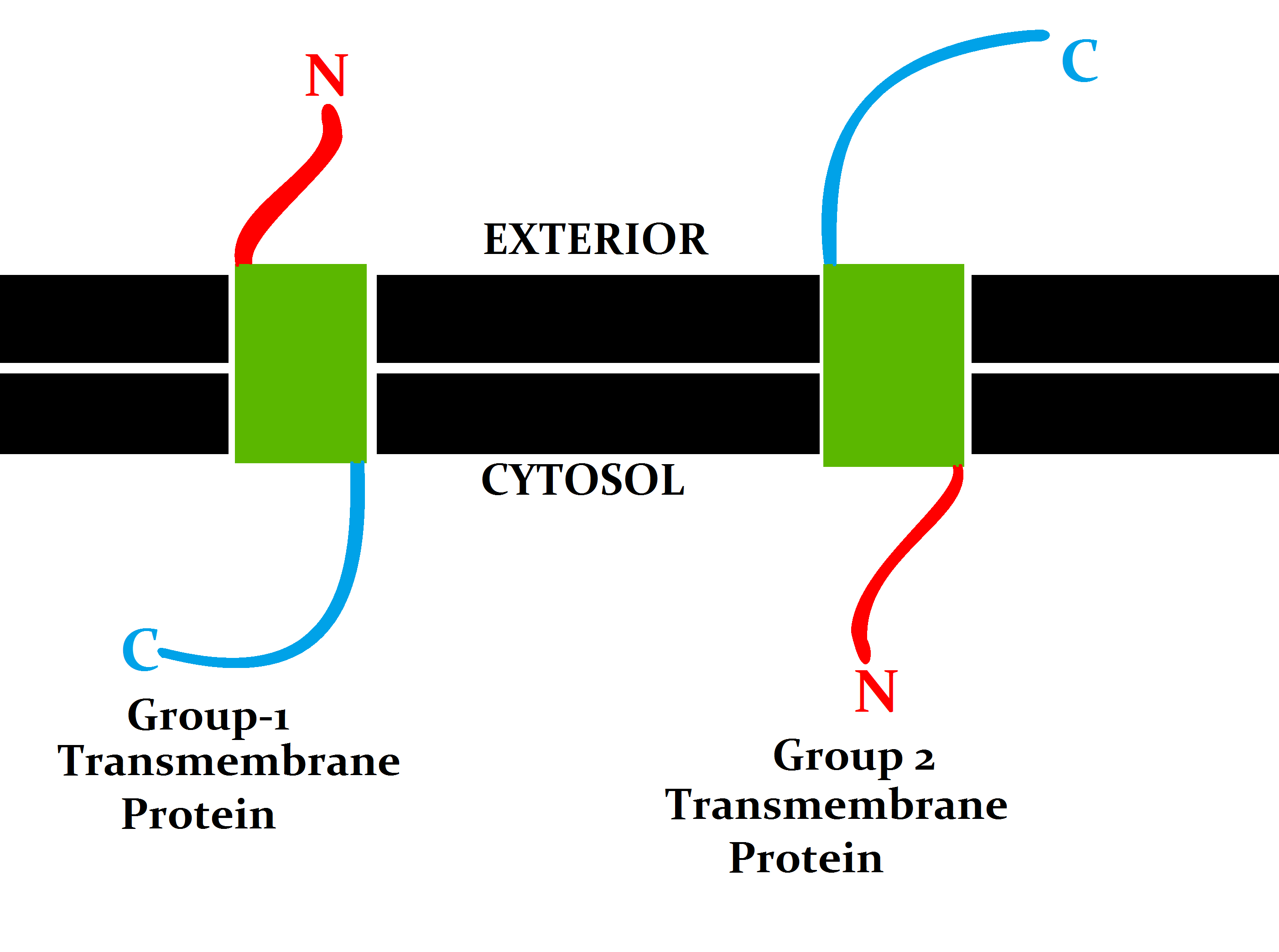
Transmembrane Protein
A transmembrane protein is a type of integral membrane protein that spans the entirety of the cell membrane. Many transmembrane proteins function as gateways to permit the transport of specific substances across the membrane. They frequently undergo significant conformational changes to move a substance through the membrane. They are usually highly hydrophobic and aggregate and precipitate in water. They require detergents or nonpolar solvents for extraction, although some of them ( beta-barrels) can be also extracted using denaturing agents. The peptide sequence that spans the membrane, or the transmembrane segment, is largely hydrophobic and can be visualized using the hydropathy plot. Depending on the number of transmembrane segments, transmembrane proteins can be classified as single-pass membrane proteins, or as multipass membrane proteins. Some other integral membrane proteins are called monotopic, meaning that they are also permanently attached to the membrane, b ... [...More Info...] [...Related Items...] OR: [Wikipedia] [Google] [Baidu] |
Visible Spectrum
The visible spectrum is the spectral band, band of the electromagnetic spectrum that is visual perception, visible to the human eye. Electromagnetic radiation in this range of wavelengths is called ''visible light'' (or simply light). The optical spectrum is sometimes considered to be the same as the visible spectrum, but some authors define the term more broadly, to include the ultraviolet and infrared parts of the electromagnetic spectrum as well, known collectively as ''optical radiation''. A typical human eye will respond to wavelengths from about 380 to about 750 nanometers. In terms of frequency, this corresponds to a band in the vicinity of 400–790 Terahertz (unit), terahertz. These boundaries are not sharply defined and may vary per individual. Under optimal conditions, these limits of human perception can extend to 310 nm (ultraviolet) and 1100 nm (near infrared). The spectrum does not contain all the colors that the human visual system can distinguish. ... [...More Info...] [...Related Items...] OR: [Wikipedia] [Google] [Baidu] |
Chromophore
A chromophore is the part of a molecule responsible for its color. The word is derived . The color that is seen by our eyes is that of the light not Absorption (electromagnetic radiation), absorbed by the reflecting object within a certain wavelength spectrum of visible spectrum, visible light. The chromophore is a region in the molecule where the energy difference between two separate molecular orbitals falls within the range of the visible spectrum (or in informal contexts, the spectrum under scrutiny). Visible light that hits the chromophore can thus be absorbed by exciting an electron from its ground state into an excited state. In biological molecules that serve to capture or detect light energy, the chromophore is the Moiety (chemistry), moiety that causes a conformational change in the molecule when hit by light. Conjugated pi-bond system chromophores Just like how two adjacent p-orbitals in a molecule will form a pi-bond, three or more adjacent p-orbitals in a molec ... [...More Info...] [...Related Items...] OR: [Wikipedia] [Google] [Baidu] |
Photopic Vision
Photopic vision is the vision of the eye under well-lit conditions (luminance levels from 10 to 108 cd/m2). In humans and many other animals, photopic vision allows color perception, mediated by cone cells, and a significantly higher visual acuity and temporal resolution than available with scotopic vision. The human eye uses three types of cones to sense light in three bands of color. The biological pigments of the cones have maximum absorption values at wavelengths of about 420 nm (blue), 534 nm (bluish-green), and 564 nm (yellowish-green). The color of the pure signal of the cones could be described as violet, blue-green, and scarlet red, respectively, but, in their wavelengths of maximum absorption other cones are activated as well. The sensitivity ranges of the conecells overlap to provide vision throughout the visible spectrum. The maximum efficacy is 683 lm/W at a wavelength of 555 nm (green). By definition, light at a frequency of hertz ha ... [...More Info...] [...Related Items...] OR: [Wikipedia] [Google] [Baidu] |


